Case Report - (2022) Volume 10, Issue 9
A Case Report on Non-Union of Lateral Condyle of Humerus Treated with Screw Fixation
Bageshree Ajit Oak*, Vijay Narasimman Reddy and Murali Krishnan
*Correspondence: Bageshree Ajit Oak, Department of Orthopedics, Sree Balaji Medical College and Hospital, India, Email:
Abstract
Lateral condyle fracture of the humerus is the most common elbow fracture that involves the growth plate and the second most common elbow fracture in children following supracondylar fracture of the humerus. These fractures are prone to go for non-union. We report a case of a 7-year-old girl who suffered a left lateral condylar fracture nonunion and gross valgus deformity. Fixation of fracture was performed by a native method 4 years ago. Now presented with limited range of motion (ROM) of the left elbow caused by the union of the lateral condylar fracture and subluxation of the radio humeral joint with gross genu valgus deformity. Surgical debridement of non-union site, the bone ends freshened, condyle fragment apposed in position, bone grafting done, and two cannulated cancellous screw fixations done. Implant excision was done 15 months later.
Keywords
Bageshree Ajit Oak, Vijay Narasimman Reddy, Murali Krishnan, A Case Report on Non-Union of Lateral Condyle of Humerus Treated with Screw Fixation, J Res Med Dent Sci, 2022, 10 (9):219-221.
Introduction
Fractures of the lateral condyle fracture (LCF) of the humerus are the second most common elbow fracture in children following supracondylar fracture of the humerus and account for 10–20% of pediatric elbow fractures [1,2]. This type of fracture is neglected many a time due to parents/clinician negligence [3]. Incorrectly treated lateral physical injury may remain unnoticed until months or years after the initial injury [4]. LCF is known for complications such as nonunion, ulnar nerve palsy, hypertrophic scar, avascular necrosis of the ossific nucleus, malunion, and angular deformity [5,6]. Lateral condyle fractures are classified by Milch classification [7] and also depending on displacement. When these fractures, especially Milch type 2 and displaced fractures, are not treated appropriately, they are likely to end up in complications like non-union, cubitus valgus with increased carrying angle, with or without tardy ulnar nerve palsy.
Case Report
A 7-year-old girl reported to the outpatient department with her mother and gave a history of slip and fall on her outstretched hand 4 years before the date of visit. She had taken native splinting in the form of immobilization with a bamboo stick. The patient’s mother noticed some deformity and came with complaints of gross progressive outward bowing (valgus deformity) of the left elbow. The patient did not have any neurological complaints or pain.
On examination, the patient had an increased carrying angle with gross valgus deformity, with lateral condyle thickened and irregular. No muscle wasting was identified. The three-point bony relation was disturbed, with the widening of intercondylar distance and with the migration of lateral epicondyle proximally. Movements at the elbow joint like flexion, extension, pronation, and supination were full and free. She did not have signs of distal neurovascular deficit.
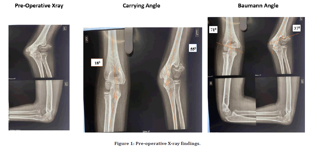
Her X-ray of the Left elbow showed non-union of the lateral humeral condyle with proximal migration of the lateral condyle. Her carrying angle on the affected side was 550 as compared to 180 on the normal side and Baumann’s angle on the affected side was 370 as compared to 710. The normal value of the Baumann angle ranges from 610-800, anything greater than this is concerning for the fracture with displacement. We diagnosed the nonunion lateral condyle of the humerus with gross valgus deformity (Figure 1).

Figure 1. Pre-operative X-ray findings.
Surgical management
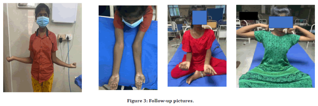
Under general anesthesia, the patient was placed supine on a radiolucent table. The affected limb was then prepped and draped free. Kocher’s approach was used in all the patients. The fracture was reduced and under C-arm, the guide wire was placed perpendicular to the fracture line. With the confirmation of proper alignment, two 4 mm screws were passed, one for lateral condyle fracture compression and another advancing from medial condyle to lateral condyle to support holding the fracture fragments. The patient was advised to do implant exit immediately after 8 months, as the valgus deformity and fracture site healing was adequate at 8 months (130, which is near normal) but due to the patient’s negligence and delay the valgus deformity again reappeared due to bone growth at 15 months (valgus angle 300) and then implant exit was done at 15 months. Functionally the patient does not have any problem as of now, but valgus deformity is present. The patient was advised to follow up every 6 months and corrective osteotomy can be done in case of severe deformity along with or without ulnar nerve involvement after attainment of skeletal maturity (Figures 2 and Figure 3).

Figure 2. Follow-up X-ray findings.

Figure 3. Follow-up pictures.
Discussion
Fracture of the lateral condyle of the humerus in children that do not unite after 12 weeks following injury, is considered as nonunion of the lateral condyle [8]. There are multiple causes of nonunion, it primarily occurs due to the pull of the supinator, long extensors of the wrist and fingers, and radial collateral ligaments. Other reasons include inadequate stabilization or fixation, failure to recognize the fracture, articular liquid penetration of the fracture site, and minor blood supply of fractured fragments because of vessels penetrating from the metaphysis, insufficient immobilization period, and fracture relocation [8]. Although LCF in children is very common, there are many reasons for its delayed presentation like lack of awareness of the parents, financial constraints, and health care facilities not available, fractures being managed by osteopaths [9]. Fracture non-union of lateral condyle of the humerus, pose a challenge to the surgeon in terms of selection of treatment modality. Operative treatment is considered for displaced fracture >2mm. The commonly used implants for fixation are Kirschner wire (K-wires) and cannulated screws (CS). The advantage of screw over K-wire is rotational stability, interfragmentary compression at the fracture site, prevents secondary fracture displacement, decreases consolidation time, and the risk of valgus deformity. Screw fixation provides superior fixation compared to pin fixation, and lower rates of lateral overgrowth, fixation loss, and infection [10,11].
Conclusion
Non-union of lateral condyle fractures of the humerus is usually associated with gross lateral condyle displacement. In such a stable elbow with non-union of lateral condyle fracture without ulnar nerve palsy, can be treated successfully by open reduction and internal fixation with cancellous screw with excellent results. Screw provides absolute stability which reduces the possibility of lateral prominence and promotes early fracture union. Absolute stability of fracture permits an early range of motion with an early return to a pre-injury state.
References
- Mizuta T, Benson WM, Foster BK, et al. Statistical analysis of the incidence of physeal injuries. J Pediatr Orthop 1987; 7:518-523.
- Jakob R, Fowles JV, Rang M, et al. Observations concerning fractures of the lateral humeral condyle in children. J Bone Joint Surg Br 1997; 57:430-436.
- Landin LA, Danielsson LG. Elbow fractures in children. An epidemiological analysis of 589 cases. Acta Orthop Scand 1986; 57:309-312.
- Beaty JH, Kasser JR. Rockwood and wilkins’ fractures in children. Philadelphia: Lippincott Williams and Wilkins; 2006.
- Skak SV, Olsen SD, Smaabrekke A. Deformity after fracture of the lateral humeral condyle in children. J Pediatr Orthop B 2001;10:142-152
- Hasler CC, von Laer L. Prevention of growth disturbances after fractures of the lateral humeral condyle in children. J Pediatr Orthop 2001; 10:123-130.
- Milch H. Frcature and fracture dislocations of humeral condyles. J Trauma 1964; 4:592.
- Singh KA, Shah H. Nonunion of the lateral condyle of humerus. J Orthop Assoc South Indian States 2022; 19:S51-856.
- Saraf SK, Khare GN. Late presentation of fractures of the lateral condyle of the humerus in children. Indian J Orthop 2011; 45:39-44.
- Loke WP, Shukur MH, Yeap JK. Screw osteosynthesis of displaced lateral humeral condyle fractures in children: A mid-term review. Med J Malaysia 2006; 61:40-44.
- Laer LV. Screw fixation of lateral condyle fractures of the humerus in children. Orthop Traumatol 1993; 2:29-35.
Indexed at, Google Scholar, Cross Ref
Indexed at, Google Scholar, Cross Ref
Indexed at, Google Scholar, Cross Ref
Indexed at, Google Scholar, Cross Ref
Indexed at, Google Scholar, Cross Ref
Indexed at, Google Scholar, Cross Ref
Indexed at, Google Scholar, Cross Ref
Author Info
Bageshree Ajit Oak*, Vijay Narasimman Reddy and Murali Krishnan
Department of Orthopedics, Sree Balaji Medical College and Hospital, Chennai, IndiaReceived: 30-Aug-2022, Manuscript No. jrmds-22-73341; , Pre QC No. jrmds-22-73341(PQ); Editor assigned: 01-Sep-2022, Pre QC No. jrmds-22-73341(PQ); Reviewed: 16-Sep-2022, QC No. jrmds-22-73341(Q); Revised: 21-Sep-2022, Manuscript No. jrmds-22-73341(R); Published: 28-Sep-2022
