Research - (2021) Volume 9, Issue 6
A Prospective and Comparative Study of Functional Outcome of Anterior Cruciate Ligament Repair Using Quadriceps and Hamstrings Tendon Grafts
Battu Dinesh Chandra and Madhukar*
*Correspondence: Madhukar, Department of Orthopaedics, Sree Balaji Medical College & Hospital Affiliated to Bharath Institute of Higher Education and Research, Chennai, Tamil Nadu, India, Email:
Abstract
The purpose of the study is to evaluate the functional outcome of arthroscopic anterior cruciate ligament reconstruction using hamstring tendon (Gracilis and semitendinosus) autograft versus Quadriceps tendon graft in individuals with ACL injuries. Arthroscopic reconstruction of the injured ACL has become the gold standard. Open reconstruction of ACL which was done earlier is not practised nowadays due to the complications associated such as increased post op pain, stiffness, and a lengthy rehabilitation phase. The “ideal graft” for ACL reconstruction is still a topic of debate. The most used grafts are Quadriceps tendon graft, bone patellar tendon bone graft and hamstring graft. Many studies have demonstrated comparable functional outcomes for different grafts.
Keywords
Anterior cruciate ligament (ACL), Arthroscope, Bone graft, Osteoarthritis, Articular cartilage, Anteromedial portal
Introduction
The knee joint is one of the body's most complicated joint. There is an increase in the occurrence of knee ligament injuries due to the ever- increasing road traffic accidents and increased involvement in sports activities. Knee joint has proximal femur bone distally tibia and fibula bone with ligaments and capsules, meniscus, and bursa. Important ligaments are Anterior cruciate ligament (ACL), Posterior cruciate Ligament (PCL), Medial collateral Ligament (MCL), Lateral collateral Ligament (LCL). The ACL together with other ligaments, capsule is the primary knee stabilizer and prevents anterior translation, and limits valgus and rotational stress to some extent.
The signs of knee instability, discomfort, and a decrease in joint function arise when an ACL injury occurs. Even though patients with less expected knee score can be treated with conservative treatment with intensive physiotherapy, bracing and lifestyle modification can be tried in symptomatic young active individuals, ACL reconstruction is necessary. Also ACL injuries are mostly associated with injury of meniscus which can to be addressed, else person can develop early onset of osteoarthritis of knee [1-19].
Mechanism of injury
The common modes of injury of ACL are
Direct contact (30%).
Non-contact (70%).
Women are more susceptible to ACL injury than men. This may be due to
- Women have a smaller intercondylar notch.
- Lesser strength and smaller size of ACL in females.
- Females have a greater Q angle compared with males.
- Hormonal factors may cause laxity of ligaments.
- Neuromuscular risk factors.
- The commonest mode of injury is non-contact deceleration with valgus and twisting movement.
Physical examination
This includes inspection, palpation, measurements, and movements of the knee joint. Then the tests for cruciate ligaments, collateral ligaments and menisci are done which help in diagnosis and further plan of management.
Materials and Methods
This a prospective study conducted in Sree Balaji Medical College and Hospital, Chromepet, Chennai from august 2017 to September 2019. There were 50 patients included in our study of which 36 patients (72%) were male and 14 (28%) were female.36 patients were operated with hamstring graft and 14 were operated with quadriceps tendon graft. The patients were followed up for an average duration of 17.6 months with minimum follow up of 7 months and maximum follow up of 18 months.
All young and middle-aged patients presenting with unilateral knee complaints and history of trauma to the knee in the orthopaedic emergency and outpatient departments in Sree Balaji Medical College and Hospital, Chromepet, Chennai were evaluated by a thorough general and local examination, the uninjured knee was first examined in a stable patient and in the supine position to determine ligament excursions after which the affected knee was examined. Injuries to the associated structures were assessed by performing the following clinical tests.
- Valgus/ varus stress test (for collateral ligaments).
- McMurray’s test/ Apley grinding test (for menisci).
- Posterior drawer test (for posterior cruciate ligament).
Routine radiographs of both knees in standing position in anteroposterior view and lateral view of the affected knee were taken. MRI of the knee was done in all ACL torn cases for confirmation.
Inclusion criteria
- Clinical /MRI evidence of symptomatic individuals with anterior cruciate ligament insufficiency.
- Patients between age 20 to 40 (skeletally matured patients).
- Associated with medial or lateral meniscus tear.
- Associated Grade I and II MCL and LCL injuries.
- No history of previous surgery in the knee.
- A normal contralateral knee.
Exclusion criteria
- Patients with systemic diseases compromising their pre- anaesthetic fitness.
- Associated with PCL tear.
- Associated Grade III MCL and LCL injuries.
- Patient with osteoarthritic knee.
- Patients with associated fracture of tibial plateau.
- Patients with local skin infections.
Pre-operative work up
Patients with ACL tear proven clinically and radiologically. Routine investigations like haemoglobin, total and differential counts, platelet count, chest X ray, ECG were taken and anaesthetic assessment for regional and general anesthesia was done.
Pre-operative rehabilitation
Pre-operative strength and range of movement of knee joint were measured and documented.
• Static and dynamic quadriceps and hamstrings exercise were taught to patients while awaiting surgery.
• All patients were enlightened on post–operative rehabilitation.
Consent
All patient in this study were explained about the injury, diagnosis, various management options, complication of non - operative treatment and operative management, per-operative and post-operative complications, donor site morbidity, injury to surrounding structures, infection, compartment syndrome, anaesthesia risks, post - operative knee pain, restriction of range of motion.
Surgical technique
The technique of single bundle ACL reconstruction was done with one tibial tunnel and one femoral tunnel with their centres corresponding to the centre of the native ACL tibial and femoral attachment sites respectively. The femoral tunnel was made using the anteromedial portal thereby creating an anatomic femoral tunnel position. The graft was fixed at the tibial side using bioscrew / titanium interference screw or endobutton and at the femoral side using end button.
Diagnostic arthroscopy
Before the harvesting of graft, diagnostic arthroscopy was done first. In 90 degrees of knee flexion, anterolateral port (viewing portal) is made using 11 blade at the level of inferior pole of patella just lateral to the patellar tendon.
Hamstring graft harvest and graft preparation
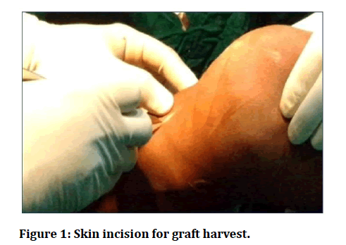
A 3cm oblique skin incision is made starting 5 cm below the medial joint line and 1 cm medial to the tibial tuberosity. The oblique incision is preferred because it gives a wider exposure of pes anserinus and there is less chance of injury to the infrapatellar branch of the saphenous nerve. It is planned to do the graft harvest and tibial tunnel drilling through the same incision.
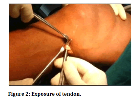
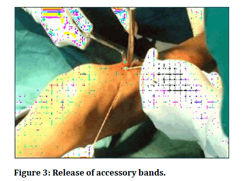
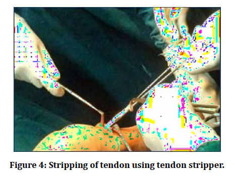
The knee is placed in 90 degrees of flexion and proximal dissection of the tendons were done using blunt dissection by fingers till the musculotendinous junction thereby releasing adhesions and accessory bands, while continuous traction was being applied through the threads.
The main band which connects the medial head of gastrocnemius is usually cut with the help of scissors. It is confirmed that as the tendon is pulled distally, there should be no dimpling posteriorly over the gastrocnemius.
The graft length to be placed inside the femoral tunnel is marked to ensure correct placement of graft within the femoral tunnel while being viewed arthroscopically.
The loop of the strand graft is tied to the posts in the graftmaster board and pretensioning is done by applying a pressure of about 15 pounds for around fifteen minutes (Figures 1 to Figure 4).
Figure 1: Skin incision for graft harvest.
Figure 2: Exposure of tendon.
Figure 3: Release of accessory bands.
Figure 4: Stripping of tendon using tendon stripper.
Evaluation
All patients were subjected to post operative anteroposterior and lateral radiographs to determine the tunnel placement and position of endobutton and interference screw. Patients were followed at 6 weeks, 6 months and 1 year and functional outcomes assessed.
The International Knee Documentation 2000 score(IKDC) and Lysholm and Gillquist Knee Scoring Scale were used for evaluation of patients.
The Subjective IKDC scale was evaluated by summing the scores for the individual items and then transforming the score to a scale that ranges from 0 to 100. To calculate the final subjective IKDC score simply add the score of each item and divide by the maximum possible score which was 87.
Subjective IKDC score = [Sum of items/Maximum possible score] × 100
The Lysholm and Gillquist Knee Scoring Scale consists of eight parameters for evaluation. The parameters evaluated are
- Limping.
- Aided walking.
- Episodes of knee locking.
- Knee instability.
- Knee pain.
- Knee swelling.
- Climbing of stairs.
- Squatting.
The individual parameters were allotted specific scores depending on the patient’s functional ability. The maximum possible knee score was.
Based on the outcome scores they were divided into Excellent, Good, Fair and Poor.
- Excellent–95–100.
- Good– 84–94.
- Fair– 65–83.
- Poor–64 or less.
Results
Fifty cases of arthroscopic ACL reconstruction were regularly followed for an average period of 17.6 months in Sree Balaji Medical College, Chrompet, Chennai. (From august 2017 to September 2017).
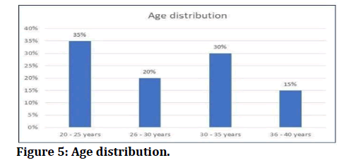
Most of the patients (35%) were in the age group of 20 to 25 years followed by 30% in the age group of 30 to 35 years (Table 1 and Figure 5).
Figure 5: Age distribution.
Table 1: Age distribution.
| Age(years) | Patients | Percentage |
|---|---|---|
| 20-25 years | 15 | 35% |
| 26-30 years | 18 | 20% |
| 30-35 years | 12 | 30% |
| 36-40 years | 5 | 15% |
| Total | 50 | 100% |
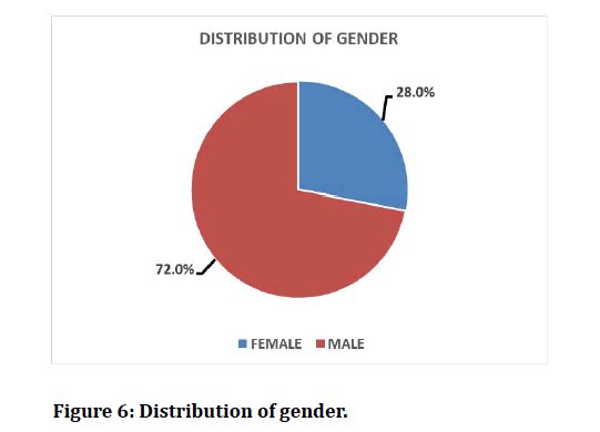
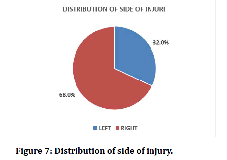
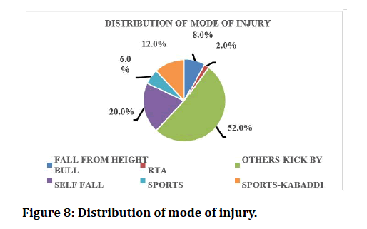
Of the 50 patients included in our study, 36 (72%) were Male patients and 14 (28%) were female (Table 2 and Figure 6). In this study, the right side was more commonly injured (68%) than the left side (32%) (Table 3 and Figure 7). he most common mode of injury in our study was Road Traffic Accidents (52%) followed by Self fall(20%), fall from height (8%) The other modes of injury in our study were self fall and kick by bull (Table 4 and Figure 8).
Figure 6: Distribution of gender.
Figure 7: Distribution of side of injury.
Figure 8: Distribution of mode of injury.
Table 2: Sex distribution.
| Gender | Number of patients | Percentage |
|---|---|---|
| Male | 36 | 72% |
| Female | 14 | 28% |
| Total | 50 | 100 |
Table 3: Side involvement.
| Side | Number of patients | Percentage |
|---|---|---|
| Right | 34 | 68% |
| Left | 16 | 32% |
| Total | 50 | 100% |
Table 4: Mode of Injury.
| Mode of injury | Distribution of mode of injury | Percentage |
|---|---|---|
| Fall from height | 4 | 8 |
| Others-kick by bull | 1 | 2 |
| Rta | 26 | 52 |
| Self-fall | 10 | 20 |
| Sports | 3 | 6 |
| Sports-kabaddi | 6 | 12 |
| Total | 50 | 100 |
Case Illustrations
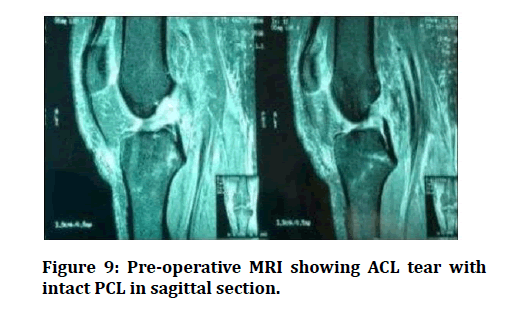
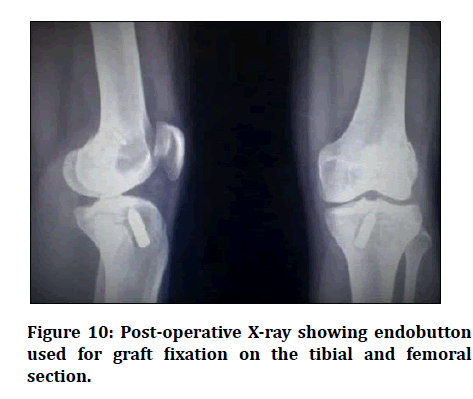
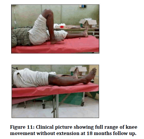
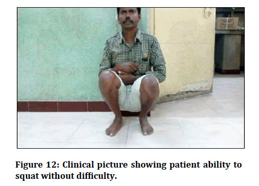
Case I: Figure 9 to Figure 12.
Figure 9: Pre-operative MRI showing ACL tear with intact PCL in sagittal section.
Figure 10: Post-operative X-ray showing endobutton used for graft fixation on the tibial and femoral section.
Figure 11: Clinical picture showing full range of knee movement without extension at 18 months follow up.
Figure 12: Clinical picture showing patient ability to squat without difficulty.
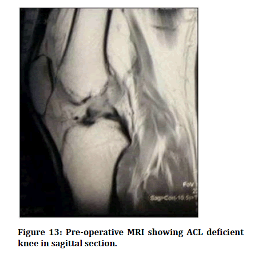
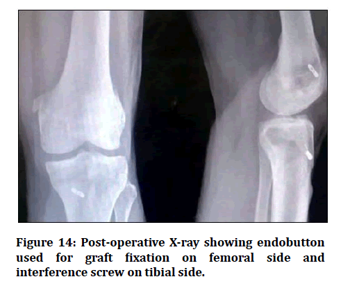
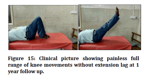
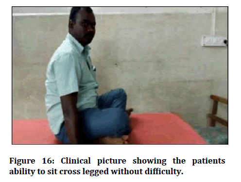
Case II: Figure 13 to Figure 16.
Figure 13: Pre-operative MRI showing ACL deficient knee in sagittal section.
Figure 14: Post-operative X-ray showing endobutton used for graft fixation on femoral side and interference screw on tibial side.
Figure 15: Clinical picture showing painless full range of knee movements without extension lag at 1 year follow up.
Figure 16: Clinical picture showing the patients ability to sit cross legged without difficulty.
Discussion
Arthroscopic reconstruction of the injured ACL has become the gold standard and is one of the most common procedures done in orthopaedics and thus it has been extensively studied and outcomes of ACL reconstruction have gained considerable attention.
The choice of graft is a topic of great debate in recent years. The various options include bone patellar tendon bone graft, hamstring auto- graft, quadriceps tendon, various synthetic grafts and allograft but the hamstring graft has been increasingly used in recent. The advantages of arthroscopic ACL reconstruction using hamstring graft include decreased surgical site morbidity, decreased occurrence of patellofemoral adhesions and reduced incidence of anterior knee pain. Though the semitendinosus tendon has only 75% and gracilis 49% of the strength of native ACL, the quadrupled semitendinosus or semitendinosus-gracilis have a greater tensile load.
Our study is to evaluate the functional outcome of arthroscopic anatomical single bundle ACL reconstruction using hamstring autograft versus quadriceps tendon graft. This prospective study was conducted in Sree Balaji Medical College and Hospital, Chrompet, Chennai to clinically evaluate the clinical results of arthroscopic ACL reconstruction between qudriceps tendon and hamstrings tendon graft. This study group comprised of 50 patients with follow up. Patients who went for ACL reconstruction with Hamstring graft are 36 and with quadriceps graft are 14 patients [20-36].
Conclusion
We conclude that ACL reconstruction gives good functional outcome with both Hamstrings and Quadriceps Graft. The post-operative functional scores improved significantly as compared to pre-operative scores. Both grafts are equally good and give good functional results. We could not find one graft being better over other. Both scores used in our study are subjective scores which limits the study design. The future scope of the study is by long-term follow up and to incorporate objective scoring system into the study design.
Funding
No funding sources.
Ethical Approval
The study was approved by the Institutional Ethics Committee.
Conflict of Interest
The authors declare no conflict of interest.
Acknowledgements
The encouragement and support from Bharath University, Chennai is gratefully acknowledged. For provided the laboratory facilities to carry out the research work.
References
- Simon D, Mascarenhas R, Saltzman BM, et al. The relationship between anterior cruciate ligament injury and osteoarthritis of the knee. Adv Orthop 2015; 2015.
- Brophy RH, Zeltser D, Wright RW, et al. Anterior cruciate ligament reconstruction and concomitant articular cartilage injury: Incidence and treatment. Arthroscopy 2010; 26:112–120.
- Galen C. On the usefulness of the parts of the body. Ithaca Cotnett University Press 1968.
- Bonnet A. Traite Des Maladies Articulaires-2nd Edn. Baillire, Paris 1853; 354-357.
- Stark J. Two cases of rupture of the crucial ligament of the knee-joint. Edinb Med Surg 1850; 74:267–271.
- Ashford WB, Kelly TH, Chapin RW, et al. Predicted quadriceps vs. quadrupled hamstring tendon graft sizing using 3-dimensional MRL. Knee 2018; 25: 1100-1
- Segond PF. Recherches cliniqueset experimentales sur les epanchementssanguins du genou par entorse. Prog med 1879; 16:297-421.
- Campbell WC. Repair of the ligaments of the knee: Report of an operation for the repair of the anterior cruciate ligament. Surg Gynecol Obstet 1936; 62:964-968.
- Macey HB. A new operative procedure for repair of ruptured cruciate ligament of the knee joint. Surg Gynecol Obstet 1939; 69:108-109.
- Jones KG. Reconstruction of the anterior cruciate ligament using the central one-third of the patellar ligament-A follow-up report. J Bone Join Surg 1970; 52A:1302-1308.
- Galway RD, Beaupre A, Macintosh DL. Pivot shift: A clinical sign of symptomatic anterior cruciate insufficiency. J Bone Join Surg 1972; 54B:763-764.
- Torg JS. Conrad W, Kalen V. Clinical diagnosis of anterior cruciate ligament instability in the athlete. Am J Sports Med 1976; 4:84-91.
- Rubin RM, Marshall JL, Wang J. Prevention of knee instability: Experimental model for prosthetic anterior cruciate ligament. Clin Orthop 1975; 113:212-236.
- Lipscomb AB, Johnston RK, Snyder RB. Evaluation of hamstring strength following use of semitendinosus and gracilis tendons to recon- struct the anterior cruciate ligament. Am J Sports Med 1982; 10:340- 342.
- Butler N. Grood Biomechanical analysis of human ligament grafts used in knee ligament repairs and reconstructions. J Bone Joint Surg 1983; 66:344-352.
- Friedman MJ. Arthroscopic semitendinosus (gracilis) reconstruction for anterior cruciate ligament deficiency. Techniques in Orthop 1988; 2:74-80.
- Paulos LE, Cherf J, Rosenberg TD. Anterior cruciate ligament reconstruction with autograft. Clin Sports Med 1991; 10:469-485.
- Sahelin AC, Weiler A. All-inside Anterior cruciate ligament reconstruction using semitendinosus tendon and soft thread-ed biodegradable interference screw fixation. Arthroscopy 1997; 13:773-779.
- Wertheiner BP. The effect of functional knee braces on strain on the anterior cruciate ligament in vivo. J Bone Joint Surg 1992; 74:1298-1312.
- Warren B. Arthrometric evaluation of knees that have a torn anterior cruciate ligament. J Bone Joint Surg 1990; 72:1299-1306.
- Zoltan. Acute tears of the anterior cruciate ligament. J Bone Joint Surg 1988; 70:1483-1488.
- Cyril F, Jackson W. Current concepts review. The science of reconstruction of the anterior cruciate ligament. J Bone Joint Surg 197; 79.
- Hamid Barzegar, Mohammadali Mohseni, Ali Sedighi, et al. Arthroscopically assisted vs. Open surgery in repairing anterior cruciate ligament avulsion. Pakistan J Biol Sci 2011; 14:496-501.
- Cameron SE, Wilson W, St Pierre P. A prospective, randomized comparison of open vs arthroscopically assisted ACL reconstruction. Orthopedics 1995; 18:249–252.
- Spindler KP, Kuhn JE, Freedman KB, et al. Anterior cruciate ligament reconstruction autograft choice: Bone-tendon-bone versus hamstring: does it really matter? A systematic review. Am J Sports Med 2004; 32:1986–1995.
- Wagner M, Kääb MJ, Schallock J, et al. Hamstring tendon versus patellar tendon anterior cruciate ligament reconstruction using biodegradable interference fit fixation: A prospective matched-group analysis. Am J Sports Med 2005; 33:1327-36.
- Kostov H, Kaftandziev I, Arsovski O, et al. Clinical outcomes of three different modes of femoral hamstring graft fixation in anterior cruciate ligament reconstruction. Macedonian Med Rev 2014; 68:44-47.
- Ma CB, Francis K, Towers J, et al. Hamstring anterior cruciate ligament reconstruction: A comparison of bioabsorbable interference screw and endobutton-post fixation. Arthroscopy 2004; 20:122–128.
- Colvin A, Sharma C, Parides M, et al. What is the best femoral fixation of hamstring autografts in anterior cruciate ligament reconstruction?: A meta-analysis. Clin Orthop Related Res 2011; 469:1075-1081.
- Shen C, Jiang SD, Jiang LS, et al. Bioabsorbable versus metallic interference screw fixation in anterior cruciate ligament reconstruction: A meta-analysis of randomized controlled trials. Arthroscopy 2010; 26:705–713.
- Kousa P, Järvinen TL, Vihavainen M, et al. The fixation strength of six hamstring tendon graft fixation devices in anterior cruciate ligament reconstruction: Part I: Femoral site. Am J Sports Med 2003; 31:174-181.
- Ma CB, Francis K, Towers J, et al. Hamstring anterior cruciate ligament reconstruction: A comparison of bioabsorbable interference screw and endobutton-post fixation. Arthroscopy: J Arthroscopic Related Surg 2004; 20:122-128.
- Musahl V, Plakseychuk A,Van Scyoc A, et al. Varying femoral tunnels between the anatomical footprint and isometric positions effect on kinematics of the anterior cruciate ligament–reconstructed knee. AJSM 2005; 33:712-8.
- Lim HC, Yoon YC, Wang JH, et al. Anatomical versus non-anatomical single bundle anterior cruciate ligament reconstruction: A cadaveric study of comparison of knee stability. Clin Orthop Surg 2012; 4:249.
- Meredick RB, Vance KJ, Appleby D, et al. Outcome of single-bundle versus double-bundle reconstruction of the anterior cruciate ligament: A meta-analysis. Am J Sports Med 2008; 36:1414-21.
- Ha JK, Lee DW, Kim JG. Single-bundle versus double-bundle anterior cruciate ligament reconstruction: A comparative study with propensity score matching. Indian J Orthop 2016; 50:505.
Author Info
Battu Dinesh Chandra and Madhukar*
Department of Orthopaedics, Sree Balaji Medical College & Hospital Affiliated to Bharath Institute of Higher Education and Research, Chennai, Tamil Nadu, IndiaCitation: Battu Dinesh Chandra, Madhukar, A Prospective and Comparative Study of Functional Outcome of Anterior Cruciate Ligament Repair Using Quadriceps and Hamstrings Tendon Grafts, J Res Med Dent Sci, 2021, 9(6): 70-77
Received: 22-Mar-2021 Accepted: 10-Jun-2021 Published: 17-Jun-2021

