Research - (2021) Volume 9, Issue 6
A Prospective Study of Diagnostic Laparoscopy in Patients Presenting with Chronic Abdominal Pain at SBMCH
Muzamil Ahmed A and Santhaseelan RG*
*Correspondence: Santhaseelan RG, Department of General Surgery, Sree Balaji Medical College & Hospital Affiliated to Bharath Institute of Higher Education and Research, Chennai, Tamil Nadu, India, Email:
Abstract
To evaluate the diagnostic and therapeutic value of laparoscopy in chronic abdominal pain. Patients with chronic abdominal pain, undiagnosed and with doubtful diagnosis by routine laboratory and imaging modality were enrolled in this study, their clinical presentation, intra operative findings, various occult etiology and clinical improvement were evaluated in this group, which will improve the awareness and importance of diagnostic laparoscopy among the surgeons. To correlate the laparoscopic findings with clinical and radiographic findings in all patients with chronic non-specific abdominal pain, the various unrevealed etiology for chronic abdominal pain and analyses the accuracy and efficacy of diagnostic laparoscopy in chronic abdominal pain. Studies show endometriosis found in 33% and pelvic adhesion in 24% of patients but no pathology was made out in 33% to 50% of the patient.
Keywords
Chronic abdominal pain, Laparoscopic, Pelvic adhesion, Pathology
Introduction
Chronic abdominal pain is a common complaint which is difficult to manage by both physician and surgeon. It is the 4th frequent cause of chronic pain syndrome in general population. worldwide. Although patients with chronic abdomen pain undergo numerous clinical & radiological diagnostic work up, definite diagnosis still remains a challenge to the surgeon. This affects the patients both physically and psychologically.
With the introduction of the diagnostic laparoscopy new tools has been added to our knowledge. Laparoscopy can identify abnormal findings and improve outcome in majority of the patients with chronic abdominal pain.
Diagnostic laparoscopy also helps the surgeon to decide whether any surgical management is mandatory and also to elicit the signs of inoperability in case of disseminated diseases such as advanced malignancy, tuberculosis etc in which the prodromal symptoms may be chronic abdominal pain.
This study is mainly designed to highlight the significance of laparoscopy in diagnosing the etiology of chronic abdominal pain , impact on the treatment and on postoperative pain relief. It also expresses diagnostic and therapeutic value of laparoscopy in chronic abdominal pain which is a most debilitating illness [1-13].
Materials and Methods
This study was conducted in patients presented with abdominal pain more than 3 months whose diagnosis was doubtful or could not be made by our routine physical, laboratory and imaging modalities. Between March 2017 and October 2018, a total number of 105 consecutive patients with chronic abdominal pain were enrolled in this prospective descriptive cross - sectional study. They were recruited from the outpatient clinic of General Surgery Department in Sree Balaji Medical College & Hospital, Chennai in the above said study period.
Inclusion criteria
Age between 15 and 65, Both males and females, Abdominal pain more than 3months.
Exclusion criteria
Known abdominal malignancy patient, Known psychiatric patient.
After getting consent from the patients, they were thoroughly interrogated and examined including per rectal and per vaginal examination and following investigations were done in all patients.
Complete haemogram with ESR, Blood Sugar, Blood Urea and Serum creatinine, Stool routine, microscopy and occult blood, Urine routine and culture, Plain X-ray abdomen, X-ray chest, Ultrasound abdomen and pelvis, CT abdomen and pelvis, Upper GI endoscopy, Colonoscopy, some patients were subjected to additional investigation according to symptoms, like Contrast gastrointestinal series, Serology for tuberculosis and Liver function test.
After undergoing thorough preoperative evaluation, their intensity of the pain was assessed by using the Verbal Rating Scale (VRS): the patient is asked to rate their pain on a five-point scale as "none, mild, moderate, severe or very severe". These patients were posted for diagnostic laparoscopy.
Results
Age and sex distribution
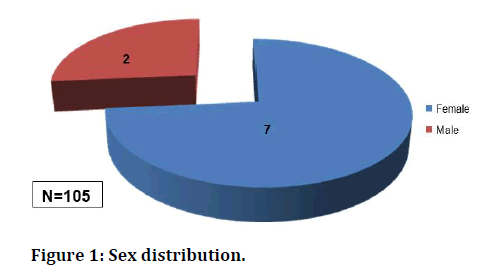
Most of them in the age group between 26 -35 in adult population (Table 1). Male Vs female ratio was in equal in study population. Females constitute more than thrice compared to male in study population (Table 2 and Figure 1).
Figure 1: Sex distribution.
Table 1: Distribution of age.
| Age | Patient |
|---|---|
| 15-25 | 33 |
| 26-35 | 38 |
| 36-45 | 14 |
| 46-55 | 11 |
| 56-65 | 9 |
| Total | 105 |
Mean age=34 years
Table 2: Sex distribution.
| Gender | Patient |
|---|---|
| Male | 27 |
| Female | 78 |
| Total | 105 |
Duration and site of pain
Mean=6 months (3-24 months). Most of the patients having pain duration around 3 months, not more than 2 years in our study.
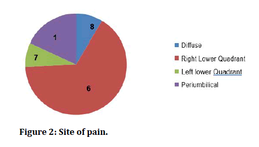
Most of the patients present with the right lower quadrant pain about 67%, particularly in the right iliac fossa region (Table 3 and Figure 2).
Figure 2: Site of pain.
Table 3: Site of pain.
| Site | Percentage |
|---|---|
| Diffuse | 9 (8%) |
| Right lower quadrant | 69(67%) |
| Left lower quadrant | 8 (7%) |
| Periumbilical | 19(18%) |
Intra operative findings
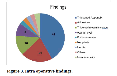
We found that appendicular pathology is the leading cause for chronic abdominal pain of unrevealed etiology and it is about 42%, followed by adhesions in about 21% (Table 4 and Figure 3).
Figure 3: Intra operative findings.
Table 4: Intra operative findings.
| Findings | Percentage |
|---|---|
| Thickened appendix | 44 (42%) |
| Adhesions | 22 (21%) |
| Enlarge mesentric nodes | 14 (13%) |
| Ovarian cyst | 9(8%) |
| Koch’s abdomen | 3(3%) |
| Neoplasia | 3 (3%) |
| Hernia | 3 (3%) |
| Others | 3(3%) |
| No abnormality | 4 (4%) |
Laparoscopic appendicectomy was done in all the patients with appendicular pathology like inflamed, thickened appendix and localized adhesion with caceum and abdominal wall. All the histopathological reports of appendix specimen showed chronic inflammation. Post operatively they recovered without any complication and all of them were pain free in the follow up of 1 month.
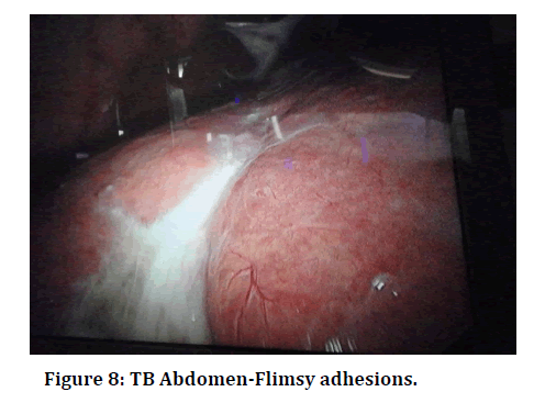
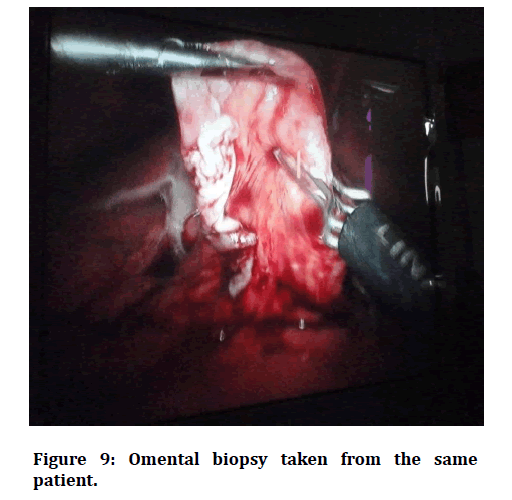
Adhesion was found in 21% (n=22), out of those 22 patients 13 patients had the history of previous surgery. Seven patients underwent LSCS (pfannenstiel scar) and other 6 had omental adhesions in midline scar. Omentum patients successfully. Other 9 patients who didn’t have the history of surgery, had the adhesion of omentum to the anterior abdominal wall with inter bowel loop adhesions, laparoscopic adhesiolysis was done in that patient successfully. Koch’s abdomen was diagnosed in 3% (n=3). Intra operative findings were multiple tubercles over the peritoneum, bowel and omentum. In one case we found that flimsy adhesion between the bowel loops and anterior abdominal wall. In all three cases minimal ascitic fluid was present. Omental and peritoneal biopsy was taken, ascitic fluid was also sent for biochemical analysis. The results confirm the tuberculous abdomen. They all were started anti tuberculous drug post operatively.
Malignancy was diagnosed in (n=3) 3% of the patient. One patient had ileo-caecal junction growth and laparoscopic right hemicolectomy was performed. Adnexal mass was found in two patients and hence salphingo- oopherectomy was performed in those two patients.
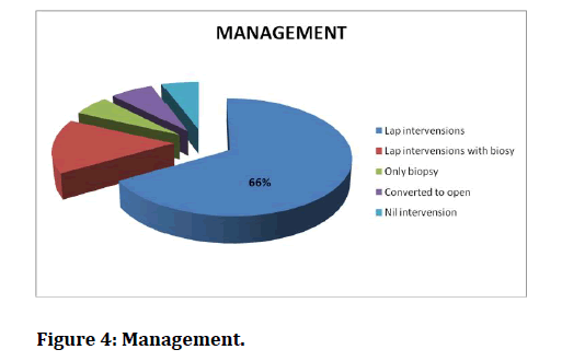
3% (n=3) of the patients had ventral hernia and underwent hernioplasty . All three patients had small defect in the paraumbilical region with omentum adherent to it. Mesh repair was done in all the three cases. Mean operating time for diagnostic laparoscopy alone is 30 minutes but it combined with therapeutic procedures it was 70 + 30 minutes. Table 5 and Figure 4 represents the management.was adherent to the anterior abdominal wall in the scar region. Laparoscopic adhesiolysis was performed in all
Figure 4: Management.
Table 5: Management.
| Lap interventions | 70(66%) |
| Lap intervention with Biopsy | 16 (15%) |
| Only biopsy | 6(6%) |
| Conversion into open | 7 (7%) |
| No interventions | 6 (6%) |
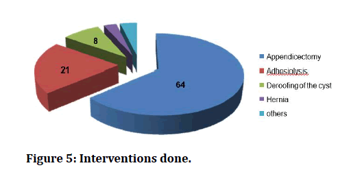
Therapeutic procedure was done in 81% (n=86) of the patients which includes appendicectomy in 42 % of patients, appendicectomy with mesenteric node biopsy in 10% of patients adhesiolysis in 22 %, and hernioplasty in 3% (Table 6 and Figure 5).
Figure 5: Interventions done.
Table 6: Interventions done.
| Lap interventions | Percentage |
|---|---|
| Appendicectomy | 67 (64%) |
| Adhesiolysis | 22 (21%) |
| Deroofing of ovarian cyst | 9(8%) |
| Hernia repair | 3 (3%) |
| Others | 4(4%) |
13% (n=14) of the patients had enlarged mesenteric nodes in the terminal ileum which was taken up for biopsy and reports showed the features of nonspecific adenitis.
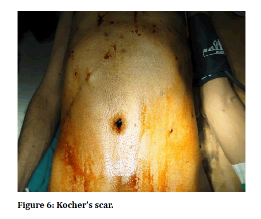
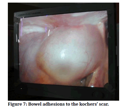
No abnormality is noted in 4% (n=4) of the patients that means negative laparoscopy present in our study. Figure 6 to Figure 9 explains further details of procedure.
Figure 6: Kocher’s scar.
Figure 7: Bowel adhesions to the kochers’ scar.
Figure 8: TB Abdomen-Flimsy adhesions.
Figure 9: Omental biopsy taken from the same patient.
Post-OP complications
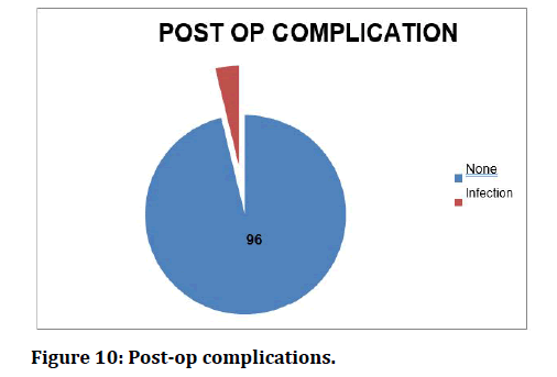
4% (n=4) of the patient had wound infection in the postoperative period which was minimal, and it was managed by appropriate antibiotics and dressing.
No other major complication was occurred in the intraoperative or post-operative period.
Mean Postoperative hospital stay was 2.5 days (Table 7 and Figure 10).
Figure 10: Post-op complications.
Table 7: Post-op complications.
| Post op complication | Percentage |
|---|---|
| None | 101 (96%) |
| Infection | 4 (4%) |
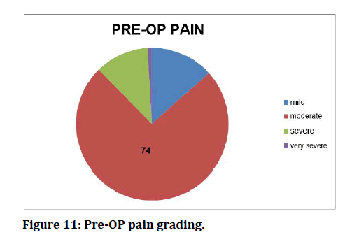
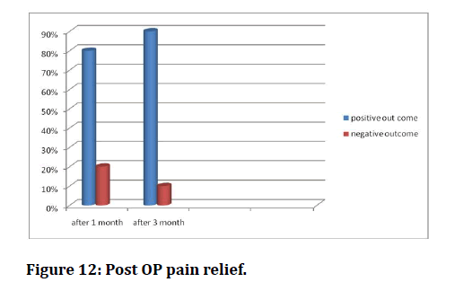
Most of the patient had moderate pain which accounts for 74% (n=78) (Table 8 and Figure 11). All patients were observed in the immediate post-operative period for pain perception and amount of analgesic were needed to treat. All of them had the follow up in 1st month and 3 rd months. Verbal Rating Scale for pain perception were analysed. At the end of 1st month 80% patients got complete pain relief and at 3rd month 90% got complete pain relief. In the remaining 10% patient there were no changes in pain grading, it may be because of the disease nature. And the patient whose laparoscopic findings were normal they also feel symptom free in the follow up. It may be due to placebo effect (Table 9 and Figure 12).
Figure 11: Pre-OP pain grading.
Figure 12: Post OP pain relief.
Table 8: Pre-OP pain grading.
| Grading | Percentage |
|---|---|
| Mild | 14(13%) |
| Moderate | 78 (74 %) |
| Severe | 12 (12%) |
| Very severe | 1(1%) |
Table 9: Post OP pain relief.
| Duration | Positive outcome | Negative outcome |
|---|---|---|
| After 1 month | 80% | 20% |
| After 3 months | 90% | 10% |
Discussion
Chronic abdominal pain is defined as continuous or intermittent pain in the abdomen for more than 3 months duration. Diagnosis and treatment of these patients is usually difficult. It is one of the most common surgical symptom and most challenging problem faced by the surgeons and physicians [14]. We evaluated the 105 consecutive patients of chronic abdominal pain with uncertain and doubtful diagnosis laparoscopically. Diagnostic laparoscopy was done in these patients because of uncertain diagnosis, to check for any concommitent pathology and mainly as an interventional procedure.
Diagnostic laparoscopy revealed normal anatomy and no pathological lesion was found in 4% of the patients. The laparoscopic study of Marana and his coworker [15] and Gowri et al. [16] who detected that laparoscopy failed to detect any abnormalities in 20% of the patients but in this study, it is 4%.Common site for chronic abdominal pain is right lower quadrant (67%) followed by periumbilical regio n (18%). Common intra operative findings were abnormal appendix (64%) followed by adhesions (22%) which requires appendicectomy and adhesiolysis respectively. Di Lorenzo and colleagues [17] reported frequency of abdominal adhesions in chronic abdominal pain were 18.6% in their study but it is 23% in this study. It was found that pain is in the site of adhesions in 90% of cases, although there was no definite correlation between extent of adhesion and severity of pain [18]. The pain in the adhesion is due to restricted mobility and distension of bowe [19]. 7% of patients required conversion into open techniques this is because of the extensive bowel adhesions. Positive outcome is 80% in the follow up of 1 month and 90% of the patients got complete pain relief in the follow up of 3 months. This figure coincides with Gouda et al. [20] study which reports, “the diagnostic laparoscopy yields 80% positive outcome in evaluation of chronic abdominal pain in the follow up of 2 months.”
Conclusion
The role of diagnostic laparoscopy in chronic abdominal pain is tremendous which increases our knowledge about various underlying abdominal disorders. Diagnostic laparoscopy can identify abnormal findings and improve the outcome in patients with chronic abdominal pain. However, it should be considered only after a complete diagnostic evaluation has been carried out. It allows effective surgical treatment of many conditions encountered at the time of diagnostic laparoscopy hence intervension can be done simultaneously. It is a safe and effective tool to establish the etiology of chronic abdominal pain and allows for appropriate interventions.
Funding
No funding sources.
Ethical Approval
The study was approved by the Institutional Ethics Committee.
Conflict of Interest
The authors declare no conflict of interest.
Acknowledgements
The encouragement and support from Bharath University, Chennai is gratefully acknowledged. For provided the laboratory facilities to carry out the research work.
References
- DeBanto JR, Varilek GW, Haas L. What could be causing chronic abdominal pain? Anything from common peptic ulcers to uncommon pancreatic trauma. Postgraduate Med 1999; 106:141-6.
- Sackier JM, Berci G, Paz-Partlow M. Elective diagnostic laparoscopy. AMJ Surg 1991; 161:326 -330
- Ruddock JC. Peritoneoscopy: A critical clinical review. Surg Clin North Am 1957; 371249-1260.
- https://journals.lww.com/dcrjournal/fulltext/2010/05000/mastery_of_surgery,_5th_ed_.21.aspx
- Gouda M El-laddan, Emad N Hokkam, The efficacy of laparoscopy in the diagnosis and management of chronic abdominal pain. J Min access Surg 2010; 6:95-99.
- Decadt B, Sussman L, Lewis MP, et al. Randomized clinical trial of early laparoscopy in the management of acute non-specific abdominal pain. J Br Surg 1999; 86:1383-1386.
- Gelbaya TA, El-Halwagy HE. Focus on primary care: Chronic pelvic pain in women. Obstetr Gynecol Survey 2001; 56:757-764.
- Wesselmann U, Czakanski PP. Pelvic pain: A chronic visceral pain syndrome. Current Pain Headache Reports 2001; 5:13-19.
- Zucker KA. Surgical laparoscopy. Lippincott Williams & Wilkins; 2001.
- https://www.jaypeebrothers.com/pgDetails.aspx?book_id=9789352708451
- https://www.worldcat.org/title/clinical-gynaecology/oclc/709560667
- https://journals.lww.com/jlgtd/fulltext/2002/04000/pelvic_pain_diagnosis_and_management_.10.aspx
- Barry S. Diagnostic laparoscopy: Surgical laparoscopy & endoscopy. 1993; 2.
- Poulin EC, Schlachta CM, Mamazza J. Early laparoscopy to help diagnose acute non-specific abdominal pain. Lancet 2000; 355:861-3.
- Hebbar S, Chawla C. Role of laparoscopy in evaluation of chronic pelvic pain. J Minimal Access Surg 2005; 1:116.
- Gowri V, Krolikowski A. Chronic pelvic pain. Laparoscopic and cystoscopic findings. Saudi Med J 2001; 22:769-70.
- Di Lorenzo N, Coscarella G, Lirosi F, et al. Impact of laparoscopic surgery in the treatment of chronic abdominal pain syndrome. Chirurgia Italiana 2002; 54:367-78.
- Mahawar KK. Laparoscopic adhesiolysis in patients with chronic abdominal pain. Lancet 2003; 361.
- Swank DJ, Van Erp WF, Van Driel OJ, et al. A prospective analysis of predictive factors on the results of laparoscopic adhesiolysis in patients with chronic abdominal pain. Surg Laparoscopy Endoscopy Percutaneous Techniques 2003; 13:88-94.
- El-Labban GM, Hokkam EN. The efficacy of laparoscopy in the diagnosis and management of chronic abdominal pain. J Minimal Access Surg 2010; 6:95.
Author Info
Muzamil Ahmed A and Santhaseelan RG*
Department of General Surgery, Sree Balaji Medical College & Hospital Affiliated to Bharath Institute of Higher Education and Research, Chennai, Tamil Nadu, IndiaCitation: Muzamil Ahmed A, Santhaseelan RG, A Prospective Study of Diagnostic Laparoscopy in Patients Presenting with Chronic Abdominal Pain at SBMCH, J Res Med Dent Sci, 2021, 9(6): 78-84
Received: 22-Mar-2021 Accepted: 10-Jun-2021 Published: 17-Jun-2021

