Research - (2021) Volume 9, Issue 4
A Prospective Study of Pre and Post Prandial Ultrasound Evaluation of Gall Bladder Volume in the Normal as well as in Diabetic Patients
D Suresh Kumar and K Kanakaraj*
*Correspondence: K Kanakaraj, Department of Radio Diagnosis, Sree Balaji Medical College & Hospital, Bharath Institute of Higher Education and Research, Chennai, Tamil Nadu, India, Email:
Abstract
To evaluate the gall bladder volume in diabetics and non-diabetic individuals by ultrasonography. To compare the gall bladder volume between diabetics and non-diabetic individual in pre and post prandial. Diabetic patients had the highest mean gallbladder volume compare to those non diabetics control group. The mean postprandial gall bladder volume was also highest in diabetics. The gallbladder ejection fraction was lowest in diabetics compare to those non diabetics groups. Longer duration of illness, doubled the prevalence of gall stone is noted in diabetics compare to those without diabetics.
Keywords
Gall bladder, Diabetic, Neuropathy, Hepatobiliary system
Introduction
Some metabolic disorders in a group are categorised in terms of hyperglycemia are known as Diabetes mellitus are resulted because of insulin action, or insulin secretion defects, or also both combined. Since long time diabetes mellitus was considered to be the commonest endocrine disorder and now a day it is on the rise due to advancing modern life style; characterized by multiple metabolic abnormalities which leads in long lasting complications in multi-organs which also involves hepatobiliary system, blood vessels, nerves, gastrointestinal tract, and kidneys, hence giving rise to serious mortality or morbidity. DAN: “Diabetic Autonomic Neuropathy” are the rarest understood and recognised diabetic complication even with its severe effects on the quality as well survival of living a healthy life [1]. One of the tiny organs of the body named gallbladder present in right side of the abdomen, just under surface of the liver. Primary function of a gallbladder is storing bile which is produced by liver and discharge the stored bile to small intestine so as to help with easy digestion. Bilirubin pigment, bile salts which are the organic detergents that helps in breaking the fat, fats, cholesterol and water all together adds up to form Bile. The crystal-like masses known as the Gallstones are formed inside the gallbladder in case the amount of bilirubin, bile salts, or cholesterol level is increased in the Bile. The formed Gallstones are of different shapes or sizes and can reach up to the size of a golf ball and can develop into multiple number [2]. Elderly people and females are more prone of getting Gallstones, and also among people who are overweight [3]. In the US around 25% people of more than 65 of age have the condition of gallstones, however not even half of them ever experience or notice any signs of it. Although the gallstones complications could be quite severe in case they are not treated on time [4]. The conditions of Gallstones are one of the grieving concerns which is diagnosed in millions of people all over the world [5]. Its occurrence is however still uncommon among developing as well as under developed societies whereas it prevails in nearly 25% among people from industrialized societies [6]. Cholesterol gallstone is mainly caused due to Diabetes mellitus and its related aspects. Patients suffering from diabetes of over twenty years the occurrence of Gallstone are estimated to be nearly 30%. However, no clear pathophysiology exists. Different researches present the occurrence of diseases related to gall bladder among diabetic patients. Such condition attributes to impaired contraction of gall bladder causing cholecystomegal, primarily because of autonomic neuropathy observed in diabetes. As for the formation of gallstone the basic pre-requisite necessity is the gallbladder stasis, whereas other factors which can lead to risk are, ileal resection, hyperlipidaemia, drugs, diet, parity, obesity, genetic factors [7-9]. The hyperlipidemia, hypertension, type II diabetes mellitus, insulin resistance, truncal obesity related metabolic syndrome are interlinked to the high secretion of hepatic cholesterol which is considered to be the highest risk aspect of cholesterol gallstones development. Gallstone disease is associated to numerous cardio metabolic risk factors, for example, obesity, unhealthy diet, sedentary lifestyle, and dyslipidemias (hypertriglyceridemia and low high-density lipoprotein cholesterol) [10-11]. Furthermore, there is expanding confirmation to recommend that parts of metabolic disorder inclusive of insulin protection, hyperinsulinemia, along with which increased triglycerides might be related to expanded hazard [12]. Epidemiological investigations on significance of gallbladder sickness in diabetes people are conflicting. A few investigations have discovered a positive relationship amongst diabetes and gallbladder illness and risks of gallstones [13]. Nevertheless, different examinations found no relationship [14]. Furthermore, the extent of the hazard assessments has changed impressively, and this could conceivably be because of frustrating by other hazard factors, for example, obesity, physical activity, or any other risk factors. Another intension was correlating the volume of gall bladder in diabetes patients in terms of parameters like, autonomic neuropathy, hyperlipidemia, parity, body mass index, sex, and age. This research was carried out to formulate a comparison in the volume of gall bladder and control in a diabetic population. For assessing the volume of gall bladder and its modality the process chosen was Ultrasonography. This is an accurate, less time consuming, inexpensive, and a safe process. Subsequently the process of Ultrasonography is being evolved rapidly and is now being used widely in every medical practice field. The results of these experiments provide the thickness of gallbladder wall, volume off gallbladder, as well as the size of the gallbladder. Usually the technique of ultrasound was applied in the form of initial technique for images for estimating the gallbladder disease in the patients. The reason behind this is the high sensitivity and specificity to detect the dimensions of gallbladder, portability, speed, as well as its real-time character. Additionally, this technique is paramount to identify the pathologies of the gallbladder such as tumours, stones, sludge, contraction, and distensions [15,16].
Materials and Methods
The present descriptive cross sectional study was conducted on 50, 25 cases as normal and 25 cases as diabetic patients enrolled from the Sree Balaji Medical College and Hospital, Chromepet, Chennai The diagnosis of diabetes in these patients was in accordance with WHO criteria i.e., fasting plasma glucose level ≥126 mg/dl, and ≥200 mg/dl plasma glucose level after 2 hr of ingestion of standardised 75 gm glucose. An informed consent was taken from all the subjects in the study and control groups. Study design and its protocol was approved by the institutional ethical committee.
Selection of study group
Total 50 patients were randomly selected for the study among patients regularly attending diabetic clinic of this hospital and following our criteria’s of selection.
Study duration
The study was conducted between December 2017 and June 2019 (18 months).
Inclusion criteria
â?? Patients with diabetes mellitus diagnosed since 2 yr or more.
â?? Functioning gall bladder with well controlled blood sugar levels.
â?? Patients of 30 to 80 yr age group.
Exclusion criteria
â?? Patients with history of pre-existing hepatobiliary or gastrointestinal disease.
â?? Those diabetic patients who are taking antihypertensive drugs also, which can interfere with autonomic functions.
â?? Obese patient with history of major cardiac arrhythmia.
â?? Pregnant females.
â?? Patient with history of cerebrovascular accident.
Statistical analysis
Statistical analysis done by using statistical Package of Social Science (SPSS Version 19; Chicago Inc., USA). Data comparison performed by applying specific statistical tests i.e. Chi Square test, Student t test were applied to find out the statistical significance of the comparisons. Qualitative variables were compared using proportions and quantitative variables were compared using mean values. Significance level was fixed at P <0.05.
Method to measure gallbladder volume
Gallbladder volume was measured in all the subjects after 12 hours overnight fasting by using a 3.5 to 5 MHz convex transducer in Siemens ACUSON S 2000 ultrasound system Siemens Medical Solutions, Mountain vie, CA, USA. The greatest length (L), maximum transverse width (W), and highest anteroposterior dimensions (H) were measured and documented. All the measurements were recorded by first author of the study on two consecutive days, and the average of the two measurements was considered as the final fasting gall bladder volume for the case and control. This protocol was adopted to ensure the reproducibility of the results.
The gall bladder volume was calculated by this formula=á´«*L*W*H 6 Where, L=Length, W=Width and H=Height of gall bladder
Many other related findings e.g. gallbladder wall thickness, presence of stones, sludge or neoplasia were searched and recorded. Gall bladder motility was observed by measuring fasting and post meal gallbladder volumes. Post meal volume was taken 90 min after giving fatty meal i.e. four slice of bread with 30 gm butter. The percentage of gallbladder contraction was calculated by the formula. Fasting Gallbladder volume - Post fatty meal Gall bladder volume * 100 Fasting gall bladder volume The results of the study have been compiled, tabulated and statistically analyzed for comparisons.
Results
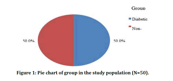
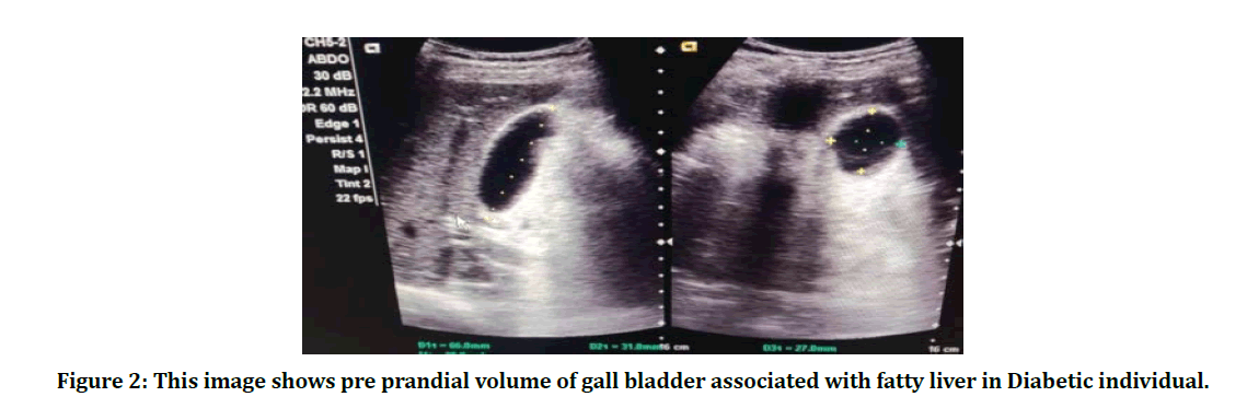
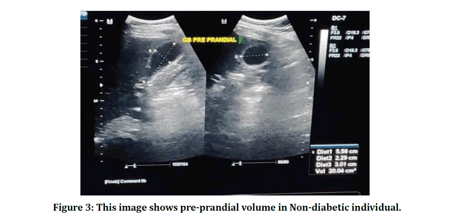
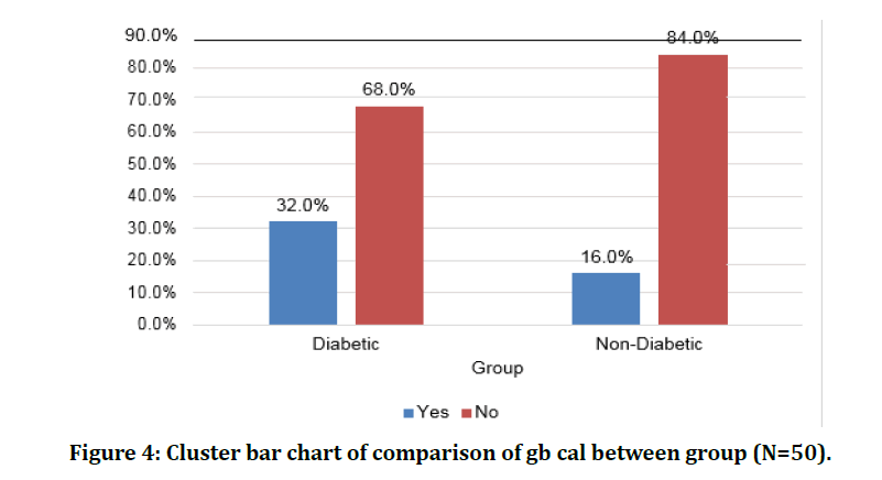
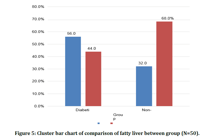
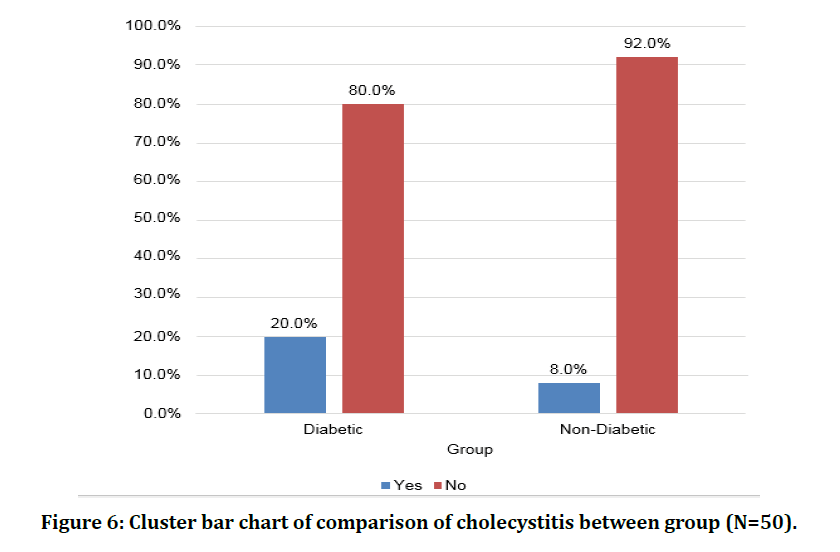
A prospective study was performed to determine the pre and post prandial ultrasonographic evaluation of gall bladder volume in the normal as well as in diabetic patients. 50 patients were studied who fulfilled the inclusion criteria. All these patients were taken from the Radiology department of Sri Balaji Medical College and Hospital. All patients were evaluated clinically and only essential investigations were performed, in addition patients underwent the diabetic test. The results obtained are as follows. Each group comprises of 25 patients and totally 50 patients were taken for this study. The mean age of patients with gall stones is 54.6 years which was statistically not significant. In the present study, the pre-prandial gall bladder volume of diabetic was21.78 ± 4.58 and non-diabetic was 22.68 ± 3.41 which was not significant.The postprandial volume of diabetic was 13.66 ± 4.4 and non-diabetic was10.48 ± 2.38 which was highly significant. The presence of gb cal was 12 (24%) and absence in 38 (76%) in both the groups as 22.68 ± 3.41 which was not significant. Fatty liver were seen in 22 (44%) patients and were not seen in 28 (56%) patients which was not significant between the group of diabetic and non-diabetic patients. In the present study, the cholecystitis was seen in 7 (14%) patients out of 50 patients that were high in diabetic 5 (20%) than non-diabetic patients 2 (8%) which was not significant (Tables 1-4 and Figures 1-6).
Table 1: Descriptive analysis of group in the study population (N=50).
| Group | Frequency | Percentages |
|---|---|---|
| Diabetic | 25 | 50.00% |
| Non-Diabetic | 25 | 50.00% |
Table 2: Comparison of mean of age between (N=50).
| Group | Frequency | Percentages |
|---|---|---|
| Diabetic | 25 | 50.00% |
| Non-Diabetic | 25 | 50.00% |
Table 3: Comparison of mean of prandial/ml and between group (N=50).
| Group (Mean± SD) | P value | ||
|---|---|---|---|
| Parameter | Diabetic (N=25) | Non-Diabetic (N=25) | |
| Age | 54.6 ± 10.24 | 50.56 ± 10.13 | 0.167 |
Table 4: Comparison of fatty liver between group (N=50).
| Group | Chi square | P Value | ||
|---|---|---|---|---|
| Fatty Liver | Diabetic (N=25) | Non-Diabetic (N=25) | ||
| Yes | 14 (56%) | 8 (32%) | 2.922 | 0.087 |
| No | 11 (44%) | 17 (68%) | ||
Figure 1: Pie chart of group in the study population (N=50).
Figure 2: This image shows pre prandial volume of gall bladder associated with fatty liver in Diabetic individual.
Figure 3: This image shows pre-prandial volume in Non-diabetic individual.
Figure 4: Cluster bar chart of comparison of gb cal between group (N=50).
Figure 5: Cluster bar chart of comparison of fatty liver between group (N=50).
Figure 6: Cluster bar chart of comparison of cholecystitis between group (N=50).
Discussion
Diabetic autonomic neuropathy of alimentary canal may take several forms: (a) gastroparesis; (b) oesophagopathy; (c) biliary tract disorders; and (d) enteropathy. Commonly cited reasons for a higher prevalence of gallstone disease in people with diabetes include; decreased motility of gall bladder, reduced postprandial cholecystokinin (CCK) release, decreased gallbladder smooth muscle sensitivity to CCK, reduced number of receptors for CCK in the wall of gallbladder, super saturation of bile, and presence of gallstones themselves [17]. Neural control of emptying of gallbladder is mediated by sympathetic and parasympathetic innervation; the former causes relaxation and the latter increases the contractility of gallbladder. Meal related release of CCK leads to contraction of gallbladder. The motility defects in patients with gallstones manifest as an increase in fasting volume, decrease in ejection fraction, decrease in rate of ejection, and an increase in residual volume of gallbladder. In our study, increased gallbladder volumes while fasting is seen in type 2 diabetics in comparison to healthy controls. Earlier studies have corroborated this finding [18]. These studies measured emptying of gallbladder as well (both its extent and rate), as a measure of motility of gallbladder and found that gallbladder emptying rate and the emptying of gallbladder emptying were also decreased in diabetics [19,20]. It was also found in all these studies, as in our study, that both an increased fasting volume of gallbladder and decreased gallbladder ejection fraction correlated with autonomic neuropathy. Due to logistical constraints, we did not study gallbladder emptying, but it would have further clarified the role of autonomic neuropathy in cases diabetic cholecystopathy [21]. Further indirect evidence establishing the effect of autonomic neuropathy leading to cholesterol gallstones can be inferred from studies that show increased prevalence of gall stones in patients with spinal cord injuries at a higher level reduced gallbladder emptying as a response to cephalic stimulation in patients with diabetes with autonomic neuropathy, a normal post-prandial CCK levels in the blood samples of diabetics and also a good response in motility to infusion of CCK and its analogues in diabetics who have autonomic neuropathy [22]. Age of these selected patients ranged from 30- 60 yrs, with mean age being 54.6 ± 10.24 in diabetic and 50.56 ± 10.13 non diabetic patients. In our study, age has a positive correlation with gallbladder volume, which can be reasoned by age related autonomic denervation that can lead to increased bile viscosity, gallbladder stasis, and a greater cholesterol content seen in the bile. Males 27 (54%) were slightly higher than female 23 (46%). Males were the predominant group. As in previous studies, no correlation was see between duration of diabetes or gender with gallbladder volume in our study [23]. On analyzing, the co- morbid factors glucose level in diabetic patients of both pre and post prandial/ ml were 21.78 and 22.68 which was high than 13.66 and 10.48 in non-diabetic patients. Cholecystitis was the predominant co- morbid factor followed by diabetes. Cholecystitis was seen in 7 (14%) patients out of 50 patients that had diabetes, 5 (20%) than in non-diabetic patients 2 (8%) which was not significant. As abnormalities of the gall bladder typically remain clinically silent in diabetics, they may suddenly present with complications such as acute cholecystitis which may require emergency cholecystectomy. In order to reduce morbidity and mortality that isassociated with emergency surgery, prophylactic cholecystectomy may be advised for asymptomatic diabetics when there is evidence of a non-functioning gallbladder demonstrated on USG [24]. In a study conducte among 100 patients with chronic diabetics 76 patients (76%) no hepatobiliary abnormality was seen, however cholelithiasis was found in 13 patients (13%), cholecystitis in 5 patients (5%) and sludge was found in six patients (6%). In the group of 100 controls 91 individuals (91%) showed no hepatobiliary pathology. Cholelithiasis was incidentally detected in 4 indiviudals (4%), cholecystitis in 2 (2%) and sludge in 3 individuals (3%). A significant difference was seen in gall bladder volume while fasting when comparing the controls and chronic diabetics (p value- 0.001). Major difference was also seen in the percentages of gall bladder contraction between controls and chronic diabetics (p value- 0.001). A higher fasting gallbladder volume and a decrease in the percentage of contraction are both seen patients with chronic diabetes mellitus, attributable to autonomic neuropathy. Prolonged biliary stasis results in complications sludge deposition, cholecystitis and cholelithiasis as late outcomes. In chronic diabetics hepatobiliary ultrasonography may be used as a screening tool for early detection of complications and as a result, avoid the serious consequences with which these patients present in emergency with a requirement for surgery [25]. In a study done which included 91 cases of diabetes mellitus and 40 healthy controls the mean gallbladder volume while fasting was 18.2 ± 2.5 ml among type1 diabetics and 25.9 ± 13.9 ml among type 2 diabetics, with the range of a maximum value of 88.0 ml and minimum value of 9.3 ml. When the type 2 diabetics were sub-grouped base on the presence or absence of autonomic neuropathy, a higher gallbladder volume was seen on average in patients having autonomic neuropathy. To a high degree, cholecystomegaly was seen in type 2 diabetics in our study. It correlated significantly with age, BMI, and severity of the autonomic neuropathy. Among males with type 2 diabetes, gallbladder volume correlated significantly LDL levels. Among females with type 2 diabetes, gallbladder volume correlated significantly with the waist-hip ratio. The volume of gallbladder also had a significant correlation with diabetic retinopathy, but not with microalbuminuria, glycemic control, duration of diabetes or hypertension [26]. This study revealed that significant number of patients with diabetes has gall bladder abnormalities ranging from increase gall bladder volume to reduced ejection fraction (GBCI) with associated increase prevalence of gall stone disease. This is seen to be worse off in diabetics with neuropathy. Association also was established between duration of diabetes and gall bladder abnormalities. Fatty liver and cholecystitis were seen mostly in diabetic patients than normal cases in this study. Ultrasonography of the gall bladder therefore is highly recommended in diabetic patient management especially in those with long duration of illness as this will aid proactive management of gall bladder complication which they are prone to, and reduce morbidity and mortality’
Conclusion
We concluded that ultrasonography is an easy and effective method for evaluation of gall bladder volume. Fasting gall bladder volume also has a weak correlation with advancing age and is statistically not significant. Thus, our study reiterates the fact that diabetic cholecystopathy, which may predispose these patients to gallstone formation, is not uncommon, especially in diabetics. This leads to increased risk of the complications of gallstone disease intrinsically, and those due to its treatment - both surgical and medical. A definite association of diabetes with gall bladder volume has been demonstrated in our study and it implies a thorough evaluation for autonomic impairment in all diabetics. All diabetics should also be evaluated for the presence of increased fasting gallbladder volumes, impaired postprandial gallbladder emptying, and gallbladder sludging; all markers of risk of progression to overt gall stone disease. The fasting gall bladder volume in the population and its clinical implications needs to be further studied on a larger group of population to get an overall insight into it. Not only diabetes, fatty liver and cholecystitis also play a role in the increase volume of gall bladder. From our study we showed there is a possible risk of the relation between diabetes patients to develop gallbladder disease, further studies should be made to make a better understanding of the potential biological mechanisms.
Funding
No funding sources.
Ethical Approval
The study was approved by the Institutional Ethics Committee.
Conflict of Interest
The authors declare no conflict of interest.
Acknowledgement
The encouragement and support from Bharath University, Chennai is gratefully acknowledged.
For provided the laboratory facilities to carry out the research work.
References
- Vinik AI, Erbas T. Recognizing and treating diabetic autonomic neuropathy. Cleve Clin J Med 2001; 68:928-44.
- Singh S, Chander R. Ultrasonographic evaluation of gall bladder disease in diabetes mellitus type 2. Inf J RadiolImag 2006; 16:4505- 508.
- Powers AC. Diabetes mellitus Harrisons principle of internal medicine 17th Edn. Pp: 2275-2310.
- Foster W, Unger RH. Diabetes mellitus. Chapter 21 in William text book of Endocrinology.
- Zaliekas J, Munson JL. Complications of gallstones: The Mirizzi syndrome, gallstone ileus, gallstone pancreatitis, complications of "lost" gallstones. Surg Clin North Am 2008; 88:1345-1368.
- Ata N, Kucukazman M, Yavuz B, et al. The metabolic syndrome is associated with complicated gallstone disease. Can J Gastroenterol 2011; 25:274–276.
- Banim PJ, Luben RN, Bulluck H, et al. The aetiology of symptomatic gallstones quantification of the effects of obesity, alcohol and serum lipids on risk. Epidemiological and biomarker data from a UK. prospective cohort study (EPIC- Norfolk). Eur J Gastroenterol Hepatol 2011; 23:733-740.
- Campbell PT, Newton CC, Patel AV, et al. Diabetes and cause-specific mortality in a prospective cohort of one million U.S. adults. Diabetes Care 2012; 35:1835-1844.
- Boland LL, Folsom AR, Rosamond WD. Atherosclerosis risk in communities (ARIC) study investigators. Hyperinsulinemia, dyslipidemia, and obesity as risk factors for hospitalized gallbladder disease. A prospective study. Ann Epidemiol 2002; 12:131-40.
- Wolson A.H. Ultrasound measurements of the gall bladder. In: Goldberg BB, Kurtz AB, eds. Atlas of Ultrasound Measurements. Chicago: Year Book Medical Publishers 1990; 108-109.
- Caroli-Bosc FX, Pugliese P, Peten EP, et al. Gallbladder volume in adults and its relationship to age, sex, body mass index, body surface area and gallstones. An epidemiologic study in a non-selected population in France. Int J Gastroenterol 1999; 60:344-348.
- Agarwal AK, Miglani S, Singla S, et al, A Goel Ultrasonographic evaluation of gall bladder volume in diabetics. JAPI 2004; 52:962-965.
- Singh S, Chander R, Singh A, et al. Ultrasonographic evaluation of gall bladder diseases in diabetes mellitus type 2 .Ind J Radiol Imag 2006; 4:505-508.
- Powers AC. Diabetes mellitus. A text book. 2275-2310.
- Ewing DJ Autonomic neuropathy its diagnosis and prognosis. Clin Endocrinol Metabol 1986; 15:855-885.
- Das AK. Diabetic autonomic neuropathy-clinical features, diagnosis and management. Novo Nordisk Update 1994; 29-37.
- American Diabetic Association: Clinical practice recommendations. Diabetes Care 2007; 27:1047.
- Diabetes action now: An initiative of WHO & international diabetes federation. Diabetes Care 2004; 27:1047.
- Masharani U. Diabetes mellitus in current medical diagnosis and treatment. Stephen J, McPhee MA Papadakis (Eds) 2008.
- Sicree R, Shaw J, Zimmet P. Diabetes and impaired glucose tolerance. In diabetes atlas, International diabetes federation. 3rd Edn 2006; 15-103.
- Wild S, Roglic G, Green A, et al. Global prevalence of diabetes mellitus: Estimates for the year 2000 & projections for 2030. Diabetes Care 2004; 27:1047-1053.
- Ahuja MMS. Epidemiological studies on diabetes mellitus in India in: Ahuja MMS Epidemiology of diabetes in developing countries Interprint New Delhi. 29-197938.
- Ramachandran A, Snehalatha C, Kapur A, et al. Diabetes epidemiology study group-DESl, high prevalence of diabetes & impaired glucose tolerance in India-National urban diabetes survey. Diabetologia 2001; 44:1094-1101.
- Fiorucci S, Bosso R, Scionti L, et al. Neurohumoral control of gallbladder motility in healthy subjects and diabetic patients with or without autonomic neuropathy. Dig Dis Sci 1990; 35:1089-1097.
- Shaw SJ, Hajnal F, Lebovitz Y, et al. Gall bladder dysfunction in diabetes mellitus. Dig Dis Sci 1993; 38:490-496.
- Dhiman RK, Arke L, Bhansali A, et al. Cisapride improves gall bladder emptying in patients with type 2 diabetes mellitus. J Gastroenterol Hepatol 2001; 16:1044-1050.
Author Info
D Suresh Kumar and K Kanakaraj*
1Department of Radio Diagnosis, Sree Balaji Medical College & Hospital, Bharath Institute of Higher Education and Research, Chennai, Tamil Nadu, IndiaReceived: 20-Mar-2021 Accepted: 06-Apr-2021 Published: 13-Apr-2021

