Research - (2021) Volume 9, Issue 4
A Study on Incidence of Helicobacter Pylori Infection in Dyspeptic Patients in Sree Balaji Medical College and Hospital
G Srinath, RNM Francis and V Ramalakshmi*
*Correspondence: V Ramalakshmi, Department of General Surgery, Sree Balaji Medical College & Hospital, Bharath Institute of Higher Education and Research, India, Email:
Abstract
Objective: Helicobacter pylori (H. pylori) infection is a major cause of various upper gastrointestinal (UGI) disorders. The aim of this study was to determine the prevalence of H. pylori among patients with dyspepsia admitted to Sree Balaji Medical college and Hospital, Chennai.
Methods: Totally, 180 patients were enrolled for the study. Their premedical history, demographic data, lifestyle habits and socioeconomic status were carefully documented. The specimen bile ducts were analysed using biochemically and microbiologically. The results were expressed using statistical Softwares.
Results: The present study showed that revealed that the infection was based on the aging and the patients and more prone in male (67%). The patients from upper middle class had higher infections (35%). Our results showed a strong correlation with H. pylori infection, urease test and biopsy. The results were statistically significant.
Conclusion: The well-described association of H. pylori with dyspepsia was confirmed by our study.
Keywords
Asymptomatic gallstone, Prevalence of gallstone disease, Urease test, Dyspepsia
Introduction
Brown et al. of Perth, Western Australia discovered H. pylori in 1983. Originally, the organism was named Campylobacter pylori because it was structurally like other Campylobacter species, such as C. jejuni [1]. Signs of H. pylori infection such as gram- negative gastric bacilli, gastric urease and epidemics of hypochlorhydria have been described since the late nineteenth century [2]. These observations could be better explained after Warren and Marshall in the early 1980’s managed to culture a bacterium that was to be designated Campylobacter pyloridis [3]. In 1989, the genus Helicobacter was created, and the bacterium received the name H. pylori [1]. H. pylori is a small (0.5 -1.0 μm in width and 2.5 to 5.0 μm in length), spiral shaped, highly mobile, gram negative rod with 4 -6 unipolar sheathed flagella [4]. The microorganism grows slowly invitro and requires special media and a microaerophilic (5% O2, 10% CO2, and 85%. N2) environment [5]. The most striking biochemical characteristic is the production of large quantities of urease. This enzyme digests urea to produce carbon dioxide and ammonia. In the presence of water this leads to the formation of ammonium hydroxide [6]. In this way, H. pylori is able to neutralize the acid in its direct environment.
H. pylori, colonizes the human stomach, can cause gastritis, is strongly associated with gastric and duodenal ulceration (DU) and has been implicated in the causation of gastric carcinoma and mucosa-associated lymphoid tissue (MALT) lymphomas. It has been reported that there is relationship between H. pylori infection and children’s gastroenterological diseases [7]. However, only 10-20% of infected individuals manifests severe complications and this selectivity in disease progression is inadequately understood [8,9]. Epidemiological studies have shown that a weakly positive correlation exists between chronic gastric infection with H. pylori and coronary heart disease [10]. Urease, vacuolating cytotoxin Vac A, and the pathogenicity island (cagPAI) gene products, are the main factors of virulence of this organism. H. pylori LPS may have an important role in autoimmunemediated damage in the gastric mucosa [11]. Half of the world’s population is estimated to be infected with H. pylori, which makes it one of the most common bacterial pathogens in humans. The prevalence of H. pylori infection worldwide is approximately 50%, it reaches as high as 80%–90% in developing countries, and about 35%–40% in the United States. Approximately 20% of persons infected with H. pylori develop related gastro duodenal disorders during their lifetime. The annual incidence of H. pylori infection is about 4%–15% in developing countries, compared with approximately 0.5% in industrialized countries. Documented risk factors include low socioeconomic status, overcrowding, poor sanitation, or hygiene, and living in a developing country.
In "Israel", the prevalence of H. pylori infection is about 60%, and the annual incidence of gastric cancer is about 15 per 100,000 populations. The prevalence of H. pylori infection was 10% among children in Egypt. In crude analyses, prevalence was associated with increasing age, non-white skin color, lower current family income, lower education level, higher size of the family, low socioeconomic conditions in childhood, higher number of siblings and attendance to day-care centers in childhood, and presence of dyspeptic symptoms. Socioeconomic conditions in childhood besides ethnicity and presence of dyspeptic symptoms were the factors significantly associated with the infection. There are several methods of diagnosing H. pylori infection including invasive procedures using mucosa biopsies taken during endoscopy (mainly culture, histology, and the rapid urease test) and non-invasive procedures. Non-invasive testing methods for detection of H. pylori or confirmation of eradication include: 1) Antibody tests (in serum, saliva, or blood); 2) Antigen tests (in stools, saliva, or urine); and 3) Radioactive or non-radioactive urea breath tests. The most interesting of the non-invasive tests is the detection of antigens in stool samples by enzyme immunoassay technique. While this test has good performance at a reasonable cost, doubts exist regarding patient and clinician compliance and actual performance, particularly about inter laboratory variability. The urea breath test is based on analysis of samples of exhaled air before and after ingestion of urea containing specially labelled carbo.
PCR is a powerful technique for the detection of target DNA in various clinical specimens, but its application to fecal specimens has been limited due to the presence of substances inhibiting the reaction. Eradication therapy of symptomatic H. pylori infection substantially reduces the recurrence of associated gastro duodenal diseases. Therapy entails complicated regimens of several antimicrobial agents for at least 2 weeks. In general, triple therapy regimens usually entail two of the following antimicrobial agents: metronidazole, amoxicillin, tetracycline, or clarithromycin in combination with a proton pump inhibitor or bismuth salts. The most common causes of treatment failure are patient noncompliance and antimicrobial resistance of the infecting H. pylori strain. Quadruple regimens are used as a salvage therapy when triple therapy regimens have failed. However, the success of treatment is usually dependent on early detection. Moreover, prevention of H. pylori infection seems to be a wise strategy. Prevention strategies require deep understanding of the transmission risks. H. pylori infection can be prevented by interrupting the transmission of the infection by improving environment, socioeconomic status, and personal hygiene. It has been shown convincingly that with the improvement of socioeconomic status in developed countries, H. pylori infection has gone down by 50% over the last 3 decades. Further studies have shown that superficial gastric ulcers in a mouse model infected with H. pylori can be prevented by the administration of a recombinant oral vaccine with E. coli heat labile toxin (LT) given as adjuvant.
Aim of the Study
✓ To study the age incidence of patient with H. pylori in dyspepsia.
✓ To study the sex incidence of patient with H. pylori in dyspepsia.
✓ To study the incidence of H. pylori with presenting complaints of dyspepsia.
✓ To study the incidence and duration of complaints patient with H. pylori presenting has dyspepsia.
✓ To study the incidence of H. pylori in endoscopic findings.
✓ To study the incidence of H. pylori in endoscopic biopsy.
✓ To study the incidence of H. pylori in rapid urease test.
✓ To study the treatment of Helicobacter pylori infection and eradication.
Materials and Methods
The materials for this study were 180 patients who were admitted in surgical wards at Sree Balaji Medical College and Hospital, Chennai from October 2013 to October 2015(period of 2yrs). All patients with dyspeptic symptoms who were referred to the Department of Gastroenterology were assessed by a consultant Surgical Gastroenterologist. Patients requiring upper gastrointestinal endoscopy were eligible for study. The procedure was explained to the patient and the need to check for Helicobacter pylori infection was explained. Informed consent was obtained from all patients.
Endoscopy was carried out with Olympus forward viewing flexible endoscope. Local pharyngeal anesthesia with 10% lignocaine spray was used in all patients as well as sedation with midazolam intravenously. All patients had evaluation of the oesophagus, stomach and proximal duodenum and abnormalities were documented. Gastric biopsies for histological examination and Rapid urease test for the presence of Helicobacter pylori organisms were performed on all patients. Five samples were taken from the stomach: two from the antrum, two from the body and one from the incisura angularis. Gastric samples for histological evaluation were sent to Department of pathology, Sree Balaji medical college and hospital. Data obtained included age, gender, occupation, socio economic status, presenting complaints, duration of symptoms, findings on endoscopy and results of gastric biopsies and rapid urease test.
Results
This study was conducted with the patients admitted to Sree Balaji Medical College and Hospital, Chennai during the period from October 2013 to October 2015. Totally, 180 cases of incidence of H. pylori in dyspepsia studied and all of them were undergone for upper gastrointestinal endoscopy, rapid urease test and biopsy. The present study revealed that the infection was based on the aging and the patients between 40 50 years were prone (39%) for the infection (Table 1 and Figure 1). The results (Table 2 and Figure 2) also showed that the male was having higher infection rate (67%) than the females (33%). Our results further showed that the occupation play a pivotal role in infection rate (Table 3 and Figure 3). The persons working in skilled area had higher rate (38%) followed by unskilled (30%), professional (25%) and unemployed (7%). The present study demonstrated the significance of socio-economic status and infection rate (Table 4 and Figure 4). The upper middle class had higher infections (35%) than middle class (25%), followed by lower middle class (14%) and lower class (6% -20%).
| Age | H. Pylori Positive | % | H. Pylori Negative | % | Total no of cases | % |
|---|---|---|---|---|---|---|
| 21–30 | 3 | 2 | 1 | 2 | 4 | 2 |
| 31–40 | 16 | 13 | 10 | 18 | 26 | 14 |
| 41–50 | 49 | 40 | 20 | 36 | 69 | 39 |
| 51–60 | 37 | 30 | 20 | 36 | 57 | 32 |
| > 60 | 19 | 15 | 5 | 10 | 24 | 13 |
| Total | 124 | 56 | 180 | 100 |
Table 1: Relationship between aging and H. pylori infection in studied patients.
| Gender | H. Pylori Positive | % | H. Pylori Negative | % | No of cases | % |
|---|---|---|---|---|---|---|
| Male | 95 | 77 | 27 | 48 | 122 | 67 |
| Female | 29 | 23 | 29 | 52 | 58 | 33 |
| TOTAL | 124 | 56 | 180 | 100 |
Table 2: Relationship between sex and H. pylori infection in studied patients.
| Occupation | H. Pylori Positive | % | H. Pylori Negative | % | No of cases | % |
|---|---|---|---|---|---|---|
| Professional | 32 | 26 | 13 | 23 | 45 | 25 |
| Skilled | 51 | 41 | 18 | 33 | 69 | 38 |
| Unskilled | 33 | 27 | 21 | 37 | 54 | 30 |
| Unemployed | 8 | 6 | 4 | 7 | 12 | 7 |
| TOTAL | 124 | 56 | 180 | 100 |
Table 3: Relationship between occupation and H. pylori infection in studied patients.
| Socio economic status | H. Pylori Positive | % | H. Pylori Negative | % | No of cases | % |
|---|---|---|---|---|---|---|
| Upper Class | 8 | 6 | 4 | 7 | 12 | 6 |
| Upper Middle Class | 49 | 40 | 13 | 23 | 62 | 35 |
| Middle Class | 28 | 23 | 16 | 29 | 44 | 25 |
| Lower Middle Class | 18 | 14 | 8 | 14 | 26 | 14 |
| Lower Class | 21 | 17 | 15 | 27 | 36 | 20 |
| Total | 124 | 56 | 180 | 100 |
Table 4: Socio economic status and H. pylori infection in studied patients.
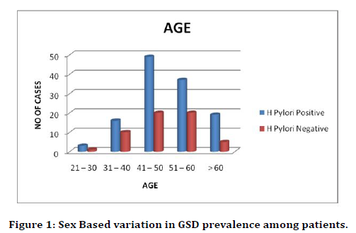
Figure 1. Sex Based variation in GSD prevalence among patients.
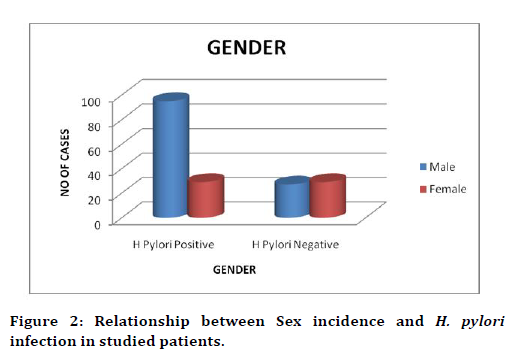
Figure 2. Relationship between Sex incidence and H. pylori infection in studied patients.
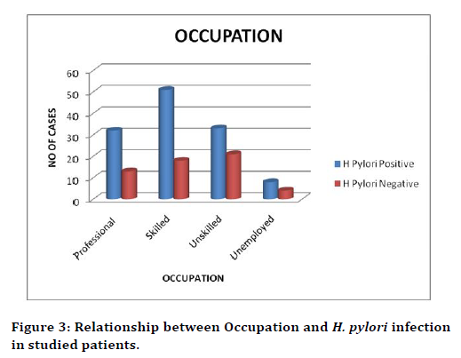
Figure 3. Relationship between Occupation and H. pylori infection in studied patients.
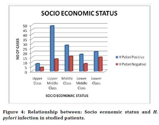
Figure 4. Relationship between: Socio economic status and H. pylori infection in studied patients.
The smokers were found to be with higher infection rate (41%) than alcoholics (32%) and interestingly, our results showed that the as both smokers + alcoholics had lower rate (7%). These results showed that the sedentary lifestyles had significant role in determining the infection rate (Table 5 and Figure 5). The patients felt the varied duration in disease symptoms (Table 6 and Figure 6). 36% of them told that the symptoms were lasting for at least 3 weeks and 37% of them felt it for 3-4 weeks. 27% registered as more than 4 weeks. The present study showed that the most common symptoms (Table 7 and Figure 7) presented by H. pylori positive was epigastric pain (30%) followed by epigastric burning (11%), early satiety (13%), nausea (11%), vomiting (16%), bloating (06%), belching (07%) Post Prandial Fullness (06%).
| Sedentary lifestyle | H. Pylori Positive | % | H. Pylori Negative | % | No of cases | % |
|---|---|---|---|---|---|---|
| Smoking | 55 | 44 | 19 | 34 | 74 | 41 |
| Alcohol | 53 | 43 | 5 | 9 | 58 | 32 |
| Smoking+Alcohol | 9 | 7 | 4 | 7 | 13 | 7 |
| Tea Totaller | 7 | 6 | 28 | 50 | 35 | 20 |
| Total | 124 | 56 | 180 | 100 |
Table 5: Sedentary lifestyle and H. pylori infection in studied patients.
| Duration of symptoms | H. Pylori Positive | % | H. Pylori Negative | % | No of cases | % |
|---|---|---|---|---|---|---|
| < 3 weeks | 49 | 40 | 16 | 29 | 65 | 36 |
| 3 to 4 weeks | 49 | 40 | 18 | 32 | 67 | 37 |
| > 4 weeks | 26 | 20 | 22 | 39 | 48 | 27 |
| Total | 124 | 56 | 180 | 100 |
Table 6: Symptom duration and H. pylori infection in studied patients.
| Symptoms | Male | % | Female | % | No of cases | % |
|---|---|---|---|---|---|---|
| Epigastric Pain | 40 | 73 | 15 | 27 | 55 | 30 |
| Epigastric Burning | 14 | 70 | 6 | 30 | 20 | 11 |
| Early Satiety | 13 | 57 | 10 | 43 | 23 | 13 |
| Nausea | 14 | 70 | 6 | 30 | 20 | 11 |
| Vomiting | 17 | 61 | 11 | 39 | 28 | 16 |
| Bloating | 8 | 72 | 3 | 28 | 11 | 6 |
| Bleching | 9 | 75 | 3 | 25 | 12 | 7 |
| Post Prandial Fullness | 7 | 64 | 4 | 36 | 11 | 6 |
| Total | 122 | 58 | 180 | 100 |
Table 7: Symptoms and H. pylori infection in studied patients.
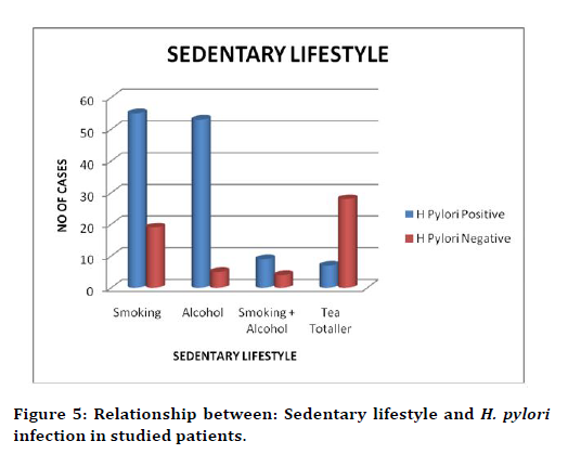
Figure 5. Relationship between: Sedentary lifestyle and H. pylori infection in studied patients.
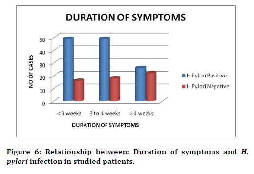
Figure 6. Relationship between: Duration of symptoms and H. pylori infection in studied patients.
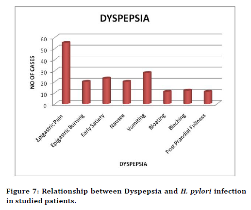
Figure 7. Relationship between Dyspepsia and H. pylori infection in studied patients.
The endoscopic finding revealed that the patients with antral erosions had higher chances (37%) of H. pylori infections. Next to them, patients with peptic ulcer (27%) were prone for infections followed by reflex oesophagitis (25%), Gastroesophageal Reflex Disease (10%). Surpringly, a single patient with gastric cancer showed lowest infection rate in the study. The results were summarized in Table. 8 and Figure. 8. A comparative analysis of H. pylori infection and Rapid Urease Test in the present study (Table 9 and Figure 9) revealed that the urease positivity could be a strong indicator for the confirmation of the infection and statistically significant (p< 0.05, Chi-square Value (χ2=17.34). In addition to, our study results also showed that biopsy could be another indicator the infection in patients (Table 10 and Figure 10). The results were statistically significant. In this study the patients who were confirmed with H. pylori positive, treatment with Triple regimen was given for a duration of 2 weeks and when they were followed up after 3 months. at the end of drug course, 95% patients with Gastroesophageal Reflex Disease and 93% of patients with Antral Erosions were cured followed by Peptic Ulcer Disease (91%), Reflex Oesophagitis (89%), of the cases were cured with respect to. However, the Gastric Malignant cases were referred to the Oncology department for further treatment (Table 11 and Figure 11).
| Endoscopic findings | Male | % | Female | % | Overall, no of cases | % |
|---|---|---|---|---|---|---|
| Peptic Ulcer Disease | 27 | 55 | 22 | 45 | 49 | 27 |
| Reflex Oesophagitis | 39 | 87 | 6 | 13 | 45 | 25 |
| Antral Erosions | 45 | 69 | 20 | 31 | 65 | 37 |
| Gastroesophageal Reflex Disease | 10 | 55 | 8 | 45 | 18 | 10 |
| Gastric Malignancy | 1 | 33 | 2 | 67 | 3 | 1 |
| Total | 122 | 58 | 180 | 100 |
Table 8: Endoscopic findings and H. pylori infection in studied patients.
| H. Pylori infection Positive | H. Pylori infection Negative | Total | ||||
|---|---|---|---|---|---|---|
| N | % | N | % | N | % | |
| Rapid Urease Test | ||||||
| Epigastric Pain | 41 | 75 | 14 | 25 | 55 | 30 |
| Epigastric Burning | 11 | 55 | 9 | 45 | 20 | 11 |
| Early Satiety | 13 | 57 | 10 | 43 | 23 | 13 |
| Nausea | 12 | 60 | 8 | 40 | 20 | 11 |
| Vomiting | 18 | 64 | 10 | 36 | 28 | 16 |
| Bloating | 6 | 55 | 5 | 45 | 11 | 6 |
| Bleching | 7 | 58 | 5 | 42 | 12 | 7 |
| Post Prandial Fullness | 6 | 55 | 5 | 45 | 11 | 6 |
| Total | 114 | 66 | 180 | 100 | ||
| Chi-square Value (χ2=17.34) P value=0.0000 (< 0.05) | ||||||
Table 9: Rapid Urease Test and H. pylori infection in studied patients.
| Biopsy | H. Pylori infection Positive | H. Pylori infection Negative | Total | |||
|---|---|---|---|---|---|---|
| N | % | N | % | N | % | |
| Peptic Ulcer Disease | 32 | 65 | 17 | 35 | 49 | 27 |
| Reflex Oesophagitis | 29 | 64 | 16 | 36 | 45 | 25 |
| Antral Erosions | 48 | 74 | 17 | 26 | 65 | 37 |
| Gastroesophageal Reflex Disease | 12 | 67 | 6 | 33 | 18 | 10 |
| Gastric Malignancy | 3 | 0 | 0 | 0 | 3 | 1 |
| Total | 124 | 100 | 56 | 100 | 180 | |
| Chi-square Value (χ2=14.25) P value=0.0015 (<0.05) | ||||||
Table 10: Biopsy and H. pylori infection in studied patients.
| H. Pylori +ve | Rx for H. Pylori Triple regimen (Clathromycin/Amoxycillin) PP1 |
Duration | Follow up after 3 months | ||
|---|---|---|---|---|---|
| Eradicated % | Not Eradicated % | ||||
| Peptic Ulcer | 32 | 32 | 14 days | 91 | 9 |
| Disease Reflex Oesophagitis | 29 | 29 | 14 days | 89 | 11 |
| Antral Erosions | 48 | 48 | 14 days | 93 | 7 |
| Gastroesophageal Reflex Disease | 12 | 12 | 14 days | 95 | 5 |
Table 11: H. pylori treatment and its eradication.
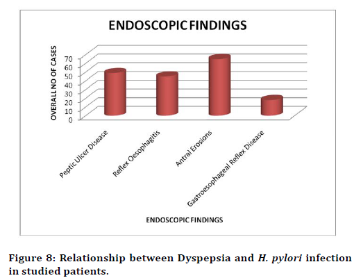
Figure 8. Relationship between Dyspepsia and H. pylori infection in studied patients.
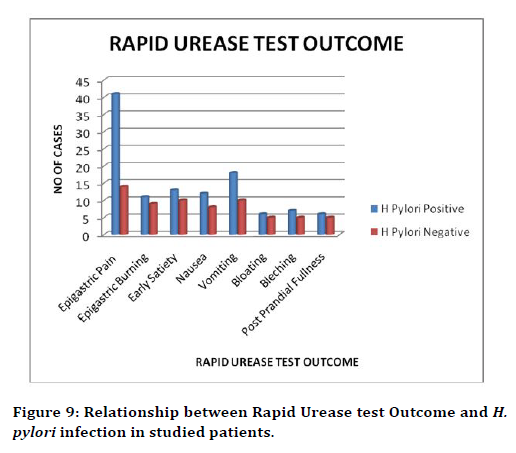
Figure 9. Relationship between Rapid Urease test Outcome and H. pylori infection in studied patients.
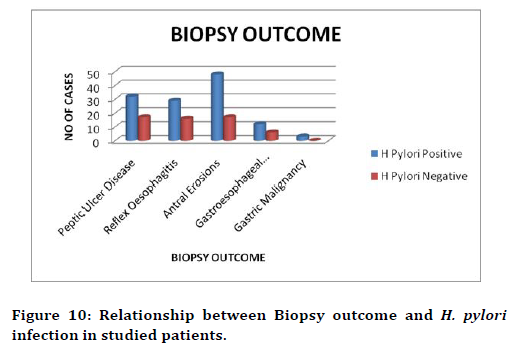
Figure 10. Relationship between Biopsy outcome and H. pylori infection in studied patients.
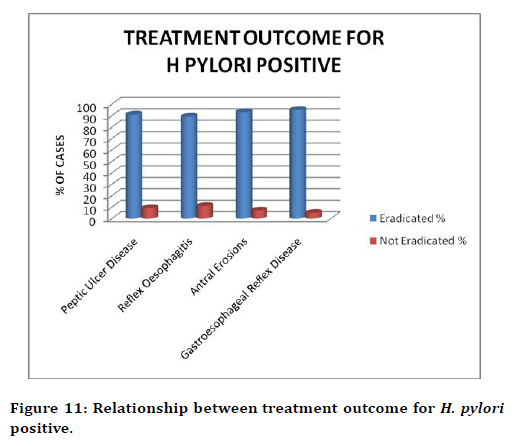
Figure 11. Relationship between treatment outcome for H. pylori positive.
Discussion
H. pylori is a spiral-shaped microaerophilic Gramnegative bacterium that colonizes the gastric mucosa of human beings. The microorganism is the major agent of gastritis and plays an important role in the pathogenesis of peptic ulcer and gastric cancer. H. pylori is believed to infect the host by the fecal-oral route and home to the gastric mucosa. Although it is acid sensitive, H. pylori can survive in the stomach for short periods by neutralizing the gastric acid. The current therapy for H. pylori infection is efficacious but the treatment regimen is complex and demanding on the patient and it does not provide resistance to future infections. The aim of this study is to determine the incidence of H. pylori infection in dyspeptic patient. In this study 180 patients undergoing upper gastrointestinal endoscopy were interviewed. Data concerning age, sex, occupational status, socio economic status, and sedentary lifestyle, duration of symptoms of dyspepsia, endoscopic findings, rapid urease test and biopsy were obtained during interviews.
The present study demonstrated that highest infection occurred at the age of 50 (39%) and this finding is agreed with similar studies in developed countries which showed that the infection begin in younger age and increasing annually with age. This is may be due to the reason that the number of participant of the older age in this study. In another study conducted in south of Brazil (2005), the author showed totally different results. Among the 563 eligible individuals, the prevalence rate of H. pylori infection was 63.4%. In crude analyses, prevalence was associated with increasing age [11]. Our results showed that the male were more prone for H. pylori infection and this accordance with previous study [11]. Several studies reported the the strong correlation between H. pylori infection and occupation [3]. The prevalence of HP infection varies among countries and within a country. The risk factors of HP infection confirmed that low socioeconomic status, crowded living conditions in childhood, low educational level of parents and unreliable well-water supply in the childhood household are associated with the infection [1]. In this work, there was significant correlation detected between socioeconomic level and HP infection. This could be attributed to a higher admission rate of patients of upper medium socioeconomic level 35%.
The present study demonstrated that the highest positive results were in 41 % are reported as smokers. In another study which agree with our result, El- Barrawy et al. [4], demonstrated that infection prevailed mostly (70%) in smokers (113 out of 161). According to odds ratio, the risk of infection was 5.3 times higher for smokers than nonsmokers, which was significant. They attributed their finding to the destructive effect of smoking on the immunity of gastric mucosa and lining layers and hence increasing its susceptibility to infection by H. pylori. Communal Shisha smoking might carry the risk of passing the infection from a diseased person to an uninfected one, as oral-to-oral infection. Tables 6 and 7 In this study most common symptoms presented by H. pylori infection were more common in patients suffering from epigastric pain (30%). Other studies added that the presence of epigastric pain, especially if associated with dyspeptic symptoms is more associated with abnormal gastric pathology. Endoscopic findings in dyspeptic patients’ Antral mucosa is the main target for HP colonization of 37% in this study. Followed by peptic ulcer disease 27%, reflex oesophagitis 25%, Gastroesophageal Reflex Disease 10% and one case of gastric cancer were positive for H.pylori. Gastritis occurs due to the inflammation of the gastric mucosa that can be caused by HP. Blaser reported that chronic active gastritis is mainly induced by HP. We have discovered a significant difference (p=0.027) between positive HP histopathology and grading of gastritis was discovered similar to Akbar et al. [9]. In healthy population, HP seroprevalence ranges from 25% in developed countries up to 80% in developing countries [6]. In this setting, it was 73.3%. This result is comparable to an earlier study by Al-Moagel et al. [8]. We must mention that our small control number (15 healthy population) should be reported as a limitation in this study. In addition, the HP seroprevalence among those individuals was mostly an indication of past HP exposure not an active infection. In this study when the comparison was made between the H Pylori patients in both the classification (positive and negative) with respect to Rapid Urease Test, P value is 0.0000 (< 0 .05) which concludes the test that the H Pylori positive cases are significant. The test which was performed on biopsy collected from patients during upper gastroscopy proved to be rapid and simple. In our study two tests were used to evaluate H. pylori infection. 1- Invasive test required endoscopy for (URUT, MB staining). Unfortunately, at present, no single test can be relied upon to detect H. pylori infection and a combination of tests is recommended as gold standard. A patient was considered an H. pylori positive if culture alone or histology plus rapid urease test (RUT) were positive. The decision by the physician to select the proper part to collect a biopsy during gastroscopy would greatly affect the outcome of the invasive tests such as methylene blue staining and Ultra rapid urease tests. A study in Yemen showed that the prevalence of H. pylori infection among 275 dyspeptic patients was 82.2%. In their study they depended on URUT for detect the infection [7]. The present study demonstrated the systemtic need of diagnosis and follow up to eradicate the H. pylori infections.
Conclusion
The present study aimed to detect incidence of H. pylori infection in dyspeptic patients and attempt to determine possible risk factors is acquisition of the microorganism. Our results showed that age, gender and socioeconomic status play a pivotal role in determining the severity of the disease. Further, we found a strong correlation with urease positivity and H. pylori. These findings certainly would be useful to prevent and eradicate the H. pylori infections.
Funding
No funding sources.
Ethical Approval
The study was approved by the Institutional Ethics Committee
Conflict of Interest
The authors declare no conflict of interest.
Acknowledgments
The encouragement and support from Bharath University, Chennai is gratefully acknowledged. For provided the laboratory facilities to carry out the research work.
References
- Brown L. Helicobacter pylori: Epidemiology and routes of transmission. Epidemiol Rev 2000; 22:83-97.
- Zmira S, Haim S, Yaron N, et al. Resistance of Helicobacter pylori isolated in Israel to metronidazole, clarithromycin, tetracycline, amoxicillin and cefixime. J Antimicrob Chemother 2002; 49:1023 -1026.
- Peters C, Schablon A, Harling M, et al. The occupational risk of Helicobacter pylori infection among gastroenterologists and their assistants. BMC Infect Dis 2011; 11:154.
- El Barrawy MA, Morad MI, Gaber M. Role of Helicobacter pylori in the genesis of gastric ulcerations among smokers and nonsmokers. Eastern Mediterranean Health J 1997; 3:316-321.
- Abo-Shadi MA, El-Shazly TA, Al-Johani MS. Clinical, endoscopic, pathological and serological findings of Helicobacter pylori infection in Saudi patients with upper gastrointestinal diseases. J Adv Med Med Res 2013; 1109-1124.
- Bardhan PK. Epidemiological features of Helicobacter pylori infection in developing countries. Clinical Infect Dis 1997; 25:973-978.
- Gunaid AA, Hassan NA, Murray-Lyon I. Prevalence and risk factors for Helicobacter pylori infection among Yemeni dyspeptic patients. Saudi Med J 2003; 24:512-517.
- Al-Moagel MA, Evans DG, Abdulghani ME, et al. Prevalence of Helicobacter (formerly Campylobacter) pylori infection in Saudia Arabia, and comparison of those with and without upper gastrointestinal symptoms. Am J Gastroenterol 1990; 85.
- Akbar DH, Eltahawy AT. Helicobacter pylori infection at a university hospital in Saudi Arabia: prevalence, comparison of diagnostic modalities and endoscopic findings. Indian J Pathol Microbiol 2005; 48:181.
- Abdollah B, Robert W, Remon A, et al. Seroepidemiology of Helicobacter pylori infection in a population of Egyptian children. Inter J Epidemiol 2000; 29:928 -932.
- Ina S, Jose B, Ari S, et al. Prevalence of Helicobacter pylori infection and associated factors among adults in Southern Brazil: a population-based cross-sectional Study. BMC Public Health. 2005; 5:1-10.
Author Info
G Srinath, RNM Francis and V Ramalakshmi*
Department of General Surgery, Sree Balaji Medical College & Hospital, Bharath Institute of Higher Education and Research, Chennai, Tamil Nadu, IndiaCitation: G Srinath, RNM Francis, V Ramalakshmi, Development, A Study on Incidence of Helicobacter pylori Infection in Dyspeptic Patients in Sree Balaji Medical College and Hospital, J Res Med Dent Sci, 2021, 9 (4): 392-399.
Received: 20-Mar-2021 Accepted: 21-Apr-2021
