Case Report - (2022) Volume 10, Issue 4
Aesthetic Rehabilitation of Missing Anterior Tooth using Loop Connector FPD to Maintain Midline Diastema: A Case Report
*Correspondence: Arun Prasad, Department of Prosthodontics, KarpagaVinayaga Institute of Dental Sciences, India, Email:
Abstract
Esthetic rehabilitation of missing anterior teeth poses a challenge in fabrication of prosthesis. Tooth loss can occur due to reasons like trauma, surgical excision of tumors, caries, periodontal problems, etc. Over a period of time, tooth loss results in loss of alveolar bone and loss of soft tissue. Restoring the combined defects of alveolar bone and soft tissue can be achieved by fixed–removable prosthesis called Andrew’s Bridge System. It consists of fixed retainer with removable pontic. This system is particularly indicated for patients with extensive loss of alveolar bone and associated soft tissue structures. This case report highlights the esthetic rehabilitation process using Andrew’s Bridge System.Keywords
Esthetics, Andrew’s bridge system
Introduction
Esthetics being the main concern for any age group of people. Unaesthetic appearance can be due to missing/ loss of anterior teeth which can be of various reasons like trauma, surgical excision of tumors, caries, periodontal problems, etc. This alveolar bone and soft tissue defects can be easily understood by Seibert’s Classification [1,2]:
Class N: Normal ridge.
Class I: A ridge with loss of facio lingual width, with normal apico coronal height.
Class II: A ridge with loss of ridge height with normal ridge width.
Class III: A ridge with loss of both height and width.
These defects can be treated by either surgical correction with extensive grafting or by prosthodontics management. The dentist and the patient mostly do not prefer surgical correction because of prolonged, complex treatment procedure and questionable prognosis. Thus, prosthodontics management is considered to be the treatment of choice for achieving esthetics by the fabricating a prosthesis that helps in supporting the soft tissues which further enhances the facial appearance and smile.
Case Report
A patient named, Mr. Jayachandran reported to Department of Prosthodontics with the chief complaint of missing teeth in lower front teeth region. Patient gave history of trauma four months back. Extra oral examination reveals a square shaped facial profile with loss of lip support. Intraoral examination reveals non vital 11, 12 and missing in relation to 31, 32, 41, 42, and 43.
Treatment options
The other treatment options which might be considered:
✓ Conventional removable partial denture.
✓ Conventional fixed partial denture.
✓ Bone graft with implant placement.
✓ Andrew’s fixed-removable partial denture.
Conventional removable partial denture was not chosen because of loss of retention. Conventional fixed partial denture was not chosen as there was too much loss of ridge defect and loss of lip support. Extensive bone graft with implant placement was not chosen as it was a time consuming and complex procedure. Andrew’s fixed–removable prosthesis was the treatment of choice because of its combined advantages of fixed and removable prosthesis which could be fabricated by enhancing soft tissue support and lip support without compromising esthetics.
Treatment procedure
Clinical steps
Endodontic treatment was done for non-vital 11, 12. IOPA revealed periodontally sound 43, 44. Intentional RCT was done for 33, 44 followed by tooth preparation for endodontically treated 11, 12, 33, and 44. Final impression was made using addition silicone (putty, light body) for fabrication of framework for Andrew’s Bridge.
Laboratory steps
Final master cast was duplicated and refractory cast was poured. Wax pattern made for retainers in 33 and 44. The bar (Rhein 83 bar attachment) was attached. Spur attached with crucible former and casting was done using Co-Cr metal to reduce the weight of the prosthesis. Metal trial was done. In order to maintain oral hygiene, gap was left in mouth to about 3-4 mm measured from crest of the ridge.
Fabrication of acrylic portion of Andrew’s bridge
Retainers were layered with porcelain and bisque trial was done by closing the gap using carding wax. Bisque trial was picked up with light body impression and master cast was poured for fabrication of acrylic portion of Andrew’s Bridge. Cast was poured and female portion of the bar was fabricated using Co-Cr. Fit was checked and adjustments made followed by which undercuts were blocked out in cast using plaster of Paris for the fabrication of denture base and teeth setting was done similar to RPD. Wax trial was done. Processing of this waxed up pontic portion was done using cold cure acrylic resin (Triplex Cold Cure). Retainers were cemented on abutments with type I GIC followed by insertion of acrylic portion of Andrew’s Bridge (Figure 1-10).
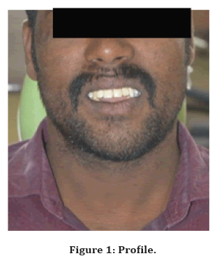
Figure 1. Profile.
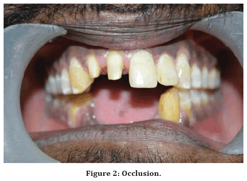
Figure 2. Occlusion.
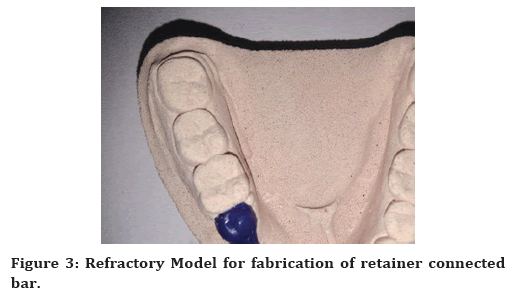
Figure 3. Refractory Model for fabrication of retainer connected bar.
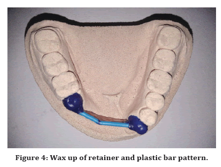
Figure 4. Wax up of retainer and plastic bar pattern.
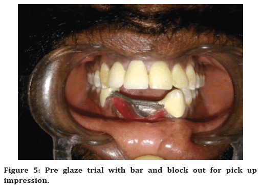
Figure 5. Pre glaze trial with bar and block out for pick up impression.
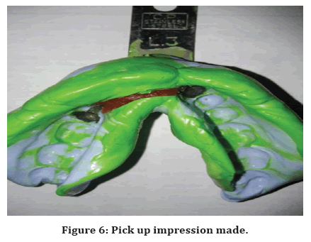
Figure 6. Pick up impression made.
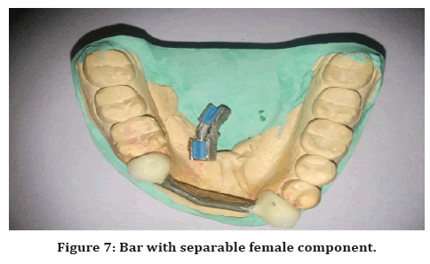
Figure 7. Bar with separable female component.
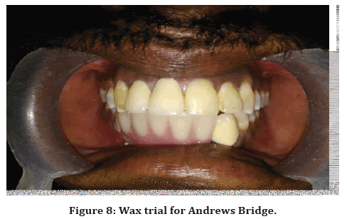
Figure 8. Wax trial for Andrews Bridge.
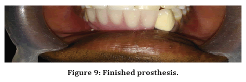
Figure 9. Finished prosthesis.
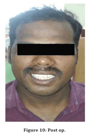
Figure 10. Post op.
Discussion
The most commonly seen defects are the combined Class III defects (56% of cases), followed by horizontal defects Class I (33 % of the cases) [3, 4]. Dr. James Andrews of Amite, Louisiana first introduced the fixed removable Andrew's system (Institute of Cosmetic Dentistry, Amite, L.A.) in 19757. The Andrews bridge consists of a metal bar and sleeve. The pontic portion of the fixedremovable partial denture is removable. This permits easy cleansing of underlying tissues. The bar is soldered to the retainers at a slight mesiodistal inclination for retention of the pontic portion. The bar is available in:
✓ Four length form to replace from one to four teeth.
✓ Four different curvatures so the correct bar can be chosen to follow the form of the residual ridge.
✓ Two widths-the standard or normal.
✓ Thin-line type.
✓ For narrow inter occlusal distance, thin-line bar is used. For adequate inter occlusal space is present, the normal-width bar is used [5].
The Andrew’s System is usually of two types based on the area of bar attachment:
✓ Pontic supported.
✓ Bone Anchored or Implant supported Andrew’s Bar System [6].
The path of withdrawal of the removable segment should be considered. If the bar has been inclined, the path of withdrawal of the flange portion should be diverged from the displacing forces exercised on the fixed portion. Though the forces exerted are minimal, the repeated removal and insertion of the removable segment in the same direction results in a broken cement bond.
The fixed removable partial denture is indicated for:
✓ Patients whose residual ridge has a relationship to the opposing dentition that would.
Prohibit the esthetic placement of the pontic of a fixed partial denture.
✓ Patients requiring diastema as to harmonize the natural dentition.
✓ Patients who have extensive alveolar bone and tissue loss [7].
The drawbacks of treating patients with Andrew’s Bridge include:
✓ Food lodgment/plaque accumulation leads to proliferation of tissues under the prosthesis.
✓ Repeated change of nylon/plastic caps in female portion of prosthesis.
The major advantage of the Andrew's System is that the pontic assembly can be removed to facilitate hygiene procedures and may be relined as the ridge resorbs. Failure of such prosthesis is found in the literature. The failures are mainly due to inadequate soldering which can be avoided by attaching retainers to the bar in a single casting [8].
Conclusion
Andrew's bridge is a good treatment of choice for Seibert’s Class III ridge defects. It permits rehabilitation of alveolar bone and soft tissue defects when conventional treatments are not feasible. The patient treated with the Andrew’s Bar System in this case report was followed-up for a period of 3 years. The patient was found to be comfortable with the prosthesis without any complaint and showed an improved esthetics and phonetics. This case report presents a technique which is simple, economical, provides better support, stability, retention, and few chair side procedure.
References
- Seibert J. Reconstruction of deformed, partially edentulous ridges, using full thickness onlay grafts, Part I. Technique and wound healing. Compend Contin Educ Dent 1983; 4:437-53.
- http://dental.downloadmedicalbook.com/2609/contemporary-fixed-prosthodontics-3rd-edition.html
- Abrams H, Kopczyk RA, Kaplan AL. Incidence of anterior ridge deformities in partially edentulous patients. J Prosthetic Dent 1987; 57:191-4.
- Bhapkar P, Botre A, Menon P, et al. Andrew's bridge System: An esthetic option. J Dent Allied Sci 2015; 4:36.
- Immekus JE, Aramany M. A fixed-removable partial denture for cleft-palate patients. J Prosthetic Dent 1975; 34:286-91.
- Soni R, Yadav H, Kumar V. Andrew's bridge system: A boon for huge ridge defect in aesthetic zone. J Oral Biol Craniofacial Res 2020; 10:138-40.
- Everhart RJ, Cavazos Jr E. Evaluation of a fixed removable partial denture: Andrews bridge system. J Prosthetic Dent 1983; 50:180-4.
- Janani T. Rehabilitation of sieberts class iii defect using fixed removable prosthesis (Andrew's bridge): A CASE REPORT. J Pharm Sci Res2016; 8:1045.
Indexed at, Google Scholar, Cross Ref
Indexed at, Google Scholar, Cross Ref
Indexed at, Google Scholar, Cross Ref
Indexed at, Google Scholar, Cross Ref
Indexed at, Google Scholar, Cross Ref
Author Info
1Department of Prosthodontics, KarpagaVinayaga Institute of Dental Sciences, Chinnakollambakkam, Madhuranthagam, Tamil Nadu, India
Citation: Arun Prasad, Nagappan, Gokul, Sameera Yusuf, Raghunathan, Visalatchi, Vinoth kumar, Aesthetic Rehabilitation of Missing Anterior Tooth using Loop Connector FPD to Maintain Midline Diastema: A Case Report, J Res Med Dent Sci, 2022, 10 (4):93-97.
Received: 30-Mar-2022, Manuscript No. JRMDS-22-59041; , Pre QC No. JRMDS-22-59041 (PQ); Editor assigned: 01-Apr-2022, Pre QC No. JRMDS-22-59041 (PQ); Reviewed: 15-Apr-2022, QC No. JRMDS-22-59041; Revised: 20-Apr-2022, Manuscript No. JRMDS-22-59041 (R); Published: 27-Apr-2022
