Research - (2021) Volume 9, Issue 1
An anatomical Pattern of the Origins of the Ventral Branches of the Abdominal Aorta: A Study Among Sudanese Cadavers
Hosam Eldeen Elsadig Gasmalla1,2, Mohammed H Karrar Alsharif3,4* and Mohamed AH Abdel Galil5
*Correspondence: Mohammed H Karrar Alsharif, Department Of Basic Medical Science, Colleges of Medicine, Prince Sattam Bin Abdulaziz University, Saudi Arabia, Email:
Abstract
The pattern of the origin of the ventral branches of abdominal aorta forms the anatomical basis for abdominal and endovascular surgeries, in this study, we inspected this pattern in Sudanese cadavers (31 males), no variations were found in the origins of the ventral branches of the abdominal aorta, the coeliac trunk, SMA and IMA were found to originate at the level of L1, L1 and L3 respectively, the bifurcation of the aorta was at the level of L4, the most constant artery regarding the site of origin was the SMA, at the level of L1, the mean distance from the coeliac trunk, SMA and IMA to the bifurcation of the aorta were 11.9 cm, 11.2 cm and 4 cm respectively, the mean distances from the coeliac trunk to SMA and from SMA to IMA were 1.2 cm and 6.8 cm respectively. To our knowledge, this is the first study to be conducted on Sudanese subjects.
Keywords
Coeliac trunk, Superior mesenteric artery, Inferior mesenteric artery, Bifurcation of the aorta
Introduction
The abdominal aorta brings the blood to the abdomen, pelvis, and lower limbs; it passes through the diaphragm to appear in the abdomen at the level of the lower border of the T12, posterior to the median arcuate ligament and ends by bifurcating into right and left common iliac arteries at the level of L4.
The ventral (anterior) branches of the abdominal aorta are in order from above downwards: coeliac, superior mesenteric and inferior mesenteric arteries.
The coeliac trunk is the artery of the foregut, it supplies the gut from the lower part of the oesophagus down to the opening of the bile duct into the duodenum, and it also supplies the foregut derivatives (liver and pancreas), it originates from the aorta at the level of T 12 vertebra.
The superior mesenteric artery arises from the front of the aorta a centimetre below the coeliac trunk at the level of L1 vertebra, and it is the artery of the midgut, starting from the duodenum below the entrance of the bile duct it supplies the rest of duodenum, jejunum, ileum, ascending colon and the proximal two-thirds of the transverse colon.
The inferior mesenteric artery arises from the front of the aorta opposite the L3 vertebra; it is much smaller than the superior mesenteric artery, it supplies the distal third of the transverse colon, descending colon, sigmoid colon and part of the rectum [1,2].
Literature Review
George et al. [3] examined 120 cadavers in his study, 115 were males, and 5 were females, he stated that the level of origin for the coeliac trunk, SMA, IMA and bifurcation of the aorta were at L1, L1, L3 and L4 respectively, he also reported the mean distances between the different origins.
The origin of the coeliac trunk is at level with T12 according to McMinn et al. and Prakash et al. [4,5] and it is at the level between T12 and L1 according to Selvaraj et al. [6]. Another study reported variable results with origins of the trunk at T12 in 34% of cases and between T12-L1 in 31% of the cases of the study [7]. It originates at the level of L1, according to Anson [8]. The origin of the SMA is at level with L1 [3-5], or between L1 and L2 [8]. The origin of IMA is at levels with L3 [3-5]. Or at L3 and intervertebral disk of L3/ L4 [8]. The incidence of additional vessel such as double coeliac trunk is about 5% [9], middle mesenteric artery, an anomalous origin of a middle colic artery is noted and arises between the superior mesenteric and inferior mesenteric arteries [10-13].
Less number of vessels my originate, the typical coeliacomesenteric trunk can be regarded as a variation of the arterial convergence at its origins progressing further between the coeliac trunk and the superior mesenteric artery [14], the occurrence of this variation is stated to be 1% - 2.7% [15], 2% according to Lippert et al. [16] and 1.5 % according to Kornafel et al. [17] many cases were reported [18,19].
the coeliac-bimesenteric trunk is a condition in which all three arteries supplying the abdominal digestive organs have converged into one trunk [14], in addition to the previous two conditions, the inferior mesenteric can arise from the superior mesenteric [20], but all the above variants are uncommon and appear as case reports. A case of right hepatic artery arising independently from the aorta also reported by Oran et al. [21].
Many studies are done to determine the relation between the origins of the ventral branches of the abdominal aorta and the vertebral column, and the relations and distances between each of them [22], examined these relationships, by taking the length of the abdominal aorta and measurements of the position of origin of the coeliac artery, SMA, IMA and renal arteries. He found that the mean level of bifurcation of the aorta was at the lower third of the body of L4, with the coeliac artery, SMA, renal arteries and IMA arising at the level of the T12/L1 intervertebral disc, upper third of the body of L1, lower third of the body of L1 and lower third of the body of L3, respectively, he concluded that the coeliac artery and SMA are both useful landmarks for determining the position of the renal arteries and the origin of the coeliac artery is considered to be the least variable, with that of the IMA showing the greatest variability, the length of abdominal aorta approximately was 12.4cm.
While the above study concentrates on using the ventral branches of the abdominal aorta in relation to renal arteries [23], made the bifurcation of the aorta as a landmark for his study on 100 specimens, 60 were males, 31 were females, and the remaining nine were not recorded, he found that the abdominal aorta is longer in males than females, and the origins of the branches were closer to the bifurcation in females, he also reported the mean distances and complete range for the origins.
General Objectives
To describe the pattern of the origin of the ventral branches of the abdominal aorta, forming an anatomical basis for abdominal and endovascular surgery.
Specific Objectives
To Detect the incidence of variations in the origin of the ventral branches of the abdominal aorta.
To Detect the relation between the origin of the ventral branches of abdominal aorta & the vertebrae.
To measure the distances between the different origins of the ventral branches of the abdominal aorta.
To measure the distances between the different origins of the ventral branches of abdominal aorta and aortic bifurcation.
Study Design
This study is a descriptive study, performed on adult male cadavers in the dissection rooms of 8 universities in Khartoum, Sudan, namely: Khartoum University, Alneelain University, University of medical sciences and technology USMT, Bahar El-Gazal University, Khartoum college of medical sciences KCMS, Ribat National University, Alzaeem Al-Azhari University and the University of Sciences & Technology.
The study included 31 cadavers, all are males, there were no great ethnic variations, data collected using ruler and string as measurement tools, the distance was measured from the centre of each vessel at its origin, the vertebral level was detected after exposing the lumbar and lower part of the vertebral column, and counting was started from the 5th lumbar vertebra and upwards.
Excluding criteria include any dissecting manipulation that may change the original anatomical position of the aorta and its ventral branches, such as cadavers with abdominal aorta being removed, cadavers with detached abdominal aorta from the vertebral column, and cadavers with ventral branches which were difficult to be identified.
Two checklists were performed by two different persons for the same cadaver for more accuracy.
Results
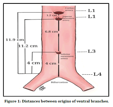
Pattern of the origin of the ventral branches of abdominal aorta forms the anatomical basis for abdominal and endovascular surgeries was studied among Sudanese cadavers (Figure 1).

Figure 1. Distances between origins of ventral branches.
In about 90% of cases, the coeliac trunk originates at the level of L1 (Table 1), the superior mesenteric artery originates at the same level L1 in all cases, (Table 2), as for the inferior mesenteric artery, the level of the origin is L3 in more than 80% of cases (Table 3). The bifurcation of the aorta is at the level of L4 in 87.1% of cases (Table 4), the mean distances from the origins of the ventral branches to the aortic bifurcation are 11.9 cm for the coeliac trunk, 11.2 cm for the superior mesenteric artery and 4 cm for the inferior mesenteric artery (Table 5). The mean distance between the coeliac trunk and the superior mesenteric artery is 1.2 cm, and from the superior to the inferior mesenteric arteries is 6.8 cm (Tables 6).
| Structure | Level | Number of cases | Percentage |
|---|---|---|---|
| Vertebra | T12 | 1 | 3.20% |
| Disc between: | T12/L1 | 2 | 6.50% |
| Vertebra | L1 | 28 | 90.30% |
Table 1: Vertebral level of the origin of the coeliac trunk (n=31).
| Structure | Level | Number of cases | Percentage |
|---|---|---|---|
| Disc between: | T12/L1 | - | - |
| Vertebra | L1 | 31 | 100% |
| Disc between: | L1/L2 | - | - |
Table 2: Vertebral level of the origin of the superior mesenteric artery (n=31).
| Structure | Level | Number of cases | Percentage |
|---|---|---|---|
| Vertebra | L3 | 26 | 83.80% |
| Disc between: | L3/L4 | 2 | 6.50% |
| Vertebra | L4 | 3 | 9.70% |
Table 3: Vertebral level of the origin of the inferior mesenteric artery (n=31).
| Structure | Level | Number of cases | Percentage |
|---|---|---|---|
| Vertebra | L4 | 27 | 87.10% |
| Disc between: | L4/L5 | 3 | 9.70% |
| Vertebra | L5 | 1 | 3.20% |
Table 4: Vertebral level of the bifurcation of the aorta (n=31).
| Mean | Range | |
|---|---|---|
| Coeliac trunk | 11.9 | 8.8 – 14 |
| SMA | 11.2 | 7 – 12.5 |
| IMA | 4 | 2 – 5 |
Table 5: Distances of the origin of the ventral branches from the aortic bifurcation. (In centimeter) (n=31).
| From | To | Mean | Range |
|---|---|---|---|
| Coeliac trunk | SMA | 1.2 | 0.5 – 2 |
| SMA | IMA | 6.8 | 05-Sep |
Table 6: Distances of the origin of the ventral branches from each other. (In centimeter) (n=31).
Discussion
Using data collected from 31 cadavers from 8 universities, it was noticed that all cadavers were males (except for one un-dissected female in the University of Khartoum). This number of available cadavers was less than that expected. Most of the other cadavers were distorted. The sample size was, therefore, limited (31 samples) and was restricted to male cadavers only. There were no variations in the origins, as stated before, most of the variations described by other authors were case reports. Two studies reporting on such variations concluded that variations in the coeliac trunk were about 5% [9], and the other one found that the incidence of a common origin for the coeliac trunk and SMA was in the range of 1% - 2.7% [15].
The origin of the coeliac trunk was at the level of L1 in 90% of samples (Table 1), it was originated at T12 according to [4], at T12/L1 intervertebral disc according to Pennington et al. [22] and at L1 as stated by Feller et al. [3,8,23]. The origin of the SMA was at level of L1 in all samples, although [8] stated that it originates from L1/L2, most of the studies agree with this finding [22] stated that the origin of the SMA is constant and can be used as a landmark for the renal arteries in surgeries. The length of the vertebral body is about 3 cm, and this can justify that the coeliac trunk and SMA can share the same origin at the level in front of the L1 vertebra. It is noted that the most constant artery regarding its site of origin is the SMA.
The origin of the IMA at L3 goes with the findings of George et al., McMinn et al., Prakash et al. and Kornafel et al. [3-5,17] however, it was originated from L3 and intervertebral disk of L3/L4 according to Anson et al. [8]. The result of this study regarding the level of the origin of the three ventral branches similar to that reported by George et al. [3].
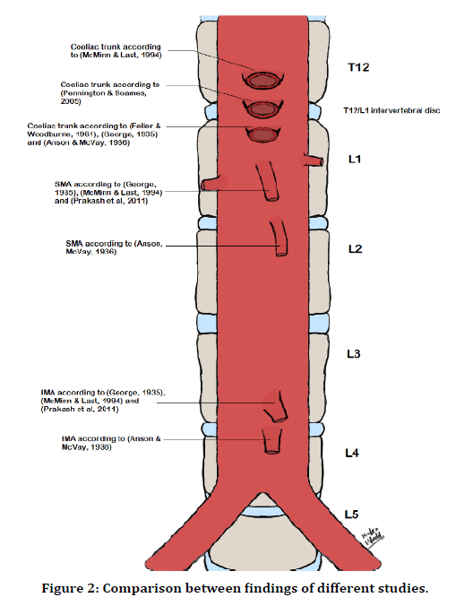
The distances between the origins were measured using the bifurcation of the aorta as a reference point because it is a landmark for many vascular variations and is easily accessible in dissection or during surgeries [3,23]. The findings regarding the distances from the coeliac trunk to the SMA and from the SMA to the IMA are in line with those reported by George et al. Feller et al. [3,23] (Tables 7 and 8). In this study aorta if found bifurcated at L5 (3.2%), and at L4/ L5 (9.7%), [24] reported 25% for the bifurcation at L5. A comparison between different studies is shown and summarized in (Figure 2).
| George | Feller-woodburne | This study | |
|---|---|---|---|
| 1935 | 1960 | ||
| Coeliac trunk | 13.2 | 12.6 | 11.9 |
| Superior mesenteric | 11.7 | 11 | 11.2 |
| Inferior mesenteric | 4.6 | 4.2 | 4 |
Table 7: Distances of the origin of the ventral branches from the aortic bifurcation. (In centimeter) Comparison of the means in three studies (n=31).
| From | To | George | Feller-woodburne | This study |
|---|---|---|---|---|
| 1935 | 1960 | |||
| Coeliac trunk | SMA | 1.6 | 1.6 | 1.2 |
| IMA | IMA | 7.1 | 6.8 | 6.8 |
Table 8: Distances of the origin of the ventral branches from each other. (In centimeter) Comparison of the means in three studies (n=31).
Figure 2. Comparison between findings of different studies.
Conclusion
No variations were found in the origins of the ventral branches of the abdominal aorta. The coeliac trunk, SMA and IMA were found to originate at the level of L1, L1 and L3 respectively, the bifurcation of the aorta was at the level of L4.
The most constant artery regarding the site of origin was the SMA, at the level of L1.
The mean distance from the coeliac trunk, SMA and IMA to the bifurcation of the aorta were 11.9cm, 11.2cm and 4 cm respectively.
The mean distances from the coeliac trunk to SMA and from SMA to IMA were 1.2cm and 6.8cm respectively.
All samples were from male cadavers.
Acknowledgments
We are grateful to prince Sattam Bin Abdulaziz University, Al-Kharj, KSA for their support and encouragement to publish Research.
Authors Contributions
Hosam Eldeen Elsadig Gasmalla: introduction, design, data collection, discussion, and manuscript review. Mohammed H. Karrar Alshari: methodology, data collection, data analysis, and manuscript editing and review. Mohamed A. H. Abdel Galil: data collection and entry, literature search and methodology, manuscript preparation, editing, and review. All the authors have read and agreed to the final manuscript.
Notes on the contributors
Hosam Eldeen Elsadig Gasmalla MBBS, M.Sc., PgDip., MHPE, PhD Anatomist, and Medical Education specialist. In addition to his experience in medical education (emphasis on students’ assessment), he has got an administrative experience as a founding director of Education Development Centers (EDC) in two institutes in Sudan. As an anatomist, he is an experienced lecturer of human anatomy and histology for more than 13 years, with a range of publications from original articles to textbooks.
Mohammed Hamid Karrar Alsharif B.Sc., M.Sc. Anatomy, Histology and Embryology specialist. In addition to his academic experience in teaching Human Anatomy, Histology, and Embryology in Saudi Arabia and many Sudanese Universities for more than 11 years. As well as his ongoing research fellowship position at the Department of Histology and Embryology, Faculty of Medicine, Ondokuz Mayis University, Turkey, he used to pursue studies on peripheral nerve regeneration; besides that, he is a Ph.D. candidate in Clinical Anatomy at Department of Anatomy, National University, Sudan. In addition, the author has publications reached over 15, especially in the Human Anatomy variant, medical education, and Radiographic Anatomy, besides much other ongoing research.
Funding
This research received no external funding.
Competing Interests
The authors declare no competing interests.
Data and Materials Availability
All data associated with this study are present in the paper.
References
- Gray H, Standring S, Ellis H, et al. Gray's anatomy: The anatomical basis of clinical practice: Elsevier Churchill Livingstone; 2005.
- Moore KL, Dalley AF, Agur AMR. Clinically oriented anatomy: Lippincott Williams & Wilkins; 2006.
- George R. Topography of the unpaired visceral branches of the abdominal aorta. J Anatomy 1935; 69:196.
- McMinn RMH, Last RJ. Last's anatomy, regional and applied: Churchill livingstone 1994.
- Prakash VM, Rajini T, Shashirekha M. The abdominal aorta and its branches: anatomical variations and clinical implications. Folia Morphol 2011; 70:282-286.
- Selvaraj L, Sundaramurthi I. Study of normal branching pattern of the coeliac trunk and its variations using CT angiography. J Clin Diagnost Res 2015; 9:AC01.
- Yang IY, Oraee S, Viejo C, et al. Computed tomography celiac trunk topography relating to celiac plexus block. Regional Anesthesia Pain Med 2011; 36:21-25.
- Anson BJ, McVay CB. The topographical positions and the mutual relations of the visceral branches of the abdominal aorta. A study of 100 consecutive cadavers. Anatomical Record 1936; 67:7-15.
- Ferrari R, De Cecco C, Iafrate F, et al. Anatomical variations of the coeliac trunk and the mesenteric arteries evaluated with 64-row CT angiography. La Radiol Med 2007; 112:988-998.
- Uluçam E, Yılmaz A, Cigali BS, et al. Middle colic artery originating directly from aorta as a middle mesenteric artery. Balkan Med J 2009; 2009.
- Lawdahl R, Keller F. The middle mesenteric artery. Radiol 1987; 165:371-372.
- Yoshida T, Suzuki S, Sato T. Middle mesenteric artery: An anomalous origin of a middle colic artery. Surg Radiol Anatomy 1993; 15:361-363.
- Pillet J. Details concerning lowering of the colon in case of unusual distribution of arteries. A case of middle mesenteric artery. La Presse Méd 1961; 69:1647.
- Katagiri H, Ichimura K, Sakai T. A case of celiacomesenteric trunk with some other arterial anomalies in a Japanese woman. Anatomical Sci Int 2007; 82:53-8.
- Cavdar S, Şehirli Ü, Pekin B. Celiacomesenteric trunk. Clin Anatomy 1997; 10:231-234.
- Lippert H, Pabst R. Arterial Variations in man: Classification and frequency: Bergmann-Verlag JF. 1985; 30-53.
- Kornafel O, Baran B, Pawlikowska I, et al. Analysis of anatomical variations of the main arteries branching from the abdominal aorta, with 64-detector computed tomography. Pol J Radiol 2010; 75:38-45.
- Foghi K, Ahmadpour S. Celiacomesenteric trunk: A case report. Eur J Anat 2014; 18:191-193.
- Bhatnagar S, Rajesh S, Jain VK, et al. Celiacomesenteric trunk: a short report. Surg Radiol Anatomy 2013; 35:979-981.
- Yi SQ, Li J, Terayama H, et al. A rare case of inferior mesenteric artery arising from the superior mesenteric artery, with a review of the review of the literature. Surg Radiol Anatomy 2008; 30:159-165.
- Oran I, Yesildag A, Memis A. Aortic origin of right hepatic artery and superior mesenteric origin of splenic artery two rare variations demonstrated angiographically. Surg Radiol Anatomy 2001; 23:349-352.
- Pennington N, Soames RW. The anterior visceral branches of the abdominal aorta and their relationship to the renal arteries. Surg Radiol Anatomy 2005; 27:395-403.
- Feller I, Woodburne RT. Surgical anatomy of abdominal aorta. Annals Surg 1961; 154:239.
- Lerona PT, Tewfik HH. Bifurcation level of the aorta: Landmark for pelvic irradiation. Radiology 1975; 115:735.
Author Info
Hosam Eldeen Elsadig Gasmalla1,2, Mohammed H Karrar Alsharif3,4* and Mohamed AH Abdel Galil5
1Department of Anatomy, Faculty of Medicine, Al-Neelain University, Khartoum, Sudan2Department of Anatomy, Faculty of Medicine, Sudan International University, Khartoum, Sudan
3Department Of Basic Medical Science, Colleges of Medicine, Prince Sattam Bin Abdulaziz University, Al-Kharj 11942, Saudi Arabia
4Histology and Embryology Department, College of Medicine, Ondokuz Maiz University, Samsun, Turkey
5Department of Anatomy, Faculty of Medicine, University of Khartoum, Khartoum, Sudan
Citation: Hosam Eldeen Elsadig Gasmalla, Mohammed H Karrar Alsharif, Mohamed AH Abdel Galil, An anatomical Pattern of the Origins of the Ventral Branches of the Abdominal Aorta: A Study Among Sudanese Cadavers, J Res Med Dent Sci, 2021, 9 (1): 219-224.
Received: 01-Dec-2021 Accepted: 26-Dec-2020

