Research - (2021) Volume 9, Issue 2
Analysis of Cranial Accessory Foramina in the Sagittal Section of Skull
Vindhiya Varshini V and Karthik Ganesh Mohanraj*
*Correspondence: Karthik Ganesh Mohanraj, Department of Anatomy, Saveetha Dental College and Hospitals, Saveetha Institute of Medical and Technical Sciences (SIMATS), Saveetha University Tamilnadu, Chennai, India, Email:
Abstract
The human skull has numerous openings that enable cranial nerves and blood vessels to exit the skull and supply or receive various structures. These openings are collectively referred to as cranial foramina. In the cranial cavity, the floor is divided into three distinct recesses: the anterior fossa, middle fossa and the posterior fossa. Each cranial fossa lodges numerous named foramina, through which various anatomical structures pass through. Apart from these named foramina occasionally some accessory foramina may also be present. The aim of the study is to analyze the cranial foramina and vascular impression in the sagittal section of the skull. In the present study, a total of 24 sagittal sections of skull bones of unknown sex and without any gross abnormality were collected and subjected for morphometrical analysis. The external and internal surface of the skull bone are examined for the presence of accessory foramina, apart from the presence of regular foramina. If present they were noted and photographed with their location. The results obtained were analysed. There were several foramina present at the various regions, like in the orbit the ethmoidal foramen, near mastoid process the accessory mastoid emissary foramina. The occasional presence of accessory foramina near these sites may pose an unwanted problem to neurovascular surgeons. Thus this study of anatomical variations of foramina of the skull may provide knowledge to neurovascular surgeons, approaching the skull.
Keywords
Accessory foramina, Emissary foramina, Sagittal section of skull, Vascular impression
Introduction
The skull is an important osseous structure that rules a network of neurovascular structures and lymphatic vessels. For these networks to communicate with the entire body, the foramina must provide passage through the skull. The morphology of the foramina creates a protective enclosure for these neurovascular and lymphatic bundles [1]. In the cranial cavity, the floor is divided into three distinct cavities such as the anterior cranial fossa, middle cranial fossa and the posterior cranial fossa. Each cranial fossa lodges numerous named foramina, through which various anatomical structures pass through. Apart from these named foramina occasionally some accessory foramina may also be present. If such variations found it is of both anatomical and clinical importance with reference to the structures passing through it.
The anterior fossa consists of three specific bones: frontal bone, ethmoid bone, and lesser wings of the sphenoid bone. Together, all these bones contribute to the shallowest recess of the cranium. The cribriform plate of the ethmoid bone is porous in its structure and allows for the passage of olfactory axons of cranial nerve (CN) I through its many foramina into the nasal mucosa. These axons of olfactory nerve are responsible for the sense of smell. Also, another foramen worth mentioning is the foramen cecum. It encompasses emissary veins that drain the nasal cavity of blood and reroutes it to the superior sagittal vein. It is largely responsible for cerebral cooling given its valve less architecture [2]. Between anterior and middle fossa there is an transition called superior orbital fissure, which includes a pair of superior orbital fissures that are situated bilaterally between the lesser wing of the sphenoid bone superiorly and the greater wing of the sphenoid bone [3] inferiorly, the fissure provides motor innervation to the ocular muscles and sensory innervation to lacrimal glands and portions of the face.
Middle fossa also comprises three specific bones namely, the sphenoid bone and the paired temporal bones. These are very important neurovascular structures and provide sensory innervation to the face: sense of vision and blood to the cranium. The temporal bone transmits the internal carotid arteries bilaterally [4] through their respective carotid canals before forming the middle cerebral arteries on either side. Which later joins the circle of Willis.
The posterior fossa also comprises three specific bones: A pair of temporal bones and an occipital bone [5]. It is the deepest of the fossae and is responsible for the several passages that contain neurovascular bundles in each region of the fossa. And comprises the largest foramen: Foramen magnum which connects the contents of the skull to various body parts [6]. Thus, present study aims to analyze the cranial foramina and vascular impressions in sagittal sections of the skull.
Materials and Methods
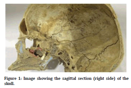
In the present study, a total of 24 sagittal sections of skull bones of known sex and without any gross abnormality were collected and subjected for morphological analysis. The external and internal features and surfaces of skull bone were examined for the presence of accessory foramina apart from the presence of regular named foramina. If presented they were noted and photographed with their location. The presence of accessory foramina were counted in the right and left sagittal sections of skull in males and females. An image of the sagittal section of the skull is shown in Figure 1. The observed data were tabulated. The results obtained were analysed for their occurrence, laterality, gender and anatomical variations.

Figure 1. Image showing the sagittal section (right side) of the skull.
Results
From the male and female sagittal sections of skulls it was observed that there were few accessory foramina present at the various regions, such as in the orbit, near the mastoid process of temporal bone and in the middle cranial fossa. In male and as well as in female skulls, the foramina which are observed are the accessory ethmoidal foramen in the orbital cavity, accessory mastoid emissary foramen in the temporal bone and foramen of Vesalius (sphenoidal emissary foramen). There were variations in the occurrence of these foramina between the right and left side and also between the male and female skulls. The foramina were predominantly more in male skulls than female skulls. The occurrence of accessory foramina in the right and left side in the sagittal section of male and female skulls are shown in Table 1.
| Location | Name of foramen | Male | Female | ||
|---|---|---|---|---|---|
| Right | Left | Right | Left | ||
| Orbit | Accessory Ethmoidal Foramen | 4 | 1 | 2 | 0 |
| Temporal bone | Accessory Mastoid Emissary Foramen | 3 | 2 | 2 | 2 |
| Middle Cranial Fossa | Foramen of Vesalius | 4 | 5 | 3 | 4 |
| Total | 11 | 8 | 7 | 6 | |
Table 1: The occurrence of accessory foramina in the right and left side in the sagittal section of male and female skulls.
Discussion
The skull bones transmit various nerves and vascular structures therefore it requires numerous foramen to pass through the skull and to supply the body. A foramen which is found between the base of skull and the complete ossified bar transmits neurovascular structures of the medial pterygoid muscles [7,8]. In the anterior compartment, the cribriform plate is the gateway to the various axons of olfactory nerve and nasal passages from inside the skull. It is interposed between the frontal and sphenoid bones. It lies horizontally with multiple foramina less than 1 mm in diameter perforating through it [9]. The mastoid emissary foramen is situated at the mastoid part of the temporal bone, nearer to the occipitomastoid suture. This foramen can also appear on the occipitomastoid suture itself and if present, it transmits an emissary vein to join the sigmoid sinus. It can have multiple occurrences [10-13]. The knowledge on emissary vein is important to understand the clinical presentation and treatments of complications such as bleeding during neurosurgical access and in cases of venous thromboembolism [14].
The parietal foramen, it is located laterally to the sagittal suture, at the boundary between the middle and posterior third of this suture. This foramen usually occurs bilaterally, however, may appear unilaterally or to be absent. Occurrence is different due to differences in ossification process of anterior fonticulus. The presence of parietal foramen is considered normal unless it is found with an excessive opening is a disorder of growth [15]. Certain studies reported the localization, incidence and variations of the parietal foramen, and even less about the possible relationship between the parietal foramen and complexity of the sagittal suture in humans [16]. However, it was found that this foramen presents clinical importance because it allows the passage of an emissary vein connecting the veins of the scalp with the superior sagittal sinus, with regard not only to the drainage of the scalp, but also with the spread infection to the sinuses of the dura mater.
The Foramen of Vesalius (sphenoidal emissary foramen), is located at anterior and medial in relation to the foramen ovale, which can be seen in the internal and the inferior view of the skull. This foramen provides the passage of an emissary vein, which communicates the structures of the face, through pterygoid plexus, with the cavernous sinus [17]. Studies have reported the incidence of the sphenoidal emissary foramen and described that it is very variable [18].
Conclusion
The occasional presence of accessory foramina near these sites near these sites may pose an unwanted damage to the structures like vessels and nerves may create a problem to neurovascular surgeons. The report of anatomical variations of foramina may provide knowledge to neurovascular surgeons, approaching the skull.
Acknowledgement
Nil.
Conflicts of Interest
The authors declare that there are no conflicts of interest in the present study.
References
- McGonnell IM, Akbareian SE. Like a hole in the head: Development, evolutionary implications and diseases of the cranial foramina. Seminars Cell Develop Biol 2019; 91:23-30.
- Cabanac M, Brinnel H. Blood flow in the emissary veins of the human head during hyperthermia. Eur J Applied Physiol Occupational Physiol 1985; 54:172–176.
- Kuta AJ, Laine FJ. Imaging the sphenoid bone and basiocciput: anatomic considerations. Seminars Ultrasound CT MR 1993; 14:146–159.
- Arey LB. The craniopharyngeal canal reviewed and reinterpreted. Anatomical Record 1950; 106:1–16.
- Akbareian SE, Pitsillides AA, Macharia RG, et al. Occipital foramina development involves localised regulation of mesenchyme proliferation and is independent of apoptosis. J Anatomy 2015; 226:560-574.
- Connor SE, Tan G, Fernando R, et al. Computed tomography pseudofractures of the mid face and skull base. Clin Radiol 2005; 60:1268-1279.
- Chouke KS. On the incidence of the foramen of civinini and the porus crotaphitico‐buccinatorius in American Whites and Negroes. I. Observations on 1544 Skulls. Am J Physical Anthropol 1946; 4:203-226.
- LeMay M. Radiology of the skull and brain. Ventricles and cisterns. Edited by Thomas NH, Gordon PD. Radiology 1979; 4:694–694.
- Abolmaali N, Gudziol V, Hummel T. Pathology of the olfactory nerve. Neuroimaging Clin North Am 2008; 18:233-242.
- Choudhari S, Thenmozhi MS. Occurrence and importance of posterior condylar foramen. Laterality 2016; 8:11-43.
- Hafeez N. Accessory foramen in the middle cranial fossa. Res J Pharm Technol 2016; 9:1880.
- Keerthana B, Thenmozhi MS. Occurrence of foramen of huschke and its clinical significance. Res J Pharm Technol 2016; 9:1841-42.
- Subashri A, Thenmozhi MS. Occipital emissary foramina in human adult skull and their clinical implications. Res J Pharm Technol 2016; 9:716.
- El Kettani C, Badaoui R, Fikri M, et al. Pulmonary oedema after venous air embolism during craniotomy. Eur J Anaesthesiol 2002; 19:846-848.
- Wysocki J, Reymond J, Skarżyński H, et al. The size of selected human skull foramina in relation to skull capacity. Folia Morphol 2006; 65:301-308.
- https://www.tandfonline.com/doi/full/10.1080/00207450802325843
- Lewis J. Other measures of quality are needed. BMJ 1995; 310:597–597.
- Reymond J, Charuta A, Wysocki J. The morphology and morphometry of the foramina of the greater wing of the human sphenoid bone. Folia Morphologica 2005; 64:188-193.
Author Info
Vindhiya Varshini V and Karthik Ganesh Mohanraj*
Department of Anatomy, Saveetha Dental College and Hospitals, Saveetha Institute of Medical and Technical Sciences (SIMATS), Saveetha University Tamilnadu, Chennai, IndiaCitation: Vindhiya Varshini V, Karthik Ganesh Mohanraj, Analysis of Cranial Accessory Foramina in the Sagittal Section of Skull, J Res Med Dent Sci, 2021, 9 (2): 265-268.
Received: 23-Sep-2020 Accepted: 15-Feb-2021
