Research - (2022) Volume 10, Issue 1
Anticariogenic Activity of Green Tea and Mint Herbal Formation
Gajapriya M1, Lakshminarayanan Arivarasu2* and Rajesh Kumar S2
*Correspondence: Lakshminarayanan Arivarasu, Department of Pharmacology, Saveetha Dental College and Hospitals, Saveetha Institutes of Medical and Technical Sciences (SIMATS), Saveetha University, Chennai, India, Email:
Abstract
Aim: The aim of this study is to evaluate the anticariogenic activity of green tea and mint herbal formulation. Introduction: Cariogenic bacteria producing or promoting the development of tooth decay leads to the breakdown of teeth. Cavities have become more common in both children and adults in recent years. Green tea leaves and mint have been used to treat numerous ailments. This study involves the preparation of green tea and mint herbal formulation. Green tea and mint are already known for its medicinal properties, of which its anticariogenic nature will be studied. Material and method: Preparation of green tea and mint herbal formulation was followed by a test for its anticariogenic activity using agar well diffusion method. Results: The green tea and mint herbal formulation exhibits significant anticariogenic activity. Conclusion: From the present study, it can be concluded that green tea and mint herbal formulation have a considerably high anticariogenic activity at high concentrations. This can be used for further investigations in employing them as less biotoxic alternatives to already existing chemically synthesised anticariogenic materials.
Keywords
Anticariogenic activity, Green tea, Medicinal properties, Mint
Introduction
Recently, herbal medicine has become a popular form of health care, globally. Herbal medicine is preferred even though few contrasts exist between herbal medicines and customary pharmacological medicines [1]. Herbal formulations have been the most effective treatment for various disease conditions and many studies have proved the efficiency of herb-herb combinations [2]. By comparing single drug herbal formulations and drug combinations, the drug combination has shown a more promising effect in the treatment of disease [3,4]. This drug combination conception has been well established in many countries and shows extraordinary success [5]. Here in this study the green tea and mint were used to prepare the herbal formulation. Green tea is a non-fermented tea. Its content of certain minerals and vitamins increases the antioxidant potential of this type of tea [6]. Green tea has the promotion of oral health and other physiological functions such as anti-hypertensive effect, body weight control, antibacterial [7-9], anti-inflammatory [10–12], antioxidant [13], anti-fibrotic properties, and neuroprotective power [14]. Mint has been used as a medicinal and aromatic plant since ancient times. This plant is mainly used as a herbal agent in the treatment of loss of appetite, bronchitis, sinusitis, common cold fever, nausea , vomiting, indigestion related problem, and it has antimicrobial, anti-inflammatory, cytotoxic activities [15]. Caries have become more prevalent worldwide both in children and adults [16]. Caries is defined as an infectious, microbiologic disease that is characterized by demineralization of the inorganic portion and the destruction of the organic substances of teeth [17]. Caries is caused by cariogenic bacteria. The cariogenic bacteria producing or promoting the development of tooth decay leads to the breakdown of teeth. [18]. Development of resistance in cariogenic bacteria against antibiotics resulted in extensive screening of natural products, particularly from plants, for anticaries activity [14]. The anticariogenic effects of green tea includes a direct bactericidal effect against S. mutans, S. aureus, S. sobrinus, E. faecalis [19, 20]. and prevention of bacterial adherence to teeth. Hence inhibition of glucosyl transferase, thus limiting the biosynthesis of sticky glucan by the bacteria [21] and involved inhibition of human and bacterial amylases [22]. Mint is also able to inhibit both caries onset and caries progression in various studies by using mint in confections [23]., toothpaste, chewing gums, mouth wash [24,25] etc. But in this study the combined action of green tea and mint against caries causing oral pathogens were accessed because the combination drug still has more beneficial effects than a single drug. Therefore, it would be of interest to know the anticariogenic activity of green tea and mint herbal formulation. Hence the aim of the study was to assess the anti-cariogenic activity of green tea and mint herbal formulation. Our team has extensive knowledge and research experience that has translate into high quality publications [20, 26-44].
Materials and Methods
Preparation of herbal formulation
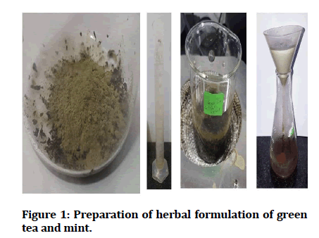
Herbal formulation was prepared using the leaf of green tea and powdered leaf of mint. To 100ml of distilled water, 1g of green tea leaf and 1g of powdered mint leaf was added and the mixture was heated for 15-20min. The product was then filtered using Whatman's filter paper. Then the filtered 70ml of mixture was again heated and concentrated to 20ml. The colour change was observed visually, and photographs were recorded (Figure 1).

Figure 1: Preparation of herbal formulation of green tea and mint.
Determination of anticariogenic activity of green tea and mint herbal formulation
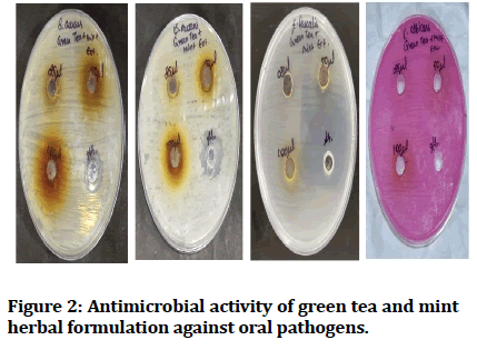
The antimicrobial activity of the prepared green tea and mint was assessed using the agar well diffusion method. The Mueller Hinton agar plates were prepared, and each plate was swabbed with four different dental pathogens such as S. aureus, E. faecalis, S. mutans, C. albicans.

Figure 2:Antimicrobial activity of green tea and mint herbal formulation against oral pathogens.
A gel puncture was used to cut four wells on each petri plates. Each well in the plate had different concentrations being 25μL, 50μL,100μL of the herbal formulation. The fourth well was a standard antibiotic (Amoxicillin) at a concentration of 10 μg/mL. The plates were incubated at 37°C for 24hrs and the diameter of the zone of inhibition formed around the well was measured and the mean values were recorded.
Results and Discussion
In the present study, a test for anticariogenic activity was carried out using the agar well diffusion method. Four agar plates for identifying the inhibitory effect over S. aureus , S. mutans, E. faecalis and C. albicans, respectively, were used.
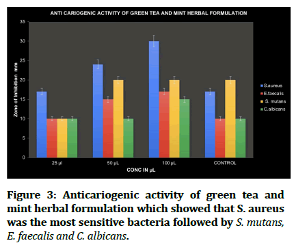
Each plate had 4 wells each with different nanoparticle concentrations being 25 μL, 50 μL, and 100 μL and while the fourth was a standard (Figure 3, Table 1) shows the zone of inhibition of four oral pathogens at different concentration of herbal formulation and compared with standard antibiotic (amoxicillin), hence against S.aureus , the diameter of the zone of inhibition of the herbal formulation at 25 μL, 50 μL, and 100 μL is observed to be 17 mm, 24 mm, and 30 mm, respectively.

Figure 3:Anticariogenic activity of green tea and mint herbal formulation which showed that S. aureus was the most sensitive bacteria followed by S. mutans, E. faecalis and C. albicans.
| Concentrations | 25μL | 50μL | 100μL | Standard |
|---|---|---|---|---|
| S. aureus | 17 | 24 | 30 | 17 |
| S. mutans | 10 | 20 | 20 | 20 |
| E. faecalis | 10 | 15 | 17 | 10 |
| C. albicans | 10 | 10 | 15 | 10 |
Table 1: Antimicrobial activity of green tea and mint herbal formulation.
With S. mutans, the diameter of the zone of inhibition of the nanoparticles at 25 μL, 50 μL, and 100 μL was obtained as 10 mm, 20 mm, and 20 mm, respectively. Against E. faecalis the diameter of the zone of inhibition at 25 μL, 50 μL, and 100 μL was observed as 10 mm, 15 mm, and 17 mm, respectively.
Against C albicans the diameter of the zone of inhibition at 25 μL, 50 μL, and 100 μL was observed as 10 mm, 10 mm, and 15 mm, respectively. Thus, maximum activity for all the three was observed at 150 μL.
Conclusion
From the present study, it can be concluded that herbal formulations of green tea and mint have significantly efficient anticariogenic activity at high concentrations. This can be used for further investigation in employing them as less biotoxic alternatives to already existing chemically synthesized drugs. Hence the herbal formulations of green tea and mint is expected to be used in future for the effective drug systems as well as for large scale commercial productions.
Acknowledgement
The authors are thankful to saveetha dental college for providing a platform to conduct the study.
Source of Funding
The present project is supported by Little Holy Angels Hr Sec School, Chennai.
References
- Mutalik S, Chetana M, Sulochana B, et al. Effect of dianex, a herbal formulation on experimentally induced diabetes mellitus. Phytother Res 2005; 19:409–15.
- Biswas NR, Gupta SK, Das GK, et al. Evaluation of ophthacare eye drops: A herbal formulation in the management of various ophthalmic disorders. Phytother Res 2001; 15:618–20.
- Pari L, Ramakrishnan R, Venkateswaran S. Antihyperglycaemic effect of Diamed, a herbal formulation, in experimental diabetes in rats. J Pharm Pharmacol 2001; 53:1139–43.
- Varshini A, Arivarasu L. Herbal sources used by the public against infections. Int J Pharm Res 2020; 84-92.
- Cabrera C, Artacho R, Giménez R. Beneficial effects of green tea—A review. J Am Coll Nutr 2006; 25:79–99.
- Yamamoto T, Juneja LR, Chu SC, et al. Chemistry and applications of green tea. CRC Press 1997; 176.
- Paul RP, Roy A, Maajida Aafreen M, et al. Antibacterial activity of white pepper oleoresin mediated silver nanoparticles against oral pathogens. J Evolution Med Dent Sci 2020; 9:2352–6.
- Rexlin PJ, Roy A, Rajeshkumar S, et al. Antimicrobial activity of coriander oleoresin mediated selenium nanoparticles against oral pathogens. J Complementary Med Res 2020; 11:35-40.
- Pranati T, Anitha R, Rajeshkumar S, et al. Preparation of silver nanoparticles using nutmeg oleoresin and its antimicrobial activity against oral pathogens. Res J Pharm Techn 2019; 12:2799–803.
- Thariny E, Arivarasu L, Rajeshkumar S. Green synthesis, antioxidant and anti-inflammatory activity of Adathoda vasica mediated copper nanoparticles. Plant Cell Biotechnol Mol Biol 2020; 32–8.
- Jaisankar AI, Arivarasu L. Free radical scavenging and anti-inflammatory activity of chlorogenic acid mediated silver nanoparticle. J Pharm Res Intern 2020; 106–12.
- Casilda Sushanthi L, Vijayashree Priyadharsini J, Hannah R, et al. A study on the variability of drug responsiveness to anti-inflammatory drugs-A pilot survey. Int J Pharm Res 2020; 12.
- Shankar SB, Arivarasu L, Rajeshkumar S. Biosynthesis of hydroxy citric acid mediated zinc nanoparticles and its antioxidant and cytotoxic activity. J Pharm Res Int 2020; 108–12.
- Moezizadeh M. Anticariogenic effect of tea: A review of literature. J Dent Oral Hyg 2013.
- Pramila DM, Xavier R, Marimuthu K, et al. Phytochemical analysis and antimicrobial potential of methanolic leaf extract of peppermint (Mentha piperita: Lamiaceae). J Med Plants Res 2012; 6:331–5.
- Selwitz RH, Ismail AI, Pitts NB. Dental caries. Lancet 2007; 369:51–9.
- Zewdu T, Abu D, Agajie M, Sahilu T. Dental caries and associated factors in Ethiopia: Systematic review and meta-analysis. Environ Health Prev Med 2021; 26:21.
- Nyvad B, Takahashi N. Integrated hypothesis of dental caries and periodontal diseases. J Oral Microbiol 2020; 12:1710953.
- Hwang JK, Shim JS, Chung JY. Anticariogenic activity of some tropical medicinal plants against Streptococcus mutans. Fitoterapia 2004; 75:596–8.
- Vairavel M, Devaraj E, Shanmugam R. An eco-friendly synthesis of Enterococcus sp.–Mediated gold nanoparticle induces cytotoxicity in human colorectal cancer cells. Environ Sci Pollut Res 2020; 27:8166–75.
- Rajeshkumar S, Tharani M, Jeevitha M, et al. Anticariogenic activity of fresh Aloe Vera gel mediated copper oxide nanoparticles. Indian J Public Health Res Develop 2019; 10.
- Yu H, Oho T, Tagomori S, et al. Anticariogenic effects of green tea. Fukuoka Igaku Zasshi 1992; 83:174–80.
- Machado C, Rivera LE, Kleinberg MWI. Clinical evaluation of the ability of Cavistat® in a mint confection to inhibit the development of dental caries in children. J Clin Dent 2008; 19:1–8.
- Al-Sum BA, Al-Arfaj AA. Antimicrobial activity of the aqueous extract of mint plant. Sci J Clin Med 2013; 2:110–3.
- Nagalingam M, Vikramathithan M, Gandhi AD, et al. Evaluation of herbal and chemical-based mouthwash against oral pathogens. Drug Invention Today 2019; 11.
- Rajeshkumar S, Kumar SV, Ramaiah A, et al. Biosynthesis of zinc oxide nanoparticles usingMangifera indica leaves and evaluation of their antioxidant and cytotoxic properties in lung cancer (A549) cells. Enzyme Microb Technol 2018; 117:91–5.
- Nandhini NT, Rajeshkumar S, Mythili S. The possible mechanism of eco-friendly synthesized nanoparticles on hazardous dyes degradation. Biocatal Agric Biotechnol 2019; 19:101138.
- Rajkumar PV, Prakasam A, Rajeshkumar S, et al. Green synthesis of silver nanoparticles using Gymnema sylvestre leaf extract and evaluation of its antibacterial activity. South African J Chem Eng 2020; 32:1-4.
- Rajasekaran S, Damodharan D, Gopal K, et al. Collective influence of 1-decanol addition, injection pressure and EGR on diesel engine characteristics fueled with diesel/LDPE oil blends. Fuel 2020; 277:118166.
- Santhoshkumar J, Sowmya B, Venkat Kumar S, et al. Toxicology evaluation and antidermatophytic activity of silver nanoparticles synthesized using leaf extract of Passiflora caerulea. S Afr J Chem Eng 2019; 29:17–23.
- Raj RK. β-Sitosterol-assisted silver nanoparticles activates Nrf2 and triggers mitochondrial apoptosis via oxidative stress in human hepatocellular cancer cell line. J Biomed Mater Res 2020; 108:1899–908.
- Saravanan M, Arokiyaraj S, Lakshmi T, et al. Synthesis of silver nanoparticles from Phenerochaete chrysosporium (MTCC-787) and their antibacterial activity against human pathogenic bacteria. Microb Pathog 2018; 117:68–72.
- Gheena S, Ezhilarasan D. Syringic acid triggers reactive oxygen species–mediated cytotoxicity in HepG2 cells. Hum Exp Toxicol 2019; 38:694–702.
- Ezhilarasan D, Sokal E, Najimi M. Hepatic fibrosis: It is time to go with hepatic stellate cell-specific therapeutic targets. Hepatobiliary Pancreat Dis Int 2018; 17:192–7.
- Ezhilarasan D. Oxidative stress is bane in chronic liver diseases: Clinical and experimental perspective. Arab J Gastroenterol 2018; 19:56–64.
- Gomathi AC, Xavier Rajarathinam SR, Mohammed Sadiq A, et al. Anticancer activity of silver nanoparticles synthesized using aqueous fruit shell extract of Tamarindus indica on MCF-7 human breast cancer cell line. J Drug Deliv Sci Technol 2020; 55:101376.
- Dua K, Wadhwa R, Singhvi G, et al. The potential of siRNA based drug delivery in respiratory disorders: Recent advances and progress. Drug Dev Res 2019; 80:714–30.
- Ramesh A, Varghese S, Jayakumar ND, et al. Comparative estimation of sulfiredoxin levels between chronic periodontitis and healthy patients - A case-control study. J Periodontol 2018; 89:1241–8.
- Arumugam P, George R, Jayaseelan VP. Aberrations of m6A regulators are associated with tumorigenesis and metastasis in head and neck squamous cell carcinoma. Arch Oral Biol 2021; 122:105030.
- Joseph B, Prasanth CS. Is photodynamic therapy a viable antiviral weapon against COVID-19 in dentistry? Oral Surg Oral Med Oral Pathol Oral Radiol 2021; 132:118–9.
- Ezhilarasan D, Apoorva VS, Ashok Vardhan N. Syzygium cumini extract induced reactive oxygen species-mediated apoptosis in human oral squamous carcinoma cells. J Oral Pathol Med 2019; 48:115–21.
- Duraisamy R, Krishnan CS, Ramasubramanian H, et al. Compatibility of Nonoriginal abutments with implants: Evaluation of microgap at the implant-abutment interface, with original and nonoriginal abutments. Implant Dent 2019; 28:289–95.
- Gnanavel V, Roopan SM, Rajeshkumar S. Aquaculture: An overview of chemical ecology of seaweeds (food species) in natural products. Aquaculture 2019; 507:1–6.
- Markov A, Thangavelu L, Aravindhan S, et al. Mesenchymal stem/stromal cells as a valuable source for the treatment of immune-mediated disorders. Stem Cell Res Ther 2021; 12:192.
Indexed at, Google Scholar, Cross Ref
Indexed at, Google Scholar, Cross Ref
Indexed at, Google Scholar, Cross Ref
Indexed at, Google Scholar, Cross Ref
Indexed at, Google Scholar, Cross Ref
Indexed at, Google Scholar, Cross Ref
Indexed at, Google Scholar, Cross Ref
Indexed at, Google Scholar, Cross Ref
Indexed at, Google Scholar, Cross Ref
Indexed at, Google Scholar, Cross Ref
Indexed at, Google Scholar, Cross Ref
Indexed at, Google Scholar, Cross Ref
Indexed at, Google Scholar, Cross Ref
Indexed at, Google Scholar, Cross Ref
Indexed at, Google Scholar, Cross Ref
Indexed at, Google Scholar, Cross Ref
Indexed at, Google Scholar, Cross Ref
Indexed at, Google Scholar, Cross Ref
Indexed at, Google Scholar, Cross Ref
Indexed at, Google Scholar, Cross Ref
Indexed at, Google Scholar, Cross Ref
Author Info
Gajapriya M1, Lakshminarayanan Arivarasu2* and Rajesh Kumar S2
1Department of Public Health Dentistry, Saveetha Dental College, Saveetha Institute of Medical and Technical Sciences (SIMATS), Saveetha University, Chennai, India2Department of Pharmacology, Saveetha Dental College and Hospitals, Saveetha Institutes of Medical and Technical Sciences (SIMATS), Saveetha University, Chennai, India
Received: 13-Dec-2021, Manuscript No. Jrmds-21-41699; , Pre QC No. Jrmds-21-41699 (PQ); Editor assigned: 15-Dec-2021, Pre QC No. Jrmds-21-41699 (PQ); Reviewed: 29-Dec-2021, QC No. Jrmds-21-41699; Revised: 03-Jan-2022, Manuscript No. Jrmds-21-41699 (R); Published: 10-Jan-2022
