Research - (2019) Volume 7, Issue 5
Assessment of Anatomical Position of Posterior Teeth and Alveolar Bone Height in Malaysian Population Based on Panoramic Radiographs
Norliza Ibrahim1*, Tameem Khuder2, Farishah Nur Binti Abd Samad1, Syahira Binti Tuan Baharom1, Muhammad Khan Asif1, Rohana Ahmad3, Norsiah Yunus4 and Samah Mohammed AL-Amery1
*Correspondence: Norliza Ibrahim, Department of Oral and Maxillofacial Clinical Sciences, Faculty of Dentistry, University of Malaya, Malaysia, Email:
Abstract
Introduction: Familiar knowledge of posterior teeth positions and alveolar bone heights of dentate maxilla and mandible and their alterations according to gender and race may serve as a reference for implant planning, orthodontic therapy, and forensic work.
The aim: The aim of this study was to determine the location of first premolar and first molar in the maxilla and the mandible from the midline followed by the assessment of the maxillary and mandibular alveolar bone heights using panoramic radiographs of the dentate Malaysian population.
Materials and Methods: Panoramic images of 153 subjects were collected and classified according to gender, race and age group. Horizontal and vertical heights of maxilla and mandible at the first premolar and first molar areas were measured using Image J software.
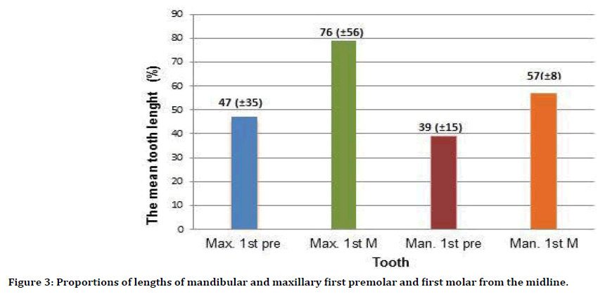
Results: There were no statistically significant differences among gender, race and age groups regarding tooth position, except for maxillary first premolars that were located more distally in females (p=0.016). Maxillary first premolars and first molars were located approximately 47% and 76%, respectively, of the horizontal length of the maxilla from the midline. Mandibular first premolars and first molars were located at 39% and 57%, respectively, of the length of the mandible from the midline. Alveolar bone heights of dentate males were greater than females. Indians have the smallest alveolar bone height compared to Malays and Chinese.
Conclusions: The positions of posterior teeth are not influenced by gender, race, and age in the Malaysian population. The alveolar bone heights of dentate maxilla and mandible are influenced by gender. However, at certain locations, the height can be influenced by race.
Keywords
Panoramic, Age group, Tooth position, Alveolar bone height, Implantation
Introduction
Bone resorption after tooth loss is a progressive and irreversible process with inevitable consequences influenced by age, gender, facial anatomy, general health, nutritional status, edentulous duration and by occlusal force distribution [1]. Reduction in residual ridge height will affect the denture’s support, retention, stability and masticatory function [2,3]. Fortunately, dental implantology has evolved greatly over the past years. Implant treatment showed 5-9 years survival rate of 81% and 91% for maxillary and mandibular arches respectively [4]. A removable prosthesis reduces patient’s function to one-sixth of the function experienced with natural dentition. While Implant-retained prostheses may return the patient’s function to near normal as the implants are able to stimulate bone thus maintaining its dimension [5].
Rehabilitation of implants prosthodontics is of great value for edentulous patients. It can be effectively accomplished through diagnostic wax or cast with clinical and radiographic examinations [4]. Radiographic examination is an essential element in implant treatment planning as it allows the clinician to assess the width, contour, and the accurate bone height. Ideally, implantation should be at the former location of a tooth in a jaw and the vertical height of the alveolar bone should be adequate to place implants at a distance of 1 to 2 mm from the adjacent structures with 1 to 1.5 mm of bone on either side [6,7].
Cephalometric radiographs are used for diagnosis and treatment planning due to high reproducibility and less magnification. However, the superimposition of the right and left sides make it impossible to distinguish between them during the radiographic assessment [8]. Panoramic radiograph (DPT) is commonly used to overcome superimpositions that occur on cephalometric radiographs. DPT has been described as a practical method to assess the residual ridge resorption for patient’s examination [9]. A previous study by Larheim et al. [10], reported that the accuracy of linear measurements in panoramic radiographs can be influenced by the patient’s position in the machine. Likewise, according to Xie et al. [11], a small range of variations in vertical measurements in the mandible and the posterior regions of the maxilla was only observed if reference lines and measured points are located in the same vertical plane or in approximately the same plane as the teeth.
Malaysia has a total population of approximately 31.7 million people. The majority of Malaysian citizens are Malays (68.6%), followed by Chinese (23.4%), Indians (7.0%) and others (1%) (Department of Statistics Malaysia: Current Population Estimates, Malaysia, 2014– 2016). The prevalence of edentulous patients in Malaysia is 55.9% with age groups of 70-80 years and older [12]. As the elderly population (>65 years old) continue to increase, an upsurge in the demand for implant-supported treatments is observed. Hence, knowledge of the residual bone status is imperative to guide the clinician in planning the implant treatment for Malaysians population. Malaysian population uniquely consists of three races (Malay, Chinese and Indian). However, studies on the position of Malaysian’s posterior teeth are still not evident in the literature. Therefore, the aim of this study was to assess the location of Malaysian posterior teeth (maxillary first premolars, maxillary first molars, mandibular first premolars and mandibular first molars) by using DPT images of fully dentate patients of the three main races in Malaysia. The second aim was to assess the alveolar bone heights of maxillary and mandibular arches using DPT images of dentate patients.
Materials and Methods
Relevant ethical approval for the use panoramic radiographic images was obtained from the institutional board of study of the faculty of dentistry [DF OS1614/0037(U)] of the University of Malaya. No letter of consent or questionnaires was required for this study.
Five hundred and two panoramic radiographic images (DPT) of fully dentate patients who attended the oral and maxillofacial imaging division, faculty of dentistry, the University of Malaya between January and July 2016 were retrieved. All DPT images were acquired using digital panoramic unit (Veraviewepocs 2D/J; Morita, Kyoto, Japan CS 9300C). Exposure parameters were set at 62 kV, 7.5 mA with 14.9 second exposure time. The magnification ratio was 1.3000. The DPT was traced based on the patient’s national identity card that contains information on patient’s gender and age. Data for patient’s race were archived from the database stored in the faculty. The inclusion criteria for this study were as follows:
1. Absence of obvious crowding of teeth.
2. Absence of facial asymmetry and abnormal morphology of the jaws.
3. Absence of pathologies including cysts and tumours.
4. Absence of fracture and surgical history.
5. Absence of history of systemic and bone diseases.
6. Clearly visible nasal septum and nasopalatine foramen.
7. Clearly visible inferior margins of the zygomatic processes.
8. Clearly visible maxillary tuberosities.
9. Clearly visible inferior and posterior borders of the mandible.
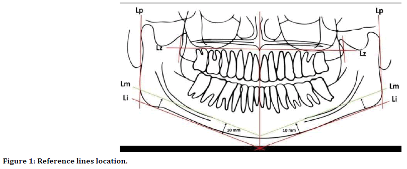
Five reference lines were drawn on each radiograph as illustrated in Figure 1. The first line is the midline across the maxilla and mandible, which is determined by a nasal septum and nasopalatine canal, followed by a line joining the most inferior borders of the two zygomatic processes (Lz) in the maxilla. In the mandible, a line passing the posterior margins of the mandibular ramus (Lp), a line tangent to the most inferior borders of the angles of the mandible and mandibular body (Li) and a line parallel and 10 mm above to Li (Lm) was drawn on both sides. This method is the adaptation of the linear measurements that was first described by [9].

Figure 1. Reference lines location.
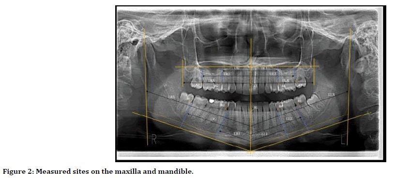
Twenty-two measurements on the maxilla and mandible were made on all images whenever possible as illustrated in Table 1 and Figure 2.
| Parameters | Description |
|---|---|
| A1 | Alveolar bone height at the maxillary midline |
| A2 | Alveolar bone height at the distal of right maxillary first premolar |
| A3 | Alveolar bone height at the distal of right maxillary first molar |
| A4 | Alveolar bone height at the distal left maxillary first premolar |
| A5 | Alveolar bone height at the distal left maxillary first molar |
| UR1 | Horizontal length measured from the maxillary midline to the distal of right maxillary first premolar |
| UR2 | Horizontal length measured from the maxillary midline to the distal of right first molar |
| URX | Horizontal length measured from the maxillary midline to the right maxillary tuberosity |
| UL1 | Horizontal length measured from the maxillary midline to the distal of left maxillary first premolar |
| UL2 | Horizontal length measured from the maxillary midline to the distal of left first molar |
| ULX | Horizontal length measured from the maxillary midline to the left maxillary tuberosity |
| B1 | Alveolar bone height at the mandibular midline |
| B2 | Alveolar bone height at the distal of right mandibular first premolar |
| B3 | Alveolar bone height at the distal of right mandibular first molar |
| B4 | Alveolar bone height at the distal of the left mandibular first premolar |
| B5 | Alveolar bone height at the distal of the left mandibular first molar |
| LR1 | Horizontal length measured along Lm from the midline to the distal of right mandibular first premolar |
| LR2 | Horizontal length measured along Lm from the midline to the distal of right first molar |
| LRX | Horizontal length measured along Lm from the midline to the most posterior end of the right mandibular ramus |
| LL1 | Horizontal length measured along Lm from the midline to the distal of left mandibular first premolar |
| LL2 | Horizontal length measured along Lm from the midline to the distal of left first molar |
| LLX | Horizontal length along Lm from the midline to the most posterior end of the left mandibular ramus |
Table 1: Measured sites on the maxilla and mandible.

Figure 2. Measured sites on the maxilla and mandible.
Statistical analysis
Intra-observer reliability in reproducing the measurements was performed using the Intraclass Correlation Coefficient (ICC). Assessment of normality using Kolmogorov-Smirnov (K-S) test was done prior to doing statistical tests. Genderbased comparison between measurements was analysed using independent t-test. One-way ANOVA test was used to analyse the influences of race and age, with Bonferroni post-hoc test. The level of significance was set at p<0.05.
Results
Three hundred and forty-nine panoramic images did not fit the criteria and subsequently excluded from this study. The remaining 153 images were then classified according to their gender (79 males and 74 females), race (58 Malays, 58 Chinese and 37 Indians) and age group (98 patients aged 20-29, 43 patients aged 30- 39 and 12 patients aged 40-49). Intra-class Correlation Coefficient (ICC) test showed high intra-observer reliability (ICC=0.98-1.00) in all parameters.
Assessment of posterior teeth positions
Proportions of lengths on maxilla and mandible indicating the relative positions of a maxillary first premolar, maxillary first molar, mandibular first premolar and mandibular first molar were obtained by calculating the means of percentage on either side (Figure 3).

Figure 3. Proportions of lengths of mandibular and maxillary first premolar and first molar from the midline.
The position of maxillary first premolar showed a statistically significant difference when measured between genders (p=0.016), and the independent t-test demonstrated that the horizontal distance of maxillary first premolar in females was located further distally from the midline compared to males (Table 2A). While no significant differences were observed when measuring the posterior teeth positions among race and age groups as shown in Tables 2B and 2C respectively.
Assessment of alveolar bone height
Independent t-test results showed that the difference in the alveolar bone height was statistically significant between genders and males show higher values of alveolar bone height as compared to females at all measured sites (Table. 3).
One-way ANOVA test showed no significant difference in alveolar bone height of the maxillary midline, maxillary first molar and mandibular midline in race-based comparison. While remaining areas showed a significant difference and Post-hoc tests demonstrated that Indians have the smallest bone height among the three races (Table 4).
The differences were not significant when measuring the alveolar bone height among age groups (Table 5).
Discussion
Dental panoramic radiograph (DPT) allows the assessment of dentition, bone height of both maxilla and mandible, the temporomandibular joints and important structures in relation to dental implantology such as the maxillary sinuses, mental foramen and the inferior alveolar nerve [13,14]. Although cone-beam computed tomography images (CBCT) are increasingly used for demonstrating the planned dental implant site [15], DPT images are used due to inadequate amount of CBCT datasets that meet the criteria of this study.
The location for posterior teeth of various populations has been successfully determined using DPT images. Studies on Finland [9] and Turkish [16] population have reported almost similar location for their posterior teeth i.e. approximately 35% (first premolar) and 55% (first molar) from the midline of the mandible. Our findings exhibited that the posterior teeth of Malaysian are located slightly further from the mandibular midline (mandibular first premolars 39% and first molars 57%) when compared to Finland and Turkish population. However, the location for Malaysian’s posterior teeth were nearer to the midline in comparison to Spanish population (mandibular first premolars 49% and first molars 61%) [17]. For maxillary teeth, the Malaysian’s first premolars were located nearer to the midline (47%) in comparison to Spanish population (55%), while molar teeth were located further posteriorly in the Malaysian population (76%) compared to (72%) in the Spanish population [17]. Thus, a specific data on teeth location is essential in guiding the clinicians in identifying the ideal implant receptor site for a patient of a different population.
There was no statistically significant difference in the positions of teeth in different age groups and races in Malaysia. Similarly, a previous study has reported small correlation coefficient between age and measurements made on DPTs [16]. However, the position of maxillary first premolars is found to be different between genders, specifically that they were further distally located from the midline in females compared to males. Alveolar bone dimensions should be evaluated when planning dental implant treatment [18]. Alp Saglam et al. [8] has reported that male’s mandibular bone height is greater than that of the female. This is in agreement with our finding. Apart from bone height, our result is also consistent with previous studies that described the influenced of gender on the facial dimension [19,20]. Unlike females, the growth of males continues until early adulthood [21]. Thus, the difference in facial dimensions might possibly due to different facial growth pattern between genders.
Variability within a population is heavily influenced by genetic alterations and the environment [22]. In Malaysian population, Malays and Chinese belong to the Mongoloid race, whereas Indians belong to the subgroup of Caucasoid called Indo-Dravidian [23]. Consequently, the present study indicated a statistically significant difference based on race in the alveolar bone height at maxillary and mandibular first premolars and mandibular molar sites when compared between the three major races in Malaysia. Although there were no significant differences observed at maxillary first molar, and at maxillary and mandibular midline measured sites, there was a trend of lower bone height in the Indian population compared to Malay and Chinese population.
The shape and dimension of mature jaws are affected by natural dentition [9]. This study was conducted on DPT of fully dentate patients to assess the ideal bone height at the implant site. Thus, no significant difference was observed in alveolar bone heights among different age groups. With the presence of natural teeth, the masticatory force will be distributed through the periodontal ligament to the alveolar bone, loss of teeth will cause alveolar bone atrophy and the masticatory force will be directed onto the bone surface instead. Crum and Rooney [24] suggested that retention of teeth and use of overdentures may help preserve the mandibular alveolar bone. This is because the discrete proprioceptive ability is maintained in presence of overdenture preventing bone resorption. Bone loss is also found to be less in implant-supported dentures than that in conventional dentures [25].
This study has few limitations which include inability in identifying the presence of malocclusion; distortion, magnification, and limitations of 2D DPT images in assessing the implant receptor sites. Thus, future studies should consider the use of CBCT in determining the location of posterior teeth and the height of alveolar bone to ensure an accurate reference for predicting implant locations for Malaysians populations. Moreover, there was limited number of DPTs for fully dentate patients who attended the Oral and Maxillofacial Imaging Division, Faculty of Dentistry, the University of Malaya. Therefore, further studies with higher number of DPTs will be needed to validate our findings.
Conclusion
In the Malaysian population, the positions of posterior teeth are not influenced by gender, race, and age. However, the alveolar bone heights of dentate maxilla and mandible are influenced by gender, and at certain locations by race.
Acknowledgment
The authors would like to express their appreciation to the radiographers at the Oral and Maxillofacial Imaging Division, Faculty of Dentistry, the University of Malaya for their assistance in archiving the panoramic images throughout this study.
References
- Khuder T, Yunus N, Sulaiman E, et al. Association between occlusal force distribution in implant overdenture prostheses and residual ridge resorption. J Oral Rehabil 2017; 398-404.
- Brodeur JM, Laurin D, Vallee R, et al. Nutrient intake and gastrointestinal disorders related to masticatory performance in the edentulous elderly. J Prosthet Dent 1993; 70:468-473.
- Soikkonen K, Ainamo A, Xie Q. Height of the residual ridge and radiographic appearance of bony structure in the jaws of clinically edentulous elderly people. J Oral Rehabil 1996; 23:470-475.
- Adell R, Lekholm U, Rockler B, et al. A 15-year study of osseointegrated implants in the treatment of the edentulous jaw. Int J Oral Surg 1981; 10:387-416.
- Misch CE. Contemporary implant dentistry: Elsevier Health Sciences 4th Edn. 2007.
- Bagchi P, Joshi N. Role of radiographic evaluation in treatment planning for dental implants: A review. J Dent Allied Sci 2012; 1:21-25.
- Juodzbalys G, Raustia AM. Accuracy of clinical and radiological classification of the jawbone anatomy for implantation: A survey of 374 patients. J Oral Implantol 2004; 30:30-39.
- Alp Saglam A. The vertical heights of maxillary and mandibular bones in panoramic radiographs of dentate and edentulous subjects. Quintessence Int 2002; 33:433-436.
- Xie Q, Wolf J, Ainamo A. Quantitative assessment of vertical heights of maxillary and mandibular bones in panoramic radiographs of elderly dentate and edentulous subjects. Acta Odontol Scand 1997; 55:155-161.
- Larheim T, Svanaes D. Reproducibility of rotational panoramic radiography: mandibular linear dimensions and angles. Am J Orthod Dentofacial Orthop 1986; 90:45-51.
- Xie Q, Soikkonen K, Wolf J, et al. Effect of head positioning in panoramic radiography on vertical measurements: an in vitro study. Dentomaxillofac Radiol 1996; 25:61-66.
- Shamdol Z, Ismail NM, Hamzah NT, et al. Prevalence and associated factors of edentulism among elderly Muslims in Kota Bharu, Kelantan, Malaysia. J Islamic Med Assoc North Am 2008; 40:143-148.
- Canger EM, Celenk P. Radiographic evaluation of alveolar ridge heights of dentate and edentulous patients. Gerodontology 2012; 29:17-23.
- Lee S, Lee S, Huh K, et al. The effects of location of alveolar crest on the vertical bone heights on panoramic radiographs. Dentomaxillofac Radiol 2014; 117-121.
- Angelopoulos C, Thomas S, Hechler S, et al. Comparison between digital panoramic radiography and cone-beam computed tomography for the identification of the mandibular canal as part of presurgical dental implant assessment. J Oral Maxillofac Surg 2008; 66:2130-2135.
- Güler A, Sumer M, Sumer P, et al. The evaluation of vertical heights of maxillary and mandibular bones and the location of anatomic landmarks in panoramic radiographs of edentulous patients for implant dentistry. J Oral Rehabil 2005; 32:741-746.
- López-Roldán A, Abad DS, Bertomeu IG, et al. Bone resorption processes in patients wearing overdentures. A 6-years retrospective study. Med Oral Patol Oral Cir Bucal 2009; 14:203-209.
- Shenoy VK. Single tooth implants: Pretreatment considerations and pretreatment evaluation. J Interdiscipl Dent 2012; 2:149.
- Kurkcuoglu A, Bahadıroglu S, Buyukberber SG, et al. Evaluation of Lower Face Heights and Ratios According to Sex. Rev Arg Anat Clin 2013; 5:213-221.
- Zhuang Z, Landsittel D, Benson S, et al. Facial anthropometric differences among gender, ethnicity, and age groups. Ann Occup Hyg 2010; 54:391-402.
- Mehta M, Saini V, Nath S, et al. CT scan images for sex discrimination: A preliminary study on Gujarati population. J Forensic Radiol Imaging 2015; 3:43-48.
- Hoffmann AA, Willi Y. Detecting genetic responses to environmental change. Nat Rev Genet 2008; 9:421-432.
- Hashirn Yaacob B, Phrabhakaran Narnbiar B. Racial characteristics of human teeth with special emphasis on the Mongoloid dentition. Malays J Pathol 1996; 18:1-7.
- Crum RJ, Rooney GE. Alveolar bone loss in overdentures: a 5-year study. J Prosthet Dent 1978; 40:610-613.
- Van Waas M, Jonkman R, Kalk W, et al. Differences two years after tooth extraction in mandibular bone reduction in patients treated with immediate overdentures or with immediate complete dentures. J Dent Res 1993; 72:1001-1004.
Author Info
Norliza Ibrahim1*, Tameem Khuder2, Farishah Nur Binti Abd Samad1, Syahira Binti Tuan Baharom1, Muhammad Khan Asif1, Rohana Ahmad3, Norsiah Yunus4 and Samah Mohammed AL-Amery1
1Department of Oral and Maxillofacial Clinical Sciences, Faculty of Dentistry, University of Malaya, Malaysia2Department of Prosthodontic, Faculty of Dentistry, Mustansiriyah University, Iraq
3Department of Prosthodontics, Centre of Studies for Restorative Dentistry, Universiti Teknologi, Malaysia
4Department of Restorative Dentistry, Faculty of Dentistry, University of Malaya, Malaysia
Citation: Norliza Ibrahim, Tameem Khuder, Farishah Nur Binti Abd Samad, Syahira Binti Tuan Baharom, Muhammad Khan Asif, Rohana Ahmad, Norsiah Yunus, Samah Mohammed AL-Amery, Assessment of Anatomical Position of Posterior Teeth and Alveolar Bone Height in Malaysian Population Based on Panoramic Radiographs, J Res Med Dent Sci, 2019, 7(5): 110-117.
Received: 03-Sep-2019 Accepted: 14-Oct-2019
