Research - (2022) Volume 10, Issue 1
Assessment of Cytotoxicity of Copper and Graphene Oxide Nano composite Synthesized Using Amla Extract Formulation-An In-Vitro Study
Gaurav N Ketkar1, Sankari Malaiappan1* and Rajeshkumar S2
*Correspondence: Sankari Malaiappan, Department of Periodontics, Saveetha Dental College and Hospital, Saveetha Institute of Medical and Technical Sciences, India, Email:
Abstract
Background: Several conventional methods are used for synthesis of nanoparticles. But toxic chemicals are required as capping agents to maintain stability, thus leading to toxicity in the environment. Thus, we need to shift to “Green Synthesis”. Hence, this study was conducted to assess the cytotoxicity of copper and graphene oxide nano composite reinforced with amla extract. Copper is well known for its intrinsic antibacterial properties, graphene oxide for its barrier and structural strength hence these were chosen to create a nanocomposite. Aim: Aim of the study was green preparation of nano copper with nano graphene oxide nanocomposite and its cytotoxicity evaluation. Material and methods: Cytotoxic effect of copper and graphene oxide nanocomposite with amla extract was assessed using Brine Shrimp Assay respectively at 5 µl, 10 µl, 20 µl, 40 µl, 80 µl. Results: The copper and graphene oxide nanocomposite are safe to be used in the dental materials till 20 µL concentration. Although at 40 µl, 80 µl concentrations cytotoxicity was observed. Conclusion: Within the limits of the study, it can be concluded that copper and graphene oxide Nano composite can be safely used in concentrations up to 20 µL as periodontal dressing or incorporated as bone grafts in periodontal regeneration.
Keywords
Copper, Characterisation, Graphene oxide, Green synthesis, nanoparticle, Nanocomposite
Introduction
Nanotechnology is an emerging technology and has led to a new revolution in every field of science. Nanoparticles have gained great importance in the research community in recent years. This technology has been used in the fields of optics, electronics, and biomedical and materials sciences [1]. Potent antimicrobial, anticancer, antioxidant agents, drug, and gene delivery, etc are some of the highlighted advantages of the nanoparticles in recent years [2–4]. Nanotechnology deals with nanoparticles that are atomic or molecular aggregates characterized by size less than 100 nm. These are basic elements derived by modifying their atomic and molecular properties [5,6].
Several conventional methods are used for synthesis of zinc oxide nanoparticles like chemical reduction [7], laser ablation [8], solvothermal, inert gas condensation [9,10], sol-gel method [11]. Even though less time is utilized for synthesizing large quantities of nanoparticles using conventional physical and chemical methods, toxic chemicals are required as capping agents to maintain stability, thus leading to toxicity in the environment. “Green synthesis” offers numerous benefits of eco friendliness and compatibility for biomedical applications, where toxic chemicals are not used for the synthesis protocol. The use of agricultural wastes [9] or plants and their parts [10], has emerged as an alternative to chemical synthetic procedures because it does not require elaborate processes such as intracellular synthesis and multiple purification steps or the maintenance of microbial cell cultures.
United States Environmental Protection Agency (US EPA) recognized copper as the first antimicrobial metal in the year of 2008. One of the most important advantages of copper as an antimicrobial agent is its low levels of resistance in the microorganisms.
Copper nanoparticles (CuNP) are superior owing to their nontoxicity, biocompatibility, use in drug and bactericidal activity [7,12,13]. Contact killing property of copper was studied widely in recent years. Studies have shown that increased bacterial intracellular oxidative stress in the bacterial cell wall due to release of ions from the copper surface which results in bacterial cell lysis [14]. Copper as material is very versatile. A study done by Dr Gaurav Ketkar et al. shows excellent intrinsic antimicrobial properties of copper over stainless steel. Similarly copper nanoparticles have good antimicrobial properties against oral anaerobic organisms [15]. Synthesis of copper nanoparticles is highly technique sensitive due to its high incidence of oxide layer formation on the nanoparticle surface which will result in reduced antibacterial property [16].
Graphene oxide is known for its excellent mechanical strength, electrical conductivity and most importantly the barrier properties, also easy step-down preparation of graphene oxide nanoparticles makes it one of the most efficient carriers of nanoparticles in any nanocomposite. It is an atomically thin, 2-dimensional (2D) sheet of sp2 carbon atoms in a honeycomb structure [15].
The objective of this study was to use amla fruit extract to synthesize copper and graphene oxide nanoparticles and to evaluate its cytotoxicity as its excellent potency against oral aerobes was already proven in the previous studies [15].
Material and Methods
The study was carried out in the multidisciplinary research lab of Saveetha dental college and hospital Chennai. Ethical and research committee approval was granted before commencing the study (IHEC Ref No: IHEC/SDC/PERIO-1901/21/48).
Preparation of amla extract
Freshly collected organic amla fruits were thoroughly washed multiple times in distilled water. Seed was taken out and the pulp was cut into small pieces using a sterile knife and was grounded into small particles by means of a mortar and pestle. Amla extract was prepared by 1 grams of amla pulp with 100 ml distilled water to make 1 molar solution of amla extract [15].
Synthesis of nanoparticles
The synthesis of copper nanoparticles was simply obtained by the reduction of copper sulphate solution. amla plant extract was used as a reducing or capping agent. 1 molar copper sulphate solution was prepared by mixing 20 millimolar CuSO4 into 60 ml water and 40 ml of the synthesised 1M amla extract was added to the solution. The prepared solution was kept overnight on an orbital shaker for homogenous mixing of all particles. Then the mixture was collected in 5 test tubes and CuNP’s were separated from solution by centrifugation for 20 minutes. Similar process was followed for preparation of the GO nanoparticles where 0.5 gm of graphene oxide powder was dissolved in 50 ml of distilled water and 50 ml of prepared extract was then added to the solution and 20 min centrifuge was used to separate the nanoparticles from the solutions.
Synthesis of CuGO nanocomposite
Nanocomposite synthesis was done by mixing 50 ml of both 1M solutions of copper and graphene oxide nanoparticles as mentioned in the previous steps. The nanocomposite solution was stirred overnight on an orbital shaker followed by a magnetic heated stirrer till colour change was observed.
UV-vis spectrometric readings were taken hourly to check the synthesis of copper-graphene oxide nano composite. The resultant mixture was centrifuged and CuGO nanocomposite was obtained [15].
Cytotoxic effect
The cytotoxicity of copper and graphene oxide nanocomposite was extract was assessed using Brine shrimp assay. 12 well ELISA plates were taken and to each plate 6-8 ml of saltwater was added; followed by adding 10 nauplii to each well.
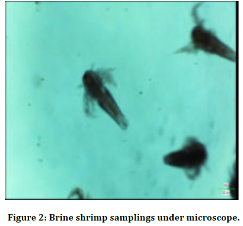
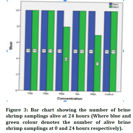
Copper and graphene oxide nanocomposite was added to each well at different concentrations (5 μl, 10 μl, 20 μl, 40 μl, 80 μl) and was then incubated for 24 hrs. After 24 hrs, the total number of live and dead nauplii was counted and the mortality rate was checked (Figures 1-3).

Figure 1. Shrimp eggs.

Figure 2. Brine shrimp samplings under microscope.

Figure 3. Bar chart showing the number of brine shrimp samplings alive at 24 hours (Where blue and green colour denotes the number of alive brine shrimp samplings at 0 and 24 hours respectively).
The current concentrations for evaluation of cytotoxicity were derived from a study done by Samikannu Kanagesan et al and Dr Rajeshkumar et al [17,18] % death=Number of dead nauplii Number of dead nauplii−number of live nauplii 100.
Results
Table 1 depicts the cytotoxicity of copper and graphene oxide nanocomposite reinforced with amla extract. Upto 20 μl concentration there was a death of 100% of nauplii samplings that were alive at the end of the test period.
| Concentration of nanocomposite (µl) | Viable napulii (24 hrs) | Death % |
|---|---|---|
| Control (0 µl) | 10 | 0 |
| 5 µl | 10 | 0 |
| 10 µl | 10 | 0 |
| 20 µl | 10 | 0 |
| 40 µl | 8 | 20 |
| 80 µl | 7 | 30 |
Table 1: Depicting the cytotoxicity of the copper and graphene oxide nano composite reinforced with amla extract
At 40 μl 20% death rate was seen and 30% subsequently at 80 μL concentration. It was seen that as the concentration increased the cytotoxicity of the nanoparticles increased.
Discussion
Nanotoxicity, the sector of vigorous research, has emerged from the increased use of nanoparticles mainly in commercially available products. The risk factors of nanomaterials are still unknown [2].
For this purpose, extensive procedures are being carried out to evaluate the toxic effects of these nanomaterials. Cell cytotoxicity refers to the ability of certain chemicals or mediator cells to destroy living cells.
By using a cytotoxic compound, healthy living cells can either be induced to undergo necrosis (accidental cell death) or apoptosis (programmed cell death). Given this information, the ability to accurately measure cytotoxicity can prove to be a very valuable tool in identifying compounds that might pose certain health risks in humans [9]. This can be of vital importance during the research phase of developing new pharmaceutical products to ensure the safety of the end-users.
Recently, many researchers have focused on brine shrimp lethality assay owing to their low cost, convenience, ideal life span and rapid screening procedure. The species of Brine shrimp, Artemia salina is commonly used nowadays in drug discovery to screen the toxic effect of different components.
Brine shrimp assay was first proposed by Michael et al. which was then developed (Figure 1).
There has been a rapid evolution of nanoparticle synthesis recently as compared to the early part of the century [19].
Earlier conventional methods were used for the synthesis of the nanoparticles. Even though less time is utilized for synthesizing large quantities of nanoparticles using conventional physical and chemical methods, toxic chemicals are required as capping agents to maintain stability. These methods resulted in toxicity in the environment due to use of toxic chemicals. To avoid the use of such chemicals, the Green Synthesis method was proposed and is widely used all over the world. It is an eco-friendly method as well as it is highly cost effective [20]. We therefore undertook this study to evaluate the cytotoxicity of the copper and graphene oxide nanocomposite reinforced with amla extract. Antibacterial properties of the same composite were found to be excellent against oral microbes in the previous studies [15].
Amla or Phyllanthus Emblica is known since ancient times for its medicinal value and is commonly used in Ayurvedic medicine. Amla is a rich source of vitamin C which is a highly essential vitamin in maintenance of epithelial homeostasis. It also acts as potent antioxidants. Emblica officinalis (Figure 1) (Amla) has a lot of medicinal properties and has been used since ancient times. A wide range of phytochemical components present in amla including alkaloids, tannins, and flavonoids are responsible for its variety of medicinal uses. It belongs to the family: Euphorbiaceae. cytoprotective, antitussive, gastroprotective, as antioxidant, immunomodulatory, antipyretic, analgesic, are some of the most important uses of the amla plant. It has beneficial effects in the treatment of cancer, heart conditions, liver treatments etc. Superoxide radicals (O2–.) have been implicated in several pathological disorders and are responsible for elevated oxidative stress. Periodontal disease can be caused by immune reaction between pathogenic bacteria and host. Periodontal tissue destruction leads to over-production of lipid peroxides, inflammatory mediator, and oxidised proteins. These products further activate macrophages, neutrophils, and fibroblasts to generate more ROS [21]. Studies have shown that amla extract acts as a very good antioxidant by scavenging the reactive oxygen species and protects the antioxidant enzymes like SOD required for the cellular defence. Amla is a rich source of vitamin C which is a highly essential vitamin in maintenance of epithelial homeostasis. Juice of amla plants have been used as chemical free mouthwash which was found highly effective in the treatment of aphthae. Keeping all these beneficial effects of amla fruit also easy availability we decided to choose amla for the green preparation of the nanocomposite in the present study [22-25].
Excellent mechanical strength, electrical conductivity and most importantly the barrier properties, also easy stepdown preparation of graphene oxide nanoparticles makes it one of the most efficient carriers of nanoparticles in any nanocomposite this is the reason behind choosing graphene oxide as one of the components in the composite. Additionally, graphene oxide plays an important role in nano drug delivery systems because of its controlling mechanisms, including targeting and stimulation with pH, chemical interactions, thermal, photo- and magnetic induction [22].
Due to the large surface area to volume ratio, copper nanoparticles have been used as potential antimicrobial agents in many biomedical applications. But the excess use of any metal nanoparticles increases the chance of toxicity to humans, other living beings, and the environment [23]. Samikannu Kanagesan et al. in a study proved copper nanoparticles to be toxic post >125mg/μL concentrations. Hence the concentrations chosen in the study were in accordance with his study [17]. Copper and graphene oxide nanocomposite has proven to be a unique mixture with reliable antimicrobial properties [16].
Limitations and Future Prospects
Although the study shows excellent results in brine shrimp assay, other methods which are more reliable can be performed. The material proved to have antimicrobial, anti-inflammatory, antioxidant, and anti-cytotoxic effects hence it can be incorporated into dental material. The material is safe to be used for human experimentation in such as local drug delivery systems and incorporation in materials.
Conclusion
Within the limits of this study, it can be concluded that copper and graphene oxide nanocomposite show no cytotoxicity upto20 μl concentration. The nanocomposite can be incorporated in dental materials or used as a coating on suture materials to reinforce their biomechanical properties.
Acknowledgements
We would like to thank our director of Saveetha University Dental College for his support and contribution during the period of the study. Also special thanks to Dr. Rajeshkumar for his expertise and guidance for the study.
Conflict of Interest
All authors in the present study declare no potential conflict of interest.
References
- Rico CM, Majumdar S, Duarte-Gardea M, et al. Interaction of nanoparticles with edible plants and their possible implications in the food chain. J Agric Food Chem 2011; 59:3485–98.
- Rajeshkumar S, Malarkodi C, Gnanajobitha G, et al. Seaweed-mediated synthesis of gold nanoparticles using Turbinaria conoides and its characterization. J Nanostructure Chem 2013; 3:44.
- Malarkodi C, Rajeshkumar S, Paulkumar K, et al. Bactericidal activity of bio mediated silver nanoparticles synthesized by Serratia nematodiphila. Drug Invention Today 2013; 5:119-25.
- Rajeshkumar S. Anticancer activity of eco-friendly gold nanoparticles against lung and liver cancer cells. J Genet Eng Biotechnol 2016; 141:195–202.
- Daniel MC, Astruc D. Gold nanoparticles: assembly, supramolecular chemistry, quantum-size-related properties, and applications toward biology, catalysis, and nanotechnology. Chem Rev 2004; 104:293–346.
- Jamdagni P, Rana JS, Khatri P, et al. Comparative account of antifungal activity of green and chemically synthesized zinc oxide nanoparticles in combination with agricultural fungicides. J Nanodimens 2018; 9:198-208.
- https://www.wiley.com/en-us/Materials+Science+and+Engineering:+An+Introduction,+10th+Edition-p-9781119405498
- Snehal Yedurkar M, Kapil Punjabi D, Chandra Maurya B, et al. Biosynthesis of Zinc oxide nanoparticles using euphorbia milii leaf extract-A green approach. Materials Today 2018; 5:22561–9.
- Mohapatra S, Leelavathi L, Rajeshkumar S, et al. Assessment of cytotoxicity, anti-inflammatory and antioxidant activity of zinc oxide nanoparticles synthesized using clove and cinnamon formulation: An in-vitro study. J Evol Med Dent Sci 2020; 9:1859.
- Chang H, Tsai MH. Synthesis, and characterization of Zno nanoparticles having prism shape by a novel gas condensation process. 2008; 18:734-743.
- Li H, Wang J, Liu H, et al. Zinc oxide films prepared by sol–gel method. J Crystal Growth 2005; 275:e943-6.
- Yusefi-Tanha E, Fallah S, Rostamnejadi A, et al. Particle size and concentration dependent toxicity of copper oxide nanoparticles (CuONPs) on seed yield and antioxidant defense system in soil grown soybean (Glycinemax cv. Kowsar). Sci Total Environ 2020; 715:136994.
- Abdolhosseinzadeh M, Khodamoradi N. Synthesis & study of nano size copper oxide particle via chemical method. Adv Mater Res 2014; 829:187-191.
- Abiodun-Solanke I, Ajayi D, Arigbede A. Nanotechnology, and its application in dentistry. Ann Med Health Sci Res 2014; 4:S171–7.
- Guidelli EJ, Ramos AP, Zaniquelli MED, et al. Green synthesis of colloidal silver nanoparticles using natural rubber latex extracted from Hevea brasiliensis. Spectrochim Acta A Mol Biomol Spectrosc 2011; 82:140–5.
- Ketkar GN, Malaiappan S. Green preparation of nano copper (cu) with nano graphene oxide (go) nano composite characterization and antimicrobial activity against oral aerobic pathogens. Plant Cell Biotechnol Mol Biol 2020; 31–9.
- Kanagesan S, Hashim M, Aziz SAB, et al. Evaluation of antioxidant and cytotoxicity activities of copper ferrite (cufe2o4) and zinc ferrite (znfe2o4) nanoparticles synthesized by sol-gel self-combustion method. Applied Sci 2016; 184.
- Rieshy V, Jeevitha M, Rajeshkumar S. Cytotoxicity of copper nanoparticles synthesized using dried ginger. Plant Cell Biotechnology Molecular Biol 2020; 1–6.
- Andersson M, Pedersen JS, Palmqvist AEC. Silver nanoparticle formation in microemulsions acting both as template and reducing agent. Langmuir 2005; 21:11387–96.
- Chandran SP, Chaudhary M, Pasricha R, et al. Synthesis of gold nanotriangles and silver nanoparticles using Aloevera plant extract.
Indexed at, Google Scholar, Cross Ref
Indexed at, Google Scholar, Cross Ref
Indexed at, Google Scholar, Cross Ref
Indexed at, Google Scholar, Cross Ref
Indexed at, Google Scholar, Cross Ref
Indexed at, Google Scholar, Cross Ref
Indexed at, Google Scholar, Cross Ref
Indexed at, Google Scholar, Cross Ref
Indexed at, Google Scholar, Cross Ref
Indexed at, Google Scholar, Cross Ref
Indexed at, Google Scholar, Cross Ref
Author Info
Gaurav N Ketkar1, Sankari Malaiappan1* and Rajeshkumar S2
1Department of Periodontics, Saveetha Dental College and Hospital, Saveetha Institute of Medical and Technical Sciences, Chennai, India2Department of Pharmacology, Saveetha Dental College and Hospital, Saveetha Institute of Medical and Technical Sciences, Chennai, India
Citation: Gaurav N Ketkar, Sankari Malaiappan, Rajeshkumar S, Assessment of Cytotoxicity of Copper and Graphene Oxide Nano composite Synthesized Using Amla Extract Formulation-An In-Vitro Study, J Res Med Dent Sci, 2022, 10(1): 227-231
Received: 10-Dec-2021, Manuscript No. Jrmds-21-44367; , Pre QC No. Jrmds-21-44367 (PQ); Editor assigned: 13-Dec-2021, Pre QC No. Jrmds-21-44367 (PQ); Reviewed: 27-Dec-2021, QC No. Jrmds-21-44367; Revised: 30-Dec-2021, Manuscript No. Jrmds-21-44367 (R); Published: 06-Jan-2022
