Research - (2021) Volume 9, Issue 8
Assessment of Gestational Age Using Ultrasound
*Correspondence: G Rajathi, Department of Anatomy, Sree Balaji Medical College & Hospital Affiliated to Bharath Institute of Higher Education and Research, India, Email:
Abstract
Determining gestational age in resource-poor settings is challenging because of limited availability of ultrasound technology and late first presentation to antenatal clinic. Last menstrual period (LMP), symphysio-pubis fundal height (SFH) and Ballard Score (BS) at delivery are therefore often used. This study explains the relationship between MSD, CRL and GA (USG) in the first trimester, BPD, HC, AC, FL and GA (USG) in the second trimester, EFW, BPD, HC, AC, FL and GA(USG) in the third trimester. From the results 3D and 4D USG will improvise the ability to assess early pregnancy viability and multiple gestations.
http://www.oajournal.org/
http://www.journalsres.org/
http://www.journalsres.com/
http://www.journalsoa.org/
http://www.journalsoa.com/
http://www.journalsci.org/
http://www.journalres.org/
http://www.journalres.com/
http://www.journaloa.org/
http://www.journalinsights.org/
http://www.jpeerreview.org/
http://www.imedresearch.com/
http://www.imedpubjournals.com/
http://www.imedpubjournal.org/
http://www.imedjournals.org/
http://www.peerreviewedjournal.org/
http://www.peerjournals.org/
http://www.peerjournals.com/
http://www.sciencesinsight.org/
http://www.scholarresearch.com/
http://www.scholarres.org/
http://www.nutritionres.com/
http://www.gastroinsights.org/
http://www.pathologyinsights.org/
http://www.echemistry.org/
http://www.echemcentral.com/
http://www.chemistryres.com/
http://www.biochemresearch.org/
http://www.biochemjournals.com/
http://www.ebusinessjournals.org/
http://www.businessjournals.org/
http://www.peerjournal.org/
http://www.oajournalres.com/
http://www.alliedres.org/
http://www.alliedjournals.org/
http://www.alliedjournal.org/
http://www.scientificres.org/
http://www.scientificres.com/
,
https://www.mongoliannutrition.com/
https://www.nsbmb.com/
https://www.arabspp.org/
https://www.arabianmultidisciplinary.com/
https://www.italystemcell.com/
https://www.traditional-medicine.org/
https://www.episportsmedicine.org/
https://www.worldmedicalassociation.org/
https://www.silaeitaly.com/
https://www.ceramicsmedicine.org/
https://www.isaddictionmedicine.org/
https://www.europeanbionetwork.com/
https://www.aarsecp.com/
https://www.edycseg.org/
https://www.europeanneurology.org/
https://www.clinicaldermepi.com/
https://www.cardiac-society.com/
https://www.psychologicalassociation.org/
https://www.indian-psychology.com/
https://www.mongoliancardiology.org/
https://www.pediatricssociety.com/
https://www.cocrt.org/
https://www.european-aesthetic.com/
https://www.sohnsb.org/
Keywords
Gestational age, UltrasoundIntroduction
Radiographic techniques were generally used to measure fetal dimensions prior to ultrasound, which had the drawback of exposing radiation to the foetus. Currently with increasing use of ultrasound, a non-invasive diagnostic procedure, there is a decrease in maternal morbidity and mortality. Ultrasonography is commonly used to estimate gestational age by measuring fetal dimensions like gestational sac diameter, crown rump length, biparietal diameter, abdominal circumference, head circumference and femur length. Hence this study aims to measure these fetal parameters and to obtain accurate gestational age [1-5].
Methodology
The study this study, 145 antenatal women were selected, observed, and underwent physical examination, lab investigations to rule out maternal diseases. patients were subjected to ultrasonogram, and the Gestational age was determined by measuring fetal parameters.
Results
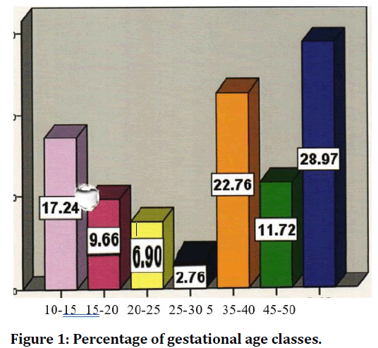
The percentage of gestational age classes are depicted in Figure1 To prove a correlation between the fetal parameters in the second and third trimester with gestational age a correlation coefficient was calculated and found to be 0.995, 0.995, 0.993, 0.997 and the values were less than 0.001, thereby showing a positive correlation between these variables. gestational age a correlation coefficient was calculated and found to be 0.995, 0.995, 0.993, 0.997 and the values were less than 0.001, thereby showing a positive correlation between these variables.

Figure 1. Percentage of gestational age classes.
Table1 explains that both CRL and MSD were contributing towards gestational age assessment and highly significant. BPD and FL were highly significant.
| GA BY USG | BPD | FL | HC | AC | ||
|---|---|---|---|---|---|---|
| Pearson Correlation | GA BY USG | 1 | 0.995 | 0.995 | 0.993 | 0.997 |
| BPD | 0.995 | 1 | 0.993 | 0.996 | 0.993 | |
| FL | 0.995 | 0.993 | 1 | 0.993 | 0.993 | |
| HC | 0.993 | 0.996 | 0.993 | 1 | 0.991 | |
| AC | 0.997 | 0.993 | 0.993 | 0.991 | 1 | |
| GA | BPD | FL | HC | AC | ||
| GA | 0.996639 | 0.996639 | 0.995293 | 0.997984 | ||
| BPD | 0.997312 | 0.997312 | 0.995293 | |||
| FL | 0.995293 | 0.995293 | ||||
| HC | 0.993946 | |||||
Table 1: CRL and MSD contribution.
HC was excluded in this model since it was not contributing.
Discussion and Conclusion
The study explains that there is a linear relationship between MSD, CRL and GA (USG) in the first trimester, BPD, HC, AC, FL, and GA(USG) in the second trimester, EFW, BPD, HC, AC, FL and GA(USG) in the third trimester. In case of abnormal measurements of fetal parameters disease conditions should be addressed. Multiple parameters should be used to assess gestational age. It is likely that the technological development of USG will continue and increases in ultrasound frequency will further improve image resolution of early pregnancies. 3D and 4D USG will also improve our ability to assess early pregnancy viability and multiple gestations [6-10].
References
- Hadlock FP, Deter RL, Harrist RB et al . Estimating fetal age: Computer-assisted analysis of multiple fetal growth parameters. Radiology 1984; 152:497-501.
- Campbell S, Warsof SL, Little D, et al. Routine ultrasound screening for prediction of gestational age. Obstet Gynecol 1985; 65:613-20.
- Shalev E, Weiner E, Zuckerman H. Assessment of gestational age by ultrasonic measurement of the femur length. Acta Obstet Gynecol Scand 1985; 64:71-4.
- Roddick. Placental thickness. J Ultrasound Med 1985; 4:479-82.
- Hadlock FP, Deter RL, Harrist RB, et al. Estimating fetal age: Computer-assisted analysis of multiple fetal growth parameters. Radiology 1984; 152:497–501.
- Group PS, Pekyi D, Ampromfi AA, et al. four artemisinin-based treatments in African pregnant women with malaria. N Engl J Med 2016; 374:913–27.
- Banoo S, Bell D, Bossuyt P, et al. Evaluation of diagnostic tests for infectious diseases: General principles. Nat Rev Microbiol 2006; 4:S20–32.
- Verhoeff FH, Milligan P, Brabin BJ, et al. Gestational age assessment by nurses in a developing country using the Ballard method, external criteria only. Ann Trop Paediatr 1997; 17:333–42.
- Wilson K, Hawken S, Potter BK, et al. Accurate prediction of gestational age using newborn screening analyte data. Am J Obstet Gynecol 2016; 214:e511–9.
- Boamah EA, Asante K, Ae-Ngibise K, et al. Gestational age assessment in the Ghana randomized air pollution and health study (GRAPHS): Ultrasound capacity building, fetal biometry protocol development, and ongoing quality control. Res Protoc 2014; 3:e77.
Author Info
Department of Anatomy, Sree Balaji Medical College & Hospital Affiliated to Bharath Institute of Higher Education and Research, Chennai, Tamil Nadu, IndiaCitation: G Rajathi, Assessment of Gestational Age Using Ultrasound , J Res Med Dent Sci, 2021, 9(8): 64-65
Received: 14-Jul-2021 Accepted: 02-Aug-2021
