Research - (2020) Volume 8, Issue 5
Assessment of Staining Quality of Curcumin as a Substitute for Eosin in Hematoxyline and Eosin Staining in Histopathology
Rubina MP1, Dr. Ashida M Krishnan2*, Riyas Basheer KB1, Mohammed Safeer TK1 and Soumya V1
*Correspondence: Dr. Ashida M Krishnan, Associate Professor (Non-Cadre), Department of Pathology, Government Medical College, Thiruvananthapuram-695011, India, Email:
Abstract
Background and objectives: Hematoxylin and Eosin staining is the globally accepted staining technique for histology and histopathology sections. Though hematoxylin is a natural dye, eosin is synthetic dye manufactured from chemicals. Eosin acts as a counterstain to hematoxylin giving sharp contrast to its blue colour. Eosin being a chemical can cause health hazards and environmental pollution. Using ecofriendly materials and going organic is the demand of this era. The present study was conducted to evaluate the staining qualities of Curcumin longa(turmeric) a natural substitute of eosin and the results of Hematoxylin and Eosin stain (H and E) was compared with Hematoxylin and Curcumin(H and C) stain. Methods: The study was conducted in the Histopathology lab of a tertiary health care centre in South India after getting Institutional Ethical Committee Clearance. For each staining method 100 sections were prepared from 20 collagen tissues, 20 epithelial tissues, 20 smooth muscle tissues, 20 bony tissues and 20 adipose tissues. The staining qualities were assessed by an experienced pathologist and the results were scored as excellent, good and poor. After entering the scores in Microsoft excel, the results were statistically analyzed. Results: H and C staining gave comparable results with H and E staining in all the five types of tissue sections studied (p value<0.05) with intense affinity on collagen and muscle fibres. Interpretation and conclusions: Curcumin is a safer and cheaper alternative to Eosin stain in histology and histopathology sections.
Keywords
Curcumin, Ecofriendly stains, Eosin substitutes, Natural dyes, Nontoxic stains, Turmeric
Introduction
Haematoxylin and Eosin staining is the globally practiced staining technique for histology and histopathology studies. Hematoxylin and eosin (H and E) stains have been used for at least a century and are still essential for recognizing various tissue types and the morphologic changes that form the basis of contemporary cancer diagnosis. In a typical tissue, nuclei are stained blue by the basic dye hematoxylin, whereas the cytoplasm and extracellular matrix have varying degrees of pink staining with acidic eosin.
Haematoxylin is a natural dye, obtained from the Mexican tree Haematoxylon campechianum while the Eosin is a synthetic dye. Synthetic dyes are often efficient but may display hazards to human and animal health. This has resulted in the withdrawal of several dyes [1]. With the worldwide concern over the use of eco-friendly and biodegradable materials, the use of natural dyes obtained from plants has again gained interest. Moreover, as many developing countries can no longer afford the everincreasing cost of synthetic dyes, the use of cheaper, naturally occurring dyes from plants is being viewed as an alternative to synthetic dyes. Based on this facts Curcuma longa was investigated as a natural dye with potential histopathological application [2].
Eosin is a name of several fluorescent acidic compounds which bind to and from salts with basic or eosinophilic proteins such as arginine and lysine. It stains them dark red or pink because of the actions of bromine on fluorescein. In addition to staining the proteins in cytoplasm, it can be used to stain collagen and muscle fibres for microscopic examination. Structures that stain readily with eosin are termed eosinophilic.
Curcumin, or diferuloylmethane, is a major chemical component of turmeric (Curcuma longa Linn) that has been consumed as a dietary spice. Chemically, curcumin is a diarylheptanoid, belonging to the group of curcuminoids, which are natural phenols responsible for turmeric's yellow colour. It contains flavonoids, which are typically polyphenolic compounds. Phenols are acidic, due to their ability to release the hydrogen from their hydroxyl group, hence the ability of C. longa to stain the basic parts of the cell [3]. C. longa was used as a counter stain for haematoxylin with the reaction like that of eosin in the haematoxylin and eosin technique except for its yellow coloration. The present study was conducted to assess the staining quality of curcumin as an eosin substitute in hematoxylin and eosin staining.
Methodology
The study design was a comparative study conducted in the Histopathology laboratory, of a tertiary health care centre in South India. The sample size was calculated as 100 for each staining technique. The period of study was six months after Institutional ethical committee approval (IEC.NO.08/11/2017/MCT dated 28/7/2017).
Methods of preparing turmeric stain
Slender dried pieces of turmeric are first powdered, and 15 gm of this powder is mixed with 100ml of 70% alcohol. The contents are mixed well and kept for an hour for sedimentation. After centrifugation, the supernatant fluid is taken for staining.
Methods of preparing eosin hematoxylin stain
1g% Alcoholic eosin is prepared by mixing 1 gram of commercially available eosin powder with 100 ml of alcohol.
Methods of preparing hematoxylin stain
Harris ’ s haematoxylin is prepared by dissolving 2.5 grams of Hematoxylin in 50 ml absolute alcohol. Alum solution is prepared by adding 50 mg of ammonium alum in hot water. The two solutions are heated up to boiling. 1.5 grams mercuric oxide is added, and mixture is cooled rapidly. Addition of 20 ml Glacial acetic acid improves nuclear staining of hematoxylin stain.
Staining method
For H and E staining deparaffinized sections are first treated with 2 changes of xylene followed by hydration of the tissue sections with descending grades of alcohol and water. After that the sections are treated with Harris hematoxylin solution for 5 minutes. This is followed by differentiation in 1% acid alcohol and Bluing in running tap water. The sections are dehydrated in 95% alcohol, counterstained with eosin for 45 seconds and again dehydrated in ascending grades of alcohol. Finally, the sections are again treated with xylene and mounted with DPX. H and C staining also follows same methods except 5 minutes staining in curcumin instead of 45 seconds eosin staining in H and E method.
The study was carried on the tissue specimen received at the histopathology lab. Five types of tissue sections were selected for this study include:
Epithelium: Skin specimens for keratinized stratified squamous epithelium (10 slides) and Intestinal specimens for glandular epithelium (10 slides).
Collagen: Keloid specimens for collagen.
Muscle: Leiomyoma specimens for Muscle.
Bone: Tissue from bony lesions for Bone.
Adipose tissue: Fat rich specimens like lipoma for Adipose tissue.
For each tissue type, 20sections stained with H and E and 20 sections stained with H and C were studied. The staining qualities were studied by a pathologist who had 8 years of experience in Histopathology. The staining quality of slides was graded into following three scores.
Score 1- P (Poor)- Refers for difficulty in appreciation of a tissue Structure.
Score 2 G (Good): Refers for sufficient appreciation of a tissue structure.
Score 3 E (Excellent): Refers for fine appreciation of a tissue structure.
The scores of the pathologist were entered in Microsoft excel and the scores were summed up for each five parameters and statistical significance was studied.
Results
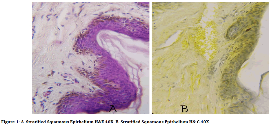
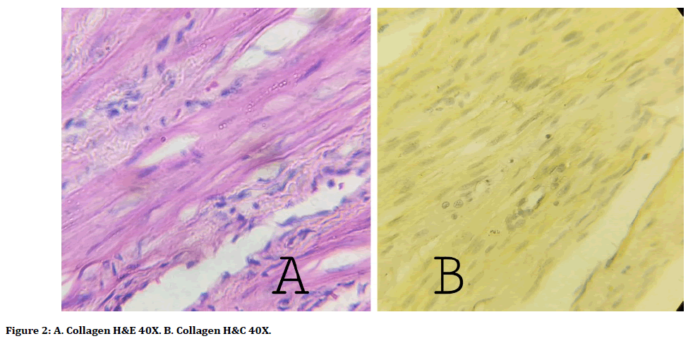
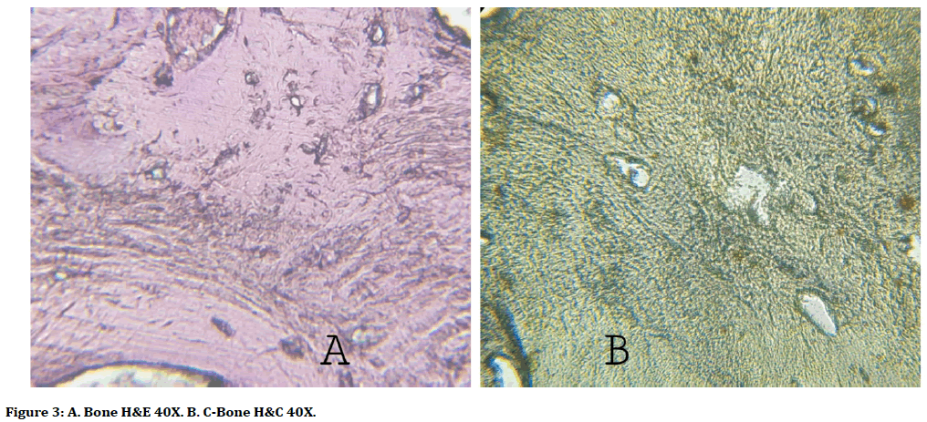
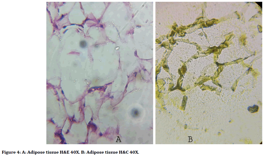
The present study evaluated the comparison between Hematoxyline and Curcumin staining and conventional Hemtoxyline and Eosin staining. The scoring of 100 tissue sections done by two observers is depicted in Table 1 and staining characters all the studied five tissue elements are depicted in Figures 1-4.
| Observer 1 | method | Type of tissue | |||||
| Collagen | Epithelium | Muscle | Bone | Adipose tissue | Total sections | ||
| H&E | 20E | 9E | 16E | 0E | 0E | 100 | |
| 0G | 10G | 4G | 5G | 20G | |||
| 0P | 1P | 0P | 15P | 0P | |||
| H&C | 0E | 0E | 0E | 0E | 0E | 100 | |
| 19G | 1G | 15G | 20G | 5G | |||
| 1P | 3P | 5P | 0P | 15P | |||
Table 1: Scoring of observer.

Figure 1. A. Stratified Squamous Epithelium H&E 40X. B. Stratified Squamous Epithelium H& C 40X.

Figure 2. A. Collagen H&E 40X. B. Collagen H&C 40X.

Figure 3. A. Bone H&E 40X. B. C-Bone H&C 40X.

Figure 4. A: Adipose tissue H&E 40X. B: Adipose tissue H&C 40X.
95% of slides stained with H and C staining were good and 100% slides are excellent with H and E staining for collagen(x2=80p<0.001). For epithelium H and E staining, 45% of slides were graded as excellent and 52.5% as good and 2.5% as poor. In epithelium - H and C staining 87.5% slides were graded as good and 12.5% as poor (x2=24.167 p<0.001). For muscle tissue H and E staining gave excellent score in75% of slides, good score in 25% of slides. For H and C staining 72.5% slides were good and remaining were graded poor in muscle tissue (x2=50.256p<0.001). Most of the slides stained with H and E and H and C staining for bone tissue were graded as poor, only 20% slides with H and E staining showed good quality(x2=10.141p=0.001). In adipose tiisue100% slides with H and E staining showed excellent staining, only 30% slides in H and C showed good score(x2=43.077p<0.001).
When used as a counter stain, C. longa stain showed a similar reaction to that of eosin in the Hematoxylin and Eosin technique except for its yellow coloration. Although statistically Eosin have proved to be better over turmeric, turmeric has shown good and comparable staining to eosin. Turmeric dye stains collagen and muscle fibres with deep yellowish orange color.
Discussion
Hematoxylin and Eosin stain (H and E stain or HE stain) is the most popular histology stain all over the world. Hematoxylin is a naturally occurring chemical and the cytoplasmic stain eosin is a manufactured from coal tar. Synthetic dyes became popular because of longer shelf life than many natural dyes. However, synthetic dyes may cause harmful effects on the environment and human beings. Eosin is an irritant for skin, eyes, and mucous membranes. Acute exposure to this synthetic dye can cause chelitis, stomaitis, dermatitis etc. There have been indefinite reports that this substance is an animal carcinogen [4]. Environmental pollution with laboratory chemicals and dyes are also reported from places where safe disposal of lab wastes is not attended well.
Replacement of chemical dyes with natural ones is truly relevant in this era of global warming and environmental pollution. Organic methods and materials gain so much global attention. The role of eosin in H and E staining is to act as a counter stain giving a sharp contrast color in cytoplasm with the hematoxylin ’ s blue nuclear color. Carmine, carcade, henna, wing fruit, china rose are few natural dyes [5]. Those natural dyes which have a contrast colour with hematoxylin can be used as a substitute for eosin. Various types of natural substitutes have been an interesting topic for many other researchers before [6].
In a similar study conducted by Sachin, et al. they observed that C.longa deeply stains collagen and muscle fibres with deep yellowish orange color [2]. ‘Curcuma longa extract as a histological dye for collagen fibres and red blood cells’’, a study conducted by Avwioro et al. concluded that the turmeric dye stains collagen and muscle fibres with deep yellowish orange colour suggesting its stronger affinity to these structures [7]. Staining qualities of Zingiber roscoe (ginger) and Curcuma longa studied in an Oral Pathology centre in 2018 concluded that ginger has better staining qualities than turmeric and both can be used as substitutes for eosin8. Lawsonia inermis (henna) extract used as eosin substitute by Lizbeth, et al. observed both henna and eosin gave comparable staining reactions [9].
The basic principle in the theory of attaining is the ionic bond between the tissue components and the dye, which is associated with electrostatic attraction between the dissimilar ions. Due to strong affinity of C. longa for the cytoplasm, it can be deduced that the C. longa extract dye is acidic in nature. The main components of turmeric are flavonoids and tannins, which are responsible for its staining properties. Flavonoids are typically polyphenolic which show acidic nature by their ability to release hydrogen from their hydroxyl group. This gives ability of C. longa to stain the basic parts of the cell. The ability of turmeric dye to stain specific tissue structures is determined by the acidity of the stain.
Traditional extraction techniques from natural sources, known as maceration and soxhlet methods were also studied and compared by few researchers. Avwioro et al., Baseey et al. conducted the study on soxhlet extracts of turmeric [7,10], while Kumar et al in 2014 did the study on tissue sections using with maceration extracts of turmeric [2]. Although turmeric stain is a natural one, it tends to achromatize when stored over a long period of time. The shelf life of turmeric was found to be inferior to eosin and Bassey et al. Avwioro et al. Kumar, et al. stated that addition of a mordant to Curcuma longa can increase the shelf life of this stain [2,7,10].
Conclusion
To go organic and ecofriendly is the necessity of modern era. Implementing ecofriendly substitutes in laboratories which are handling harmful chemical substances contributes so much to this noble concept of environment protection. The tissue sections, in the absence of stains appear colorless and make morphological features indiscernible. In our study we compared the staining qualities of eosin with its natural substitute C.longa. When C. longa was used as a counter stain for Haematoxylin; collagen, muscle fibers and epithelium stained better than adipose and bone tissue. The nuclei took the blue colouration which enabled a clear contrast to be made between the different structures of the cells. Turmeric showed a comparable staining quality to that of eosin. The strong affinity of turmeric to collagen and muscle fibres needs to be studied further. With further modifications and standardization, natural dye Curcumin can be used as a histological stain in place of synthetic and toxic eosin dye. Curcumin is also cheap and readily available.
References
- Fisher HA, Jacobson KA, Rose J, et al. et al, Hematoxylin and eosin staining of cell and tissue sections. CSH Protoc 2008; 2008:4986.
- Kumar S, Singh NN, Singh A, et al. Use of Curcuma longa L. extract to stain various tissue samples for histological studies. Ayu 2014; 35:447–451.
- Horobin RW, Kiernan JA , Conn's biological stains. A handbook of dyes stains and fluorochromes for use in biology and medicine. 10th Edn. Oxford 2002.
- Bordoloi B, Jaiswal R, Siddique S, et al. Health hazards of special stains. Saudi J Pathol Microbiol 2017; 2:175-178.
- Akinloye AJ, Illoh HC, Olagoke AO. Screening of some indigenous herbal dyes for use in plant histological staining. J For Res 2010; 21:81-84.
- Ramamoorthy A, Ravi S, Jeddy N, et al. Natural alternatives for chemicals used in histopathology lab–A literature review. J Clin Diagn Res 2016; 10:e01-e04.
- Avwioro G, Onwuka SK, Moody JO, et al. Curcuma longa extract as a histological dye for collagen fibres and red blood cells. J Anat 2007; 210:600–603.
- Sudhakaran A, Hallikeri K, Babu B. Natural stains Zingiber officinale Roscoe (ginger) and Curcuma longa L. (turmeric)–A substitute to eosin. Ayu 2018; 39:220.
- Raju L, Nambair S, Augustine S, et al. Lawsonia inermis (henna) extract: A possible natural substitute to eosin stains. J Interdis Histopathol 2018; 6:54–60.
- Bassey R, Oremosu A, Osinubi A. Curcuma longa: Staining effect on histomorphology of the testis. Macedonian J Med Sci 2012; 5:26-29.
Author Info
Rubina MP1, Dr. Ashida M Krishnan2*, Riyas Basheer KB1, Mohammed Safeer TK1 and Soumya V1
1Research Scholar, Department of Allied Health Sciences, Srinivas University, Mangalore, Karnataka-574146, India2Associate Professor (Non-Cadre), Department of Pathology, Government Medical College, Thiruvananthapuram-695011, India
Citation: Rubina MP, Ashida M Krishnan, Riyas Basheer KB, Mohammed Safeer TK, Soumya V, Assessment of Staining Quality of Curcumin as a Substitute for Eosin in Hematoxyline and Eosin Staining in Histopathology, J Res Med Dent Sci, 2020, 8(5): 146-150
Received: 17-Jul-2020 Accepted: 24-Aug-2020
