Research - (2022) Volume 10, Issue 1
Association between Occlusal Forces and Periodontal Conditions in Untreated Chronic Periodontitis Using T-Scan: A Retrospective Study
Subasree Soundarajan* and Priyalochana Gajendran
*Correspondence: Subasree Soundarajan, Department of Periodontics, Saveetha Dental College and Hospitals, Saveetha Institute of Medical and Technical Sciences Saveetha University, India, Email:
Abstract
Objective: To determine the association between occlusal forces and periodontal conditions in untreated chronic periodontitis patients and elaborate the relevant clinical implications Methodology: Thirty subjects (15 males & 15 females) with moderate to severe chronic periodontitis were recruited from the department of periodontology at Saveetha dental college and hospital from October 2020 to April 2021. Parameters assessed were - a) Probing depth at 6 sites per tooth using a William’s periodontal probe, b) Clinical attachment loss, c) Bleeding on probing (%), d) Miller’s tooth mobility index 1950, e) High occlusal forces (HOF) using T scan & f) Level of alveolar bone loss using periapical radiograph. Results: A total of 812 teeth (352 anterior teeth & 460 posterior teeth) from 30 patients (12 females & 18 males) with untreated chronic periodontitis were included in the study. The Chi square test showed that there is no significant association of occlusal forces with probing depth, and clinical attachment loss, where the p value was > 0.05. Whereas, Association of occlusal forces with teeth type, level of alveolar bone and tooth mobility showed statistical significance with a p value<0.05.Therefore, mandibular posteriors showed high occlusal forces followed by mandibular anterior, maxillary posteriors and maxillary anterior. Teeth taking up high occlusal forces showed bone loss upto apical third, whereas teeth with low occlusal forces showed bone loss upto cervical third. Teeth taking up high occlusal forces showed grade III mobility, whereas teeth with low occlusal forces showed grade I mobility. Among all the teeth groups, mandibular posteriors (31.58%) showed high occlusal forces, followed by mandibular anterior (28.07%), maxillary posteriors (22.81%), and maxillary anterior (17.54%). Conclusion: Within the limitations of this research, the current findings indicate that posterior teeth with high occlusal force in patients with untreated chronic periodontitis could represent occlusal trauma-related periodontal conditions, which may increase the risk of further periodontal destruction. The current findings can help clinicians better diagnose occlusal trauma and related periodontal conditions for better patient management and treatment outcomes.
Keywords
Periodontitis, Traumatic occlusion
Introduction
The periodontium has its own ability to respond to the stresses exerted on the tooth by the occlusal forces. While occlusal trauma is not an etiological factor in periodontitis, its existence may worsen the disease's severity. For premature interactions or interferences, occlusal changes in teeth have been suggested [1]. The magnitude, direction, length, and frequency of these occlusal forces on the periodontium determine their effect [2]. The intraarch relationship, or the relationship of the teeth within each arch to a smoothly curving line of occlusion, and the interact relationship, or the relationship of the teeth within each arch to a smoothly curving line of occlusion, are two major characteristics that define an individual's occlusal status [3].
The noxious inputs to the neuromuscular system are often increased by the disharmonies in these occlusal relationships, as a result of stress-induced clenching, which causes increased muscle activity and muscle spasms. Occlusal contacts that occur too early are called premature occlusal contacts. They are commonly found in people who have chronic periodontitis, and its magnitude was found to be substantially associated with the severity of the periodontal disease. The posterior teeth with high occlusal forces in patients with untreated chronic periodontitis may reflect occlusal trauma associated periodontal conditions. It is possible that this will raise the likelihood of further periodontal destruction [4].
The occlusal factors acting on the teeth are analysed in current practise using articulating paper marks, waxes, pressure indicator paste, and other instruments to measure the forces of the occlusion and to determine the occlusal contacts. However, these techniques have the disadvantage of not being able to detect simultaneous interaction or quantifying time and force.
There is no scientific correlation between the depth of the colour of the mark, its surface area, amount of force, or the contact timing sequence that results as the articulation paper marks is used [5,6]. This approach is solely based on the interaction of dental articulating paper and patient sensations.
A computer-aided video system, a photo occlusion tool, and the T–Scan IITM system are all popular quantitative occlusal approaches [7,8]. Bio Research Associates, Inc. and Tekscan, Inc. have recently released T-Scan IIITM, a commercially available device that overcomes the established limitations of articulation paper marks [9]. It measures and shows relative occlusal force so that the clinician can minimize repeated errors of incorrect occlusal contact selection. There are numerous upcoming computer based treatment modalities, but many clinicians have yet to overcome the limitations of conventional approaches for examining the patients. This diagnostic device has been increasingly applied in the field of prosthodontics and implantology for full mouth rehabilitation cases. Until now it has rarely been used in periodontal research and clinical management of periodontally related occlusal problems. Therefore, the aim of the present study is to evaluate the distribution of high occlusal forces (HOF) using T scan systems and assess its association with the quantity of alveolar bone loss in upper and lower dentition. The current findings could aid in enhancing our understanding of implication of occlusal trauma in periodontal disease, and thus help in improving clinical management of periodontal patients with occlusal problems.
Materials and Method
Study participants
Thirty subjects (15 males & 15 females) with moderate to severe chronic periodontitis were recruited from the department of periodontology at Saveetha dental college and hospital from October 2020 to April 2021. The study protocol was approved by the Ethical committee of the institution, and written informed consent was obtained from all the subjects prior to the study (IHEC/SDC/ PERIO-1902/20/58). Sample size was determined using the standard deviation values of the previous study, at a power of 90% (G power 3.0 software) , and was estimated to be 30 [4]. A total of 32 patients were assessed initially for the study. Out of which, 2 patients were excluded as they were not willing to participate in the study. Thus, 30 patients were included in the study.
Inclusion criteria
(A) Age: 30 to 70 years,
(B) At least 3 teeth with probing depth ≥ 6 mm, clinical attachment loss ≥ 6 mm and alveolar bone loss ≥ 30% of the root length on periapical radiograph.
(C) Presence of the first or second molar in each quadrant and no more than 3 teeth missing in upper or lower jaw.
(D) No prior periodontal treatment in the past 2 years and e) no history of temporomandibular joint.
Exclusion criteria
(A) Having complicated dentures, and
(B) History of occlusal adjustment or orthodontic treatment.
Parameters assessed
For each patient, all the teeth were grouped into 4.
• Maxillary anterior.
• Maxillary posteriors.
• Mandibular anterior.
• Mandibular posteriors.
Periodontal assessment
The following clinical parameters were assessed:
(A) Probing depth at 6 sites per tooth using a William’s periodontal probe.
(B) Clinical attachment loss.
(C) Bleeding on probing (%).
(D) Miller’s tooth mobility index 1950.
Occlusal examination
The High occlusal force (HOF) was determined using the Novus T scan system. An electric sensor strip in the form of a dental arch was placed inside the sensor holder, and connected to the handle into the patient's oral cavity. A computer recorded and analysed the occlusal force detected. All subjects were trained to bite on the sensor strip twice for practice before proper analysis was done. Patients were asked to bite in maximum intercuspation, lateral excursion and protrusive movements. The percentage of the teeth with high occlusal forces in various occlusal positions were calculated. All the T scan recordings were performed by a single examiner.
Radiographic examination
A full mouth series of periapical radiographs were taken by a single experienced technician using Radiovisiography (RVG) Casestream 5200. These digital radiographs were evaluated on the computer screen and the amount of alveolar bone loss (cervical third, middle third or apical third) was recorded.
Statistical analysis
The periodontal parameters were described as mean and standard deviation. Chi square tests were performed to assess the association between occlusal forces with (A) Teeth type (Maxillary anteriors, Maxillary posteriors, Mandibular anteriors, and Mandibular posteriors).
(B) Probing depth.
(C) Clinical attachment loss.
(D) Tooth mobility.
(E) Level of alveolar bone loss (Cervical, middle & apical third). All statistical analyses were performed using SPSS version 23.0 (Statistical Package for the social sciences). P <0.05 was considered as statistically significant.
Results
A total of 812 teeth (352 anterior teeth & 460 posterior teeth) from 30 patients (12 females & 18 males) with untreated chronic periodontitis were included in the study.
Table 1 shows the characteristics of all study participants and their periodontal conditions.
| Variables | Mean ± Standard deviation |
| Age (years) | 42.0 ± 12.1 |
| Gender (Female: male) | 12:18 |
| Total number of teeth present (anterior: posterior) | 812 (352:460) |
| Probing depth (mm) | 4.3 ± 1.6 |
| Clinical attachment loss (mm) | 5.2 ± 1.8 |
| BOP (%) | 81.0 ± 27.0 |
Table 1: Characteristics of study participants and their periodontal conditions.
The Chi square test showed that there is no significant association of occlusal forces with probing depth, and clinical attachment loss, where the p value was > 0.05.
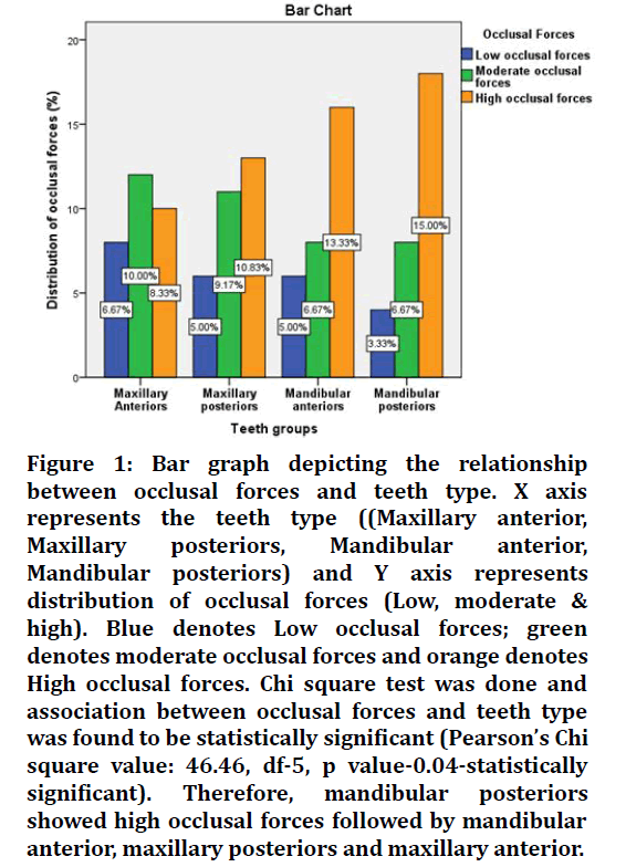
Association between occlusal forces and teeth type showed statistical significance with a p value of 0.04 (Figure 1).

Figure 1. Bar graph depicting the relationship between occlusal forces and teeth type. X axis represents the teeth type ((Maxillary anterior, Maxillary posteriors, Mandibular anterior, Mandibular posteriors) and Y axis represents distribution of occlusal forces (Low, moderate & high). Blue denotes Low occlusal forces; green denotes moderate occlusal forces and orange denotes High occlusal forces. Chi square test was done and association between occlusal forces and teeth type was found to be statistically significant (Pearson’s Chi square value: 46.46, df-5, p value-0.04-statistically significant). Therefore, mandibular posteriors showed high occlusal forces followed by mandibular anterior, maxillary posteriors and maxillary anterior.
Therefore, mandibular posteriors showed high occlusal forces followed by mandibular anteriors, maxillary posteriors and maxillary anterior.
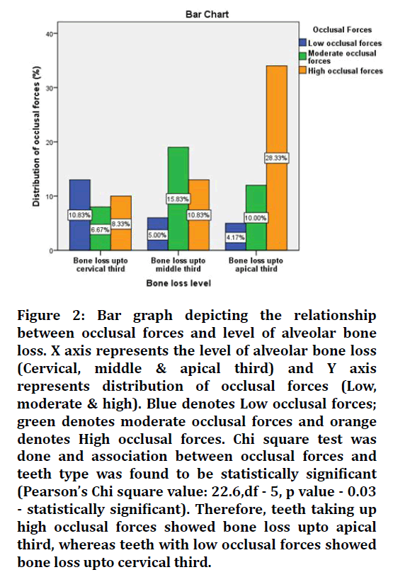
Whereas, association between occlusal forces and level of alveolar bone loss showed statistical significance with a p value of 0.03, as shown in Figure 2.

Figure 2. Bar graph depicting the relationship between occlusal forces and level of alveolar bone loss. X axis represents the level of alveolar bone loss (Cervical, middle & apical third) and Y axis represents distribution of occlusal forces (Low, moderate & high). Blue denotes Low occlusal forces; green denotes moderate occlusal forces and orange denotes High occlusal forces. Chi square test was done and association between occlusal forces and teeth type was found to be statistically significant (Pearson’s Chi square value: 22.6,df - 5, p value - 0.03 - statistically significant). Therefore, teeth taking up high occlusal forces showed bone loss upto apical third, whereas teeth with low occlusal forces showed bone loss upto cervical third.
Hence, teeth taking up high occlusal forces showed bone loss upto apical third, whereas teeth with low occlusal forces showed bone loss upto cervical third.
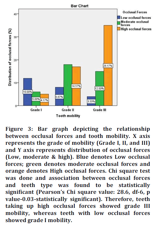
Tooth mobility index showed significant association with occlusal forces with a p value of 0.03, as depicted in Figure 3.

Figure 3. Bar graph depicting the relationship between occlusal forces and tooth mobility. X axis represents the grade of mobility (Grade I, II, and III) and Y axis represents distribution of occlusal forces (Low, moderate & high). Blue denotes Low occlusal forces; green denotes moderate occlusal forces and orange denotes High occlusal forces. Chi square test was done and association between occlusal forces and teeth type was found to be statistically significant (Pearson’s Chi square value: 28.6, df-6, p value-0.03-statistically significant). Therefore, teeth taking up high occlusal forces showed grade III mobility, whereas teeth with low occlusal forces showed grade I mobility.
Therefore, teeth taking up high occlusal forces showed grade III mobility, whereas teeth with low occlusal forces showed grade I mobility.
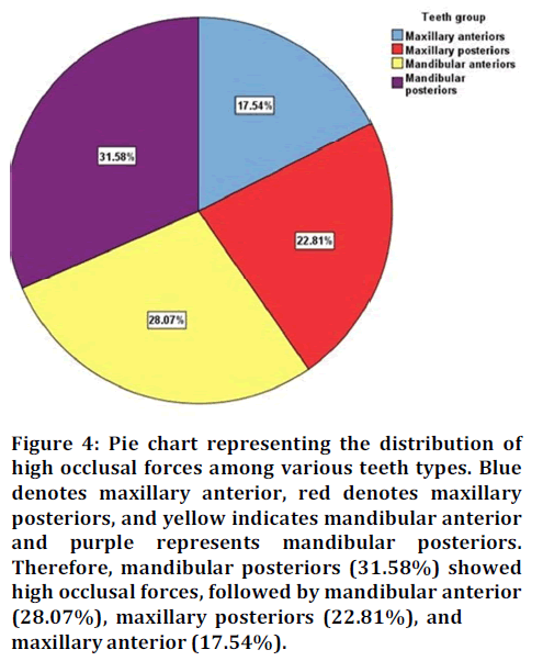
Among all the teeth groups, mandibular posteriors (31.58%) showed high occlusal forces, followed by mandibular anterior (28.07%), maxillary posteriors (22.81%), and maxillary anterior (17.54%) as in shown in Figure 4.

Figure 4. Pie chart representing the distribution of high occlusal forces among various teeth types. Blue denotes maxillary anterior, red denotes maxillary posteriors, and yellow indicates mandibular anterior and purple represents mandibular posteriors. Therefore, mandibular posteriors (31.58%) showed high occlusal forces, followed by mandibular anterior (28.07%), maxillary posteriors (22.81%), and maxillary anterior (17.54%).
Discussion
Occlusal trauma jeopardises periodontal health and makes treatment more difficult. Occlusal differences, in particular, balancing interferences have been linked to periodontal breakdown [1]. Teeth that have occlusal inconsistencies were found to have a substantially higher probing depth and a poor prognosis [10]. Treating them through selective grinding and or occlusal appliances during periodontal therapy is considered as an integral part of overall treatment of periodontal disease. The evolution of T-Scan from 1st generation to 8th generation (1847-2015) has revolutionized the concept of occlusal analysis. The “T-Scan I” was invented 25 years ago, and since then, the entire system has undergone hardware, sensor, and software revisions, such that today’s “T-Scan III” system (version 7.0) is vastly improved over the earlier systems [11].
To our knowledge, the present study is probably the first to report the association between periodontal conditions and occlusal force recorded by the T scan system in Indian population. Similar study was performed among the Chinese population [4]. The present data showed that high occlusal forces are mainly distributed in posteriors. This result was similar to that of the study done by Zhou et al [4].Takeuchi and Yamamoto investigated the association of biting force with periodontal status and found that occlusal force is positively associated with attachment loss [12]. This was not in accordance with our study results where there was no association found between occlusal forces and clinical attachment loss.
The present study showed significant association between occlusal forces and tooth mobility. This was in accordance with the study done by Harrel et al [13]. They focussed at the tooth level relationship between different occlusal contacts and probing depth, as well as the width of keratinized gingiva, and found that protrusive contacts on anterior teeth are usually not harmful. Despite the fact that the protrusive contacts in their sample may vary from the high occlusal forces observed by the T scan III device in our study, the exact effects of occlusal force on periodontal conditions in anterior teeth need more research.
Just a few tooth-level studies on the relationship between occlusal trauma and periodontal disease have been published. Various forms of premature contacts have been shown to be substantially associated with the risk of periodontal destruction [14]. Our study results show that there is significant association between occlusal forces and level of alveolar bone loss. This is in accordance with a study that showed that teeth with significant occlusal trauma showed alveolar bone resorption than the controls [15]. The present study shows that high occlusal forces are distributed more on the mandibular posterior teeth. These results are consistent with a previous study that the T scan system can reliably assess the distribution of occlusal contact in maximum intercuspation [16].
Conclusion
Within the limitations of this research, the current findings indicate that posterior teeth with high occlusal force in patients with untreated chronic periodontitis could represent occlusal trauma-related periodontal conditions, which may increase the risk of further periodontal destruction. The current findings can help clinicians better diagnose occlusal trauma and related periodontal conditions for better patient management and treatment outcomes. To confirm the current results and explain the clinical implications, further longitudinal research should be conducted.
References
- Reinhardt RA, Killeen AC. Do mobility and occlusal trauma impact periodontal longevity?. Dent Clin 2015; 59:873-83. 3
- Newman MG, Takei H, Klokkevold PR, et al. Carranza's clinical periodontology. Elsev heal sci 2011.
- Proffit WR. On the Aetiology of malocclusion. Br J Orthod 1986; 13:1–11.
- Zhou SY, Mahmood H, Cao CF, et al. Teeth under high occlusal force may reflect occlusal trauma-associated periodontal conditions in subjects with untreated chronic periodontitis. Chin J Dent Res 2017; 20:19-26.
- Schelb E, Kaiser DA, Brukl CE. Thickness and marking characteristics of occlusal registration strips. J prosth dent 1985; 54:122-6.
- Halperin GC, Halperin AR, Norling BK. Thickness, strength, and plastic deformation of occlusal registration strips. J Prosth Dent 1982; 48:575-8.
- Millstein PL. A method to determine occlusal contact and noncontact areas: Preliminary report. J prosth dent 1984; 52:106-10.
- Arcan M, Zandman F. A method for in vivo quantitative occlusal strain and stress analysis. J biomec 1984; 17:67-79.
- Kerstein RB. Combining technologies: A computerized occlusal analysis system synchronized with a computerized electromyography system. Cranio 2004; 22:96-109.
- Leib AM. The effect of occlusal discrepancies on periodontitis. I. Relationship of initial occlusal discrepancies to initial clinical parameters. J Periodontol 2001; 72:1291-4.
- Trpevska V, Kovacevska G, Benedeti A, et al. T-scan III system diagnostic tool for digital occlusal analysis in orthodontics-a modern approach. Prilozi 2014; 35:155-60.
- Takeuchi N, Yamamoto T. Correlation between periodontal status and biting force in patients with chronic periodontitis during the maintenance phase of therapy. J Clin Periodontol 2008; 35:215-20.
- Harrel SK, Nunn ME. The association of occlusal contacts with the presence of increased periodontal probing depth. J Clin Periodontol 2009; 36:1035-42.
- Pihlstrom BL, Anderson KA, Aeppli D, et al. Association between signs of trauma from occlusion and periodontitis. J periodontol 1986; 57:1-6.
- Jin LJ, Cao CF. Clinical diagnosis of trauma from occlusion and its relation with severity of periodontitis. J Clin Periodontol 1992; 19:92-7.
- García VG, Cartagena AG, Sequeros OG. Evaluation of occlusal contacts in maximum intercuspation using the T-Scan system. J Oral Rehabilit 1997; 24:899-903.
Indexed at, Google Scholar, Cross Ref
Indexed at, Google Scholar, Cross Ref
Indexed at, Google Scholar, Cross Ref
Indexed at, Google Scholar, Cross Ref
Indexed at, Google Scholar, Cross Ref
Indexed at, Google Scholar, Cross Ref
Indexed at, Google Scholar, Cross Ref
Indexed at, Google Scholar, Cross Ref
Indexed at, Google Scholar, Cross Ref
Indexed at, Google Scholar, Cross Ref
Indexed at, Google Scholar, Cross Ref
Indexed at, Google Scholar, Cross Ref
Indexed at, Google Scholar, Cross Ref
Author Info
Subasree Soundarajan* and Priyalochana Gajendran
Department of Periodontics, Saveetha Dental College and Hospitals, Saveetha Institute of Medical and Technical Sciences Saveetha University, Chennai, Tamil Nadu, IndiaCitation: Subasree Soundarajan, Priya lochana Gajendran, Association between Occlusal Forces and Periodontal Conditions in Untreated Chronic Periodontitis Using T-Scan: A Retrospective Study, J Res Med Dent Sci, 2022, 10(1): 461-465
Received: 21-Dec-2021, Manuscript No. JRMDS-22-50239; , Pre QC No. JRMDS-22-50239 (PQ); Editor assigned: 23-Dec-2021, Pre QC No. JRMDS-22-50239 (PQ); Reviewed: 06-Jan-2022, QC No. JRMDS-22-50239; Revised: 10-Jan-2022, Manuscript No. JRMDS-22-50239 (R); Published: 17-Jan-2022
