Research - (2019) Volume 7, Issue 3
Beta and Gamma EEG Oscillatory Waves of the Frontal Cortex Increase After Wet Cupping Therapy in Healthy Humans
Faruk Abdullahi1, Cevat Unal2, Menizibeya O Welcome1, Emelda Nwenendah Mpi1, Nafisa Umar1, Afaf Muhammed1, Salma U Faruk1, Hauwa Kazaure1 and Senol Dane1*
*Correspondence: Senol Dane, Department of Physiology, College of Health Sciences, Nile University of Nigeria, Nigeria, Email:
Abstract
Introduction: Wet cupping therapy is a traditional complementary therapy that alleviates pain, stress and the symptoms of several ailments in humans. However, the effect of wet cupping therapy as recorded with the electroencephalography (EEG) has not been reported. The aim of this study was to investigate the effect of compare beta and gamma activities in the EEG before and after wet cupping therapy in healthy subjects.
Materials and Methods: The EEG tracing was recorded from eight healthy right-handed volunteers according to the standard international 10/20 system, using PowerLab 26T (AD Instruments, Bella Vista, Australia) before and after wet cupping therapy. Wet cupping was performed only once per person on 5 points of the posterior neck, bilateral perispinal areas of the neck, and thoracic spine. 3 ml-5 ml of blood was removed per cupping site.
Results: In the present study, both beta and gamma activities were significantly higher after wet cupping therapy, compared to the activities of these oscillatory waves recorded before therapy.
Conclusion: Wet cupping therapy increases beta and gamma activity in healthy subjects (pË0.05). Beta and gamma activities can be used as a measure of frontal activation in wet cupping therapy. This increase in oscillatory beta and gamma cortical waves may be mediated by stimulation of the peripheral nervous system, and possibly, corresponding increase in secretion of certain neurohumoral factors.
Keywords
Wet cupping therapy, Beta activity, Gamma activity, EEG
Introduction
Wet cupping therapy (also known as Al-Hijamah) is a percutaneous pressure- and size- dependent non-specific blood filtration technique that enables the excretion of pathological substances through the fenestrated skin capillaries upon application of negative suction pressure using sucking cups [1]. This complementary therapy has been reported to treat many diseases of differing etiology and pathogenesis [1]. For example, wet cupping therapy has been shown to clear toxic substances such as heavy metals from blood and interstitial fluids [2-4]. Wet cupping therapy has been successfully applied to treat other human maladies, including atherosclerosis, hyperlipidemia, hypertension, coronary heart diseases, gout, hepatitis, and thalassemia [5]. Study by Shekarforoush et al. [6] on animals has also shown promise of application of this therapy in life threatening cardiac diseases [7,8].
This complementary therapy may have positive effects on brain functions. However, the effect of wet cupping therapy on cortical functions as recorded with electroencephalogram (EEG) in healthy humans has not been reported. The EEG is a non-invasive recording that is used to study cerebral activities in health and disease. The EEG waves are the result of action potentials and multiple excitatory and inhibitory postsynaptic potentials mostly of the neurons of the subcortical and cortical regions of the brain [9-11]. The major EEG waves are delta or δ, theta or θ, alpha or α, beta or β and gamma or γ frequency bands. These frequency bands represent cerebral signal processing in sensorimotor and cognitive domains [9,12,13].
In a recent study, it was reported that foot reflexotherapy [14] and footbath therapy [15] increases beta and gamma activities of the EEG in young healthy humans. Wet cupping therapy can hypothetically affect the different EEG waves. Unfortunately, however, there is a lack of study regarding the effect of wet cupping therapy on EEG waves. While the effects of wet cupping therapy on different maladies of humans have been investigated, [1-8] the effects of this therapy on cerebral informationprocessing or brain oscillations as recorded on EEG in healthy humans are not known. This study was to investigate the effect of wet cupping therapy on the EEG waves in the healthy humans.
Materials and Methods
Ethical statement
The experimental protocol was in line with the Declaration of Helsinki and approved by the Research Ethics Committee/Institutional Review Board (REC/IRB) on the Use of Human Subjects (NUN/PHS/REC/IRB-UH/ 018/09/015). Permission was obtained from the concerned authorities before the participants were approached for possible involvement in the study.
Participants
Apparently healthy males who worked at different Faculties (Arts, Science, Engineering, and Health Sciences) of the Nile University of Nigeria, Abuja, were involved in the study. Within a period of one week, 51 males who were present in their various faculties were randomly approached for possible involvement in the study. Of the 51 males, 38 agreed to participate in the study. Of these, 2 males were on medication as directed by their physician, but did not disclose the illness for which medication was taken. So these 2 males were excluded from the study. Thirty six healthy right handed males (mean age ± standard deviation, 22.61 ± 11.34 years) participated in this study. All participants were right-handed, according to self-report and confirmed by Edinburgh Handedness Inventory [16]. They had comparable education level (15 years-17 years).
Inclusion criteria
1. Willingness to participate.
2. Absence of any health problem based on recent medical examination. The absence or presence of chronic illnesses and other habits was determined through their past medical history.
3. Total abstinence from drugs. Participants were non-smokers, and were not on medications.
Exclusion criteria
1. Unwillingness to participate in the study.
2. Presence of health problems such as psychiatric, respiratory, metabolic, cardiac or central and autonomic nervous system disease, which may affect EEG tracing. The Mini-Mental State Examination (MMSE) was initially used to screen participants of cognitive deficit. No participant had cognitive deficit on this test.
3. Individuals using medications or drugs were not considered for participation. Those who fail the drug abuse test were not involved in the study.
Procedure
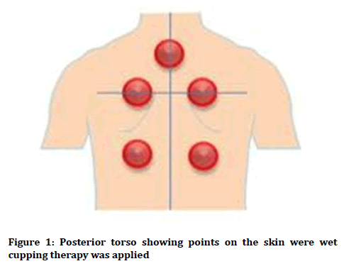
Wet cupping therapy was conducted according to the British Cupping Society and Natural Health Institutes [3,17]. For the cupping therapy, sterile disposable cups 5 cm in diameter were used. Five points of the posterior neck, bilateral perispinal areas of the neck, and thoracic spine were selected for treatment (Figure 1). These points are classic wet cupping points chosen for all cupping therapies. Application areas were cleaned with antiseptic solutions. Cups (Sharon Xie Jiangmen Xinli Medical Apparatus And Instruments Co., Ltd., China) were placed on these points, and negative pressure was applied by a cupping pump (Sharon Xie Jiangmen Xinli Medical Apparatus and Instruments Co., Ltd., China) on the middle level to withdraw blood and intercellular fluid enough. The cups were removed after 2 minutes to 3 minutes. Then, the skin within the cupping sites was punctured to a 2 mm depth by using a 26-gauge disposable lancet. Then, vacuum pumping was applied three times and 3 ml to 5 ml of blood was drained per cupping site. Application sites were covered with sterile pads.

Figure 1: Posterior torso showing points on the skin were wet cupping therapy was applied.
EEG recording
The EEG signal was recorded according to the standard international 10/20 system, with a sampling rate of 1 kHz. EEG data was recorded by two channel bipolar montage; F4-F8 (right brain hemisphere) and F3-F7 (left brain hemisphere). The digital EEGs were recorded by using PowerLab 26T (AD Instruments, Bella Vista, Australia), a device used for multimodal monitoring of bio-signals. In this study, the EEG was recorded in all participants at baseline (before commencement of wet cupping therapy) and after wet cupping therapy. EEG frequency bands considered for analysis were (1-3 Hz), theta waves (4-7 Hz), alpha waves (8-12 Hz), beta waves (13-30 Hz), gamma1 waves (31-40 Hz), gamma2 waves (41-50 Hz) and gamma3 waves (60-80 Hz).
The electroencephalographic signal processing analyses were performed in MATLAB. The changes in frequency and amplitude of the EEG were calculated by means of power spectral analysis, measured as total power in microvolts-squared divided by frequency (μV2/Hz) [17]. We used Discrete Fourier Transform (DFT) to calculate the power spectrum of time domain discrete EEG signal. Power spectral density (PSD) is frequency response of a periodic or random signal. PSD shows distribution of signal strength depending on the frequency. The EEG data were recorded is a time domain discrete signal.
Power spectral density can be expressed with Equation 1;

Where N is the number of samples and xi () is the Discrete Fourier Transform (DFT) of the time domain discrete i () signal. i ()is calculated as shown in equation 2;
width="322" height="52"
Statistical analysis
The SPSS statistical software package (SPSS, version 18.0 for windows) was used to perform all statistical calculations. Distributions were evaluated by using one sample Kolmogorov Smirnov test. A two-tailed paired t test (Student’s t test) was used for comparisons. Results are expressed as mean ± standard deviation (SD). Differences were considered statistically significant at p˂0.05.
Results
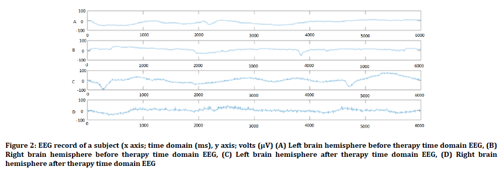
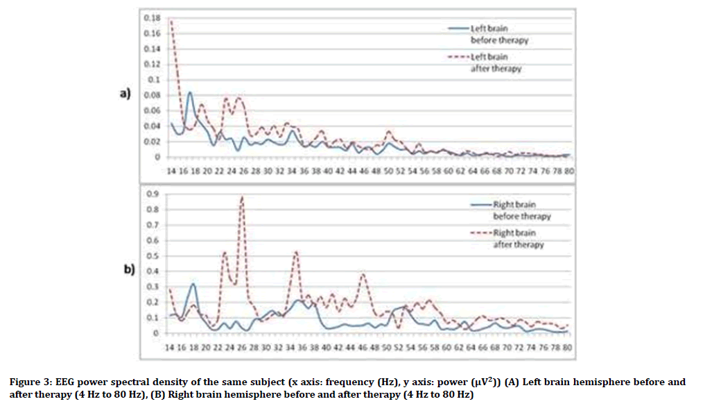
In the present study, EEG data of the F4-F8 (the right brain hemisphere) revealed that the powers of beta, gamma1, gamma2 and gamma3 waves (μv2) were increased after wet cupping therapy compared to the EEG tracing of corresponding waves before therapy (beta: t=2.21, p=0.04; gamma1: t=2.37, p=0.04; gamma2: t=2.48, p=0.03; gamma3: t=2.51, p=0.02). Comparison between the powers (μv2) of other waves (theta, delta and alpha) before and after therapy did not show any significant value. Similarly, percentages of all waves before therapy were not significantly different from their corresponding wave values after therapy (Table 1, Figures 2, 3 and 4).
| Parameters | Before Therapy | After Therapy | t | p | ||
|---|---|---|---|---|---|---|
| Mean | SD | Mean | SD | |||
| Total Power (μv2) | 75.91 | 52.56 | 51.06 | 21.12 | 2.71 | 0.01 |
| Delta Power (μv2) | 67.29 | 51.47 | 39.39 | 25.52 | 3.12 | 0.004 |
| Theta Power (μv2) | 2.78 | 1.38 | 2.67 | 1.07 | 0.45 | NS |
| Alpha Power (μv2) | 1.41 | 1.03 | 1.35 | 0.46 | 0.45 | NS |
| Beta Power (μv2) | 2.78 | 2.29 | 4.61 | 3.66 | 4.71 | 0 |
| Gamma1 Power (μv2) | 0.96 | 0.94 | 1.76 | 1.69 | 4.41 | 0 |
| Gamma2 Power (μv2) | 0.68 | 0.76 | 1.28 | 1.32 | 4.39 | 0 |
| Gamma3 Power (μv2) | 0.35 | 0.38 | 0.79 | 0.91 | 3.36 | 0.002 |
| Delta Power % | 84.3 | 13.94 | 70.56 | 16.42 | 4.21 | 0 |
| Theta Power % | 4.51 | 1.55 | 6.39 | 2.67 | 3.44 | 0.002 |
| Alpha Power % | 2.56 | 1.96 | 3.85 | 2.21 | 3.54 | 0.001 |
| Beta Power % | 5.54 | 7.19 | 11.95 | 9.66 | 3.69 | 0.001 |
| Gamma1 Power % | 1.77 | 2.64 | 4.32 | 4.16 | 3.82 | 0.001 |
| Gamma2 Power % | 1.32 | 2.31 | 2.93 | 2.67 | 3.51 | 0.001 |
| Gamma3 Power % | 0.64 | 1.08 | 1.84 | 2.06 | 3.45 | 0.001 |
Table 1: Powers and percentages of EEG bands before and after wet cupping therapy in the right brain (F4-F8) hemisphere
In the F3-F7 (left hemisphere) EEG, the powers of beta, gamma1, gamma2 and gamma3 waves (μv2) were increased after wet cupping therapy compared to corresponding EEG tracing recorded before therapy (beta: t=2.45, p=0.03; gamma1: t=2.27, p=0.04; gamma2: t=2.19, p=0.04; gamma3: t=2.45, p=0.03). Also, there was significant increase in percentages of gamma1, gamma2 and gamma3 (gamma1: t=2.71, p=0.03; gamma2: t=2.18, p=0.018; gamma3: t=2.87, p=0.02). The powers of theta, delta and alpha and percentages of theta, delta, alpha and beta waves were not statistically significant (Table 2, Figures 2, 3 and 4).
| Parameters | Before Therapy | After Therapy | t | p | ||
|---|---|---|---|---|---|---|
| Mean | SD | Mean | SD | |||
| Total Power (μv2) | 86.14 | 62.28 | 68.06 | 27.14 | 1.77 | NS |
| Delta Power (μv2) | 75.48 | 58.99 | 55.49 | 26.09 | 2.14 | 0.04 |
| Theta Power (μv2) | 3.77 | 1.09 | 3.57 | 1.39 | 0.72 | NS |
| Alpha Power (μv2) | 1.91 | 1.21 | 1.46 | 0.53 | 3.15 | 0.003 |
| Beta Power (μv2) | 3.24 | 2.34 | 4.44 | 2.55 | 4.28 | 0 |
| Gamma1 Power (μv2) | 1.02 | 0.84 | 1.86 | 1.46 | 4.82 | 0 |
| Gamma2 Power (μv2) | 0.73 | 0.57 | 1.24 | 0.85 | 4.05 | 0 |
| Gamma3 Power (μv2) | 0.37 | 0.32 | 0.72 | 0.67 | 3.48 | 0.001 |
| Delta Power % | 81.42 | 7.89 | 77.43 | 8.34 | 4.42 | 0 |
| Theta Power % | 5.94 | 2.57 | 5.83 | 2.04 | 0.24 | NS |
| Alpha Power % | 3.26 | 3.21 | 2.81 | 1.48 | 1.09 | NS |
| Beta Power % | 4.86 | 2.82 | 8.24 | 4.27 | 6.19 | 0 |
| Gamma1 Power % | 1.47 | 0.99 | 3.36 | 2.31 | 6.15 | 0 |
| Gamma2 Power % | 1.05 | 0.69 | 2.32 | 1.66 | 5.47 | 0 |
| Gamma3 Power % | 0.52 | 0.37 | 1.34 | 1.27 | 4.63 | 0 |
Table 2: Powers and percentages of EEG frequency bands before and after foot reflexotherapy in the left brain (F3-F7) hemisphere
Figure 2: EEG record of a subject (x axis; time domain (ms), y axis; volts (μV) (A) Left brain hemisphere before therapy time domain EEG, (B) Right brain hemisphere before therapy time domain EEG, (C) Left brain hemisphere after therapy time domain EEG, (D) Right brain hemisphere after therapy time domain EEG.
Figure 3: EEG power spectral density of the same subject (x axis: frequency (Hz), y axis: power (μV2)) (A) Left brain hemisphere before and after therapy (4 Hz to 80 Hz), (B) Right brain hemisphere before and after therapy (4 Hz to 80 Hz).
Figure 4: The powers of different EEG bands (x axis: band, y axis: power (μV2)) (A) Left brain hemisphere before and after therapy, (B) Right brain hemisphere before and after therapy.
Discussion
Though EEG is a non-routine investigation, it remains one of the most widely used objective methods of investigating brain activities in both normal and pathology [18-26].
This is the first study to report differences in frontal brain oscillatory beta and gamma waves of the EEG in healthy humans before and after wet cupping therapy. The powers of beta, gamma1, gamma2 and gamma3 waves of the EEG tracing increased in both the right and left hemispheres after cupping therapy compared with corresponding data recorded before cupping therapy. Also, the percentages of all gamma waves in EEG were increased after wet cupping therapy especially in right brain. These results indicate that this complementary therapy might have helpful effects on central nervous system functions in humans.
Similar to our previous results on the effects of reflexotherapy [14] and footbath therapy [15] on cortical functions, this present study also showed that application of wet cupping therapy resulted to frontal cortex activation as evidenced by increase mainly in beta and gamma waves. This suggests that the effect of wet cupping on cortical functions may be due to the skin puncture and vacuum pumping effects. The procedure stimulates cutaneous sensory receptors that consequently generate signals, which are sent to the central nervous system to activate the respective frontal areas [19]. Though wet cupping is widely known to enhance removal of toxins from the body, its effects on the brain is largely due to the local stimulation of cutaneous receptors produced by skin puncture and vacuum pumping. Beta wave is associated with normal waking consciousness, and is normally observed in the fronto-central regions of EEG recording [20]. Beta wave represents sensory motor rhythm and is related to cognitive processing and motor control [21]. Gamma waves are important for perceptual and cognitive processes [22]. Therefore an increase in beta and gamma waves indicates enhanced higher mental functions [23- 25,27]. Though the underlying mechanisms of generation of these waves are not exactly clear, animal models suggest a possible role of GABA-A system in the hippocampus, dentate gyrus, and CA1-CA3 system [23]. Indeed, these brain regions are involved in long-term memory formation and cognitive performance [9,14].
Our study suggests that beta and gamma waves can be used as functional markers of cortical activation in wet cupping therapy. This complementary therapy may serve as a means of increasing cognitive functions in humans with cognitive deficit. Indeed wet cupping therapy is currently used as an adjuvant in some neurological diseases such as chronic non-specific neck pain [28], lumbar disc herniation, cervical spondylosis [29], brachialgiaparaestheticanocturna [30], fibrositis [31], fibromyalgia [32], chronic knee osteoarthritis [33] and other pain conditions [34,35]. Interestingly, a recent study reported that stimulation of the left cerebral hemisphere with wet cupping therapy decreased the autistic symptoms in children with autism [36,37].
However, in contrast to our previous study on foot reflexotherapy, in which delta power for both frontal brain was decreased [14], in this present study, delta power was significantly increased in both hemispheres following wet cupping therapy. Delta wave is a low frequency wave that is associated with sleep, but not related to cognitive activities. Though sleep quality was not assessed in our study before and after cupping therapy, the result suggests that wet cupping therapy may enhance sleep quality or duration. Indeed, Cikar et al. showed that wet cupping therapy significantly improved sleep quality in healthy men and women [36].
Cupping therapy relieves the symptoms in a variety of diseases or clinical conditions through its effects on the regional acupoint areas. For example, Tham et al. showed that cupping maybe capable of stimulating individual acupuncture points [38]. Among the stimuli, the negative pressure applied during cupping is one of the main factors that induce therapeutic effects. Though the underlying mechanism of therapeutic effects of the negative pressure from cupping is not completely understood, it may be associated with the release of neurohumoral factors resulting from or due to stimulation of the peripheral and central nervous system. For instance, stimulation of superficial and deep tactile and pain receptors can result to the secretion of bendorphin and adrenocortical hormone into the circulation, and are transported to their receptive sites, where they exert a variety of influences [1,38].
It is important to mention that factors influencing EEG waves, including disease conditions, physiological states, age, gender, as well as artifacts [39-42] were greatly minimized in our study. Importantly, the influence of gender on EEG tracing is increasingly been reported in the literature [43-45]. Consequently we considered only males in the study, due to the gender differences in EEG tracing, and also, the effects of certain physiological state (e.g. menstrual cycle) and hormones on EEG waves [46-48].
Conclusion
Wet cupping therapy increases the frontal brain beta and gamma activities of EEG. The beta and gamma waves can be used as important functional measures of frontal brain activation in wet cupping therapy. This increase in oscillatory beta and gamma cortical waves may be mediated by stimulation of the peripheral nervous system, and possibly, corresponding increase in secretion of certain neurohumoral factors.
Future Directions
Since the present study consisted of only males, whether these results apply to females or children are not known. Therefore, it is imperative for future studies to investigate the cortical electrophysiology of wet cupping therapy on females and children as well as different categories of patients with neurodevelopmental, neurodegenerative and psychiatric disorders.
Conflict of Interest
The authors declare that there is no conflict of interest regarding the publication of this manuscript.
References
- El-Sayed SM, Al-Quliti AS, Mahmoud HS, et al. Therapeutic benefits of Al-hijamah: In light of modern medicine and prophetic medicine. Am J Med Biol Res 2014; 2:46-71.
- Tagil SM, Celik HT, Ciftci S, et al. Wet-cupping removes oxidants and decreases oxidative stress. Complement Ther Med 2014; 22:1032-6.
- Gok S, Kazanci FH, Erdamar H, et al. Is it possible to remove heavy metals from the body by wet cupping therapy (Al-hijamah)? India J Tradit Knowle 2016; 15:700-4.
- El Sayed SM, Mahmoud HS, Nabo MM. Methods of wet cupping therapy (Al-Hijamah): In light of modern medicine and prophetic medicine. Altern Integr Med 2013; 1-6.
- Niasari M, Kosari F, Ahmadi A. The effect of wet cupping on serum lipid concentrations of clinically healthy young men: A randomized controlled trial. J Altern Complement Med 2007; 13:79-82.
- Shekarforoush S, Foadoddini M, Noroozzadeh A, et al. Cardiac effects of cupping: Myocardial infarction, arrhythmias, heart rate and mean arterial blood pressure in the rat heart. Chin J Physiol 2012; 55:253-8.
- Arslan M, Yesilçam N, Aydin D, et al. Wet cupping therapy restores sympathovagal imbalances in cardiac rhythm. The Journal of Alternative and Complementary Medicine. 2014 Apr 1;20(4):318-21.
- Isik B, Aydin D, Arslan M, et al. Reflexological therapy induces a state of balance in autonomic nervous system. Clin Invest Med (Online) 2015; 38:E244.
- Kirschstein T, Köhling R. What is the source of the EEG?. Clin EEG Neurosci 2009; 40:146-9.
- Buzsáki G, Anastassiou CA, Koch C. The origin of extracellular fields and currents-EEG, ECoG, LFP and spikes. Nat Rev Neurosci 2012; 13:407.
- Schwartz AB, Cui XT, Weber DJ, et al. Brain-controlled interfaces: Movement restoration with neural prosthetics. Neuron 2006; 52:205-20.
- Demiralp T, Herrmann CS, Erdal ME, et al. DRD4 and DAT1 polymorphisms modulate human gamma band responses. Cerebral Cortex 2006; 17:1007-19.
- Hughes JR. Gamma, fast, and ultrafast waves of the brain: Their relationships with epilepsy and behavior. Epilepsy Behavior 2008; 13:25-31.
- Unal C, Welcome MO, Salako M, et al. The effect of foot reflexotherapy on the dynamics of cortical oscillatory waves in healthy humans: An EEG study. Complement Ther Med 2018; 38:42-7.
- Olanipekun A, Alhassan AK, Musa FH, et al. The effect of foot bath therapy on the dynamics of cortical oscillatory waves in healthy humans: An EEG study. J Res Med Dent Sci 2019; 7:57-61.
- Oldfield RC. The assessment and analysis of handedness: The Edinburgh inventory. Neuropsychologia 1971; 9:97-113.
- Ahmedi M, Siddiqui MR. The value of wet cupping as a therapy in modern medicine-An Islamic perspective. Webmed Central Altern Med 2014; 5:WMC004785.
- Binienda KZ, Beaudoin AM, Thorn TB, et al. Analysis of electrical brain waves in neurotoxicology: Gamma-hydroxybutyrate. Curr Neuropharmacol 2011; 9:236-9.
- Dane S, Welcome MO. A case study: Effects of foot reflexotherapy on ADHD symptoms and enuresis nocturia in a child with ADHD and enuresis nocturia. Complement Ther Clin Pract 2018; 33:139-41.
- Badrakalimuthu VR, Swamiraju R, De Waal H. EEG in psychiatric practice: To do or not to do? Adv Psychiatr Treat 2011; 17:114-21.
- Zaepffel M, Trachel R, Kilavik BE, et al. Modulations of EEG beta power during planning and execution of grasping movements. PloS One 2013; 8:e60060.
- Demiralp T, Herrmann CS, Erdal ME, et al. DRD4 and DAT1 polymorphisms modulate human gamma band responses. Cerebral Cortex 2006; 17:1007-19.
- Hughes JR. Gamma, fast, and ultrafast waves of the brain: Their relationships with epilepsy and behavior. Epilepsy Behavior 2008; 13:25-31.
- Staresina BP, Michelmann S, Bonnefond M, et al. Hippocampal pattern completion is linked to gamma power increases and alpha power decreases during recollection. Elife 2016; 5:e17397.
- Gold I. Does 40-Hz oscillation play a role in visual consciousness?. Conscious Cogn 1999; 8:186-95.
- Unal C, Natapraja R, Nurhayati GE, et al. Right sided lateralization of gamma activity of EEG in young healthy males. J Res Med Dent Sci 2018; 6:13-9.
- Singer W, Gray CM. Visual feature integration and the temporal correlation hypothesis. Annu Rev Neurosci 1995; 18:555-86.
- Lauche R, Langhorst J, Dobos GJ, et al. Clinically meaningful differences in pain, disability and quality of life for chronic nonspecific neck pain–A reanalysis of 4 randomized controlled trials of cupping therapy. Complement Ther Med 2013; 21:342-7.
- Cao H, Li X, Liu J. An updated review of the efficacy of cupping therapy. PloS One 2012; 7:e31793.
- Lüdtke R, Albrecht U, Stange R, et al. Brachialgia paraesthetica nocturna can be relieved by “wet cupping”-Results of a randomised pilot study. Complement Ther Med 2006; 14:247-53.
- Zhang HL. Blood-letting puncture and cupping therapies combined with acupuncture for treatment of 140 cases of fibrositis. J Tradit Chin Med 2009; 29:277-8.
- Cao H, Hu H, Colagiuri B, et al. Medicinal cupping therapy in 30 patients with fibromyalgia: A case series observation. Complement Med Res 2011; 18:122-6.
- Teut M, Kaiser S, Ortiz M, et al. Pulsatile dry cupping in patients with osteoarthritis of the knee–A randomized controlled exploratory trial. BMC Complement Altern Med 2012; 12:184.
- Sultana A, ur Rahman K, Farzana MU, et al. Efficacy of hijamat bila shurt (dry cupping) on intensity of pain in dysmenorrhoea-A preliminary study. Anc Sci Life 2010; 30:47.
- Zhang SJ, Liu JP, He KQ. Treatment of acute gouty arthritis by blood-letting cupping plus herbal medicine. J Tradit Chin Med 2010; 30:18-20.
- Cikar S, Ustundag G, Haciabdullahoglu S, et al. Wet cupping (hijamah) increases sleep quality. Clin Invest Med 2015; 38:E258.
- Canbal M, Kaya H, Kocoglu A, et al. The effects of evoked left hemisphere stimulations in autistic children. Clin Invest Med 2015; 38:E242.
- Tham LM, Lee HP, Lu C. Cupping: From a biomechanical perspective. JBiomech 2006; 39:2183-93.
- Neufeld MY, Drory VE, Korczyn AD. Duration and factors affecting electroencephalogram slowing after generalized tonic-clonic seizures. J Epilepsy 1995; 8:193-6.
- Bell MA, Cuevas K. Using EEG to study cognitive development: Issues and practices. J Cogn Dev 2012; 13:281-94.
- Puce A, Hämäläinen M. A review of issues related to data acquisition and analysis in EEG/MEG studies. Brain Sci 2017; 7:58.
- Campbell IG. EEG recording and analysis for sleep research. Curr Protoc Neurosci 2009; 49:10-2.
- Kim M, Sowndhararajan K, Kim T, et al. Gender differences in electroencephalographic activity in response to the earthy odorants geosmin and 2-methylisoborneol. Appl Sci 2017; 7:876.
- Sprecher KE, Riedner BA, Smith RF, et al. High resolution topography of age-related changes in non-rapid eye movement sleep electroencephalography. PloS One 2016; 11:e0149770.
- Lithari C, Frantzidis CA, Papadelis C, et al. Are females more responsive to emotional stimuli? A neurophysiological study across arousal and valence dimensions. Brain Topogr 2010; 23:27-40.
- de Zambotti M, Willoughby AR, Sassoon SA, et al. Menstrual cycle-related variation in physiological sleep in women in the early menopausal transition. J Clin Endocrinol Metab 2015; 100:2918-26.
- Brötzner CP, Klimesch W, Doppelmayr M, et al. Resting state alpha frequency is associated with menstrual cycle phase, estradiol and use of oral contraceptives. Brain Res 2014; 1577:36-44.
- Peng F, Zhang L. Prolonged menstruation and increased menstrual blood with generalized d electroencephalogram power: A case report. Exp Ther Med 2014; 7:728-30.
Author Info
Faruk Abdullahi1, Cevat Unal2, Menizibeya O Welcome1, Emelda Nwenendah Mpi1, Nafisa Umar1, Afaf Muhammed1, Salma U Faruk1, Hauwa Kazaure1 and Senol Dane1*
1Department of Physiology, College of Health Sciences, Nile University of Nigeria, Abuja, Nigeria2Faculty of Engineering, Department of Electrical and Electronics Engineering, Nile University of Nigeria, Abuja, Nigeria
Citation: Faruk Abdullahi, Cevat Unal, Menizibeya O Welcome, Emelda Nwenendah Mpi, Nafisa Umar, Afaf Muhammed, Salma U Faruk, Hauwa Kazaure, Senol Dane, Beta and Gamma EEG Oscillatory Waves of the Frontal Cortex Increase After Wet Cupping Therapy in Healthy Humans, J Res Med Dent Sci, 2019, 7(3): 95-102.
Received: 03-Apr-2019 Accepted: 30-May-2019


