Research - (2020) Volume 8, Issue 6
Cephalometric Features of Anterior Open Bite in a Sample of Sudanese Patients
Ejlal Hassan, Amal H Abuaffan and Marwa M Hamid*
*Correspondence: Marwa M Hamid, Department of Orthodontics, Pedodontics and Preventive Dentistry, Faculty of Dentistry, University of Khartoum, Sudan, Email:
Abstract
Worldwide, the prevalence of anterior open bite varies from (1% to 37.4%), the highest being in the French population whereas the lowest is in the Malta population. In the United States, the prevalence ranges between (16%) in black ethnicity and (4%) in whites. A similar study in Sudanese school children showed a much lower prevalence (1.1%). Open bites are multifactorial in etiology; they result from interactions between hereditary and environmental factors, and are divided into, dental open bite; without craniofacial malformations, and skeletal open bite involving craniofacial dysplasia. Skeletal open bite is characterized by a long anterior face height with the palatal plane tipped upward anteriorly, longer lower anterior face height, shorter upper anterior face height, and short posterior face height.
The present study was designed to investigate these features as well as comparing them to Sudanese norms. This, hopefully, will assist in establishing a sound ground for orthodontists and maxillofacial surgeons to guide them to the proper diagnosis and treatment planning for anterior open bite.
Keywords
Open bite Sudanese Cephalograms ODI
Introduction
Edward Angle’s definition of malocclusion was limited to the anteroposterior relationship; “the mesiobuccal cusp of the upper first permanent molar occludes in the buccal groove of lower first permanent molar”. This definition clearly did not consider occlusion in the three dimensions [1], later Ackerman and Proffit added the transverse and vertical dimensions to describe malocclusions [2].
Open bite is defined as the lack of vertical contact between opposing segments [3]. It can present as an anterior open bite or posterior open bite or a combination [4].
Worldwide, the prevalence of anterior open bite varies from (1% to 37.4%), the highest being in the French population whereas the lowest is in the Malta population [5,6]. In the United States, the prevalence ranges between (16%) in black ethnicity and (4%) in whites [6]. A similar study in Sudanese school children showed a much lower prevalence (1.1%) [7].
open bites are multifactorial in etiology; they result from interactions between hereditary and environmental factors [3], and are divided into, dental open bite; without craniofacial malformations, and skeletal open bite involving craniofacial dysplasia.
Skeletal open bite is characterized by a long anterior face height with the palatal plane tipped upward anteriorly, longer lower anterior face height, shorter upper anterior face height, and short posterior face height. The gonial angle is usually obtuse and the mandibular plane is steep notched. The presence of two separate occlusal planes is a characteristic feature. Dentoalveolar heights are often normal except for the mandibular molar dentoalveolar height which is noticeably shorter [8]. The literature revealed that anterior open bite patients can display both dental and skeletal features of open bite [9,10].
Anterior open bite is one of the most challenging orthodontic problems that need a proper evaluation to reach the optimal treatment outcomes. To our knowledge, no data are available concerning the skeletal and dental features of anterior open bite among the Sudanese population. Therefore, the present study was designed to investigate these features as well as comparing them to Sudanese norms. This, hopefully, will assist in establishing a sound ground for orthodontists and maxillofacial surgeons to guide them to the proper diagnosis and treatment planning for anterior open bite.
Materials and Methods
This comparative cross-sectional study was conducted on 62 lateral cephalograms of patients with anterior open bite (11 males and 51females) and 88 lateral cephalograms of individuals with normal inclusion (51 females and 37 male). All radiographs were taken from the Orthodontic department of a public university. Ethical approval was obtained from the research committee of the university. All patients’ cephalometric radiographs in the period from January 2013 to March 2017 were examined.
The selection criteria were as follows:
Sudanese nationality.
18-30 years old
Incisor vertical overlap ≤ 0 mm.
No previous or active orthodontic treatment.
No facial deformities.
Non-distorted, good quality radiographs.
Whereas the inclusion criteria for the “normal occlusion” group were as follows:
Sudanese nationality.
18-30 years old.
No previous or active orthodontic treatment.
well-balanced face with class I occlusion, average overbite, and overjet.
No facial deformity.
No or less than 2 mm irregularity.
Non-distorted, good quality radiographs.
All lateral cephalometric radiographs were taken in the natural head position and traced manually using a transparent 0.003-inch acetate paper a 0.5 mm 3H pencil. Landmarks and reference planes used in this study are described in Figures 1 and 2. The twenty-two measurements to describe the dental and skeletal patterns are described in Figures 3 to 6.
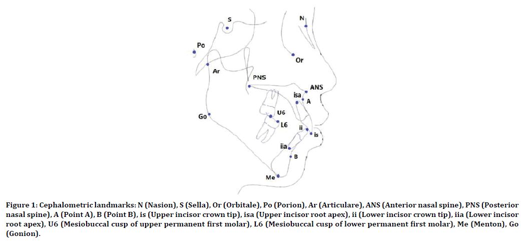
Figure 1. Cephalometric landmarks: N (Nasion), S (Sella), Or (Orbitale), Po (Porion), Ar (Articulare), ANS (Anterior nasal spine), PNS (Posterior nasal spine), A (Point A), B (Point B), is (Upper incisor crown tip), isa (Upper incisor root apex), ii (Lower incisor crown tip), iia (Lower incisor root apex), U6 (Mesiobuccal cusp of upper permanent first molar), L6 (Mesiobuccal cusp of lower permanent first molar), Me (Menton), Go (Gonion).
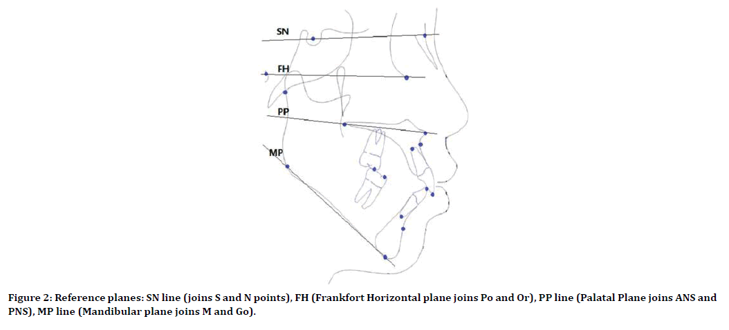
Figure 2. Reference planes: SN line (joins S and N points), FH (Frankfort Horizontal plane joins Po and Or), PP line (Palatal Plane joins ANS and PNS), MP line (Mandibular plane joins M and Go).
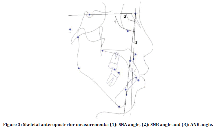
Figure 3. Skeletal anteroposterior measurements: (1): SNA angle, (2): SNB angle and (3): ANB angle.
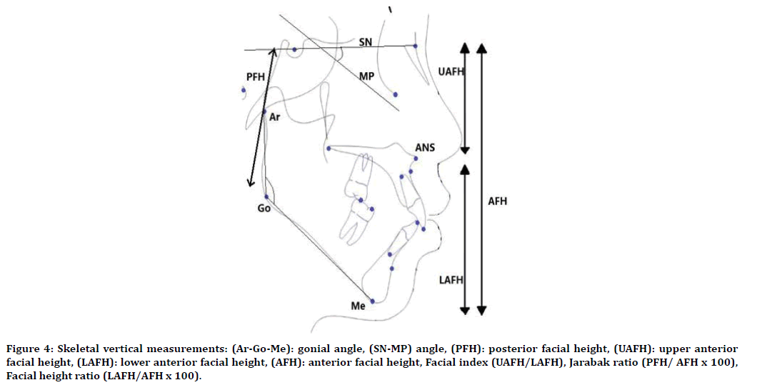
Figure 4. Skeletal vertical measurements: (Ar-Go-Me): gonial angle, (SN-MP) angle, (PFH): posterior facial height, (UAFH): upper anterior facial height, (LAFH): lower anterior facial height, (AFH): anterior facial height, Facial index (UAFH/LAFH), Jarabak ratio (PFH/ AFH x 100), Facial height ratio (LAFH/AFH x 100).
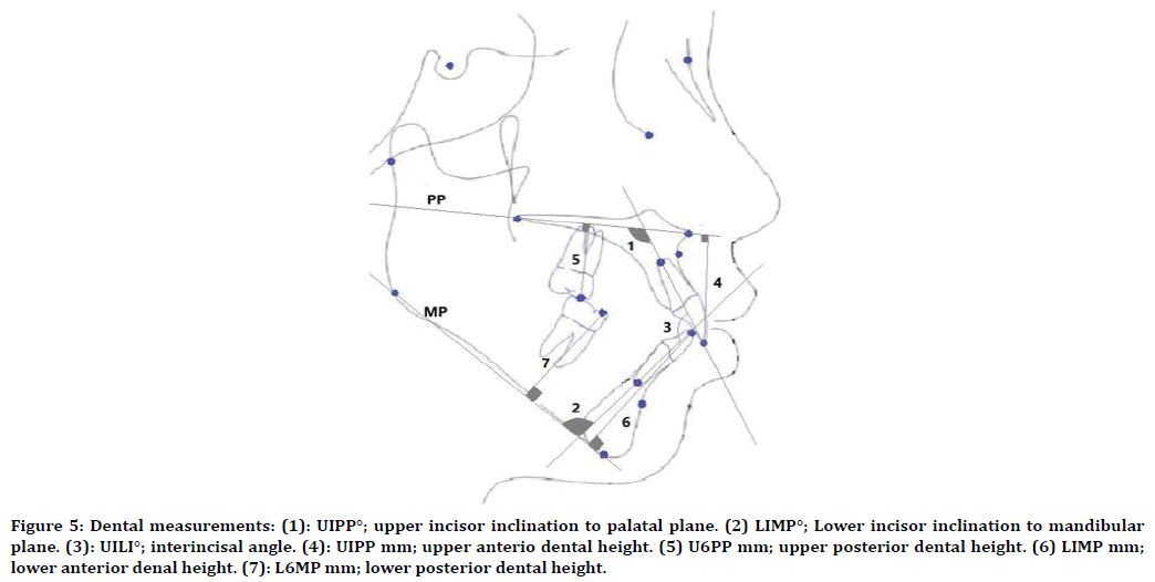
Figure 5. Dental measurements: (1): UIPP°; upper incisor inclination to palatal plane. (2) LIMP°; Lower incisor inclination to mandibular plane. (3): UILI°; interincisal angle. (4): UIPP mm; upper anterio dental height. (5) U6PP mm; upper posterior dental height. (6) LIMP mm; lower anterior denal height. (7): L6MP mm; lower posterior dental height.
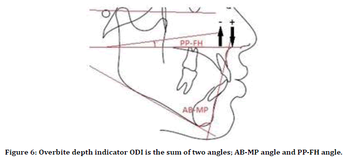
Figure 6. Overbite depth indicator ODI is the sum of two angles; AB-MP angle and PP-FH angle.
Data were collected, cleaned, and analyzed using the Statistical Package for Social Science version 22 (SPSS) software. The 95% confidence interval was considered significant with a P value of ≤ 0.05. To test the Intra-examiner reliability of the measurements, 15% of the total sample (13 radiographs of the normal occlusion group and 9 of the anterior open bite group) were randomly selected and analyzed by the main investigator and retraced after two weeks. The intraclass correlation coefficient (ICC) showed no statistically significant difference between the two readings for all study variables (Table 1).
| Variables | Interclass Correlation Coefficient (ICC) | P value* |
|---|---|---|
| SNA | 0.97 | 0 |
| SNB | 0.986 | 0 |
| ANB | 0.977 | 0 |
| Gonial angle | 0.982 | 0 |
| SN-MP angle | 0.986 | 0 |
| Facial index | 0.968 | 0 |
| Jarabak ratio | 0.985 | 0 |
| Lower Facial height ratio | 0.966 | 0 |
| UIPP angle | 0.978 | 0 |
| LIMP angle | 0.964 | 0 |
| UILI angle | 0.971 | 0 |
| U1PPmm | 0.89 | 0 |
| U6PPmm | 0.931 | 0 |
| L1MPmm | 0.922 | 0 |
| L6MPmm | 0.951 | 0 |
| UAFH | 0.958 | 0 |
| LAFH | 0.982 | 0 |
| PFH | 0.99 | 0 |
| AFH | 0.981 | 0 |
| AB-MP | 0.983 | 0 |
| PP-FH | 0.738 | 0 |
| ODI | 0.933 | 0 |
| *P ≤ 0.05 is significant | ||
Table 1: Intraclass correlation coefficient for all study variables.
Results
A total of 62 lateral cephalograms (11 males and 51females) of anterior open bite patients were analyzed and compared to 88 cephalograms (51 females and 37 male) of individuals with normal occlusion. Independent t-test was applied to test for statistically significant differences between the two groups in the following measurements: skeletal anteroposterior measurements, Skeletal vertical measurement, dental measurement, and ODI (Tables 2 to 5).
| Variable | Anterior Open bite (n=62) | Normal occlusion (n=88) | P value* | ||
|---|---|---|---|---|---|
| Mean | SD | Mean | SD | ||
| SNA | 81.74 | 3.9 | 82.97 | 4 | 0.064 |
| SNB | 76.87 | 4.5 | 79.46 | 3.7 | 0 |
| ANB | 4.87 | 2.9 | 3.5 | 2.1 | 0.002 |
| *P ≤ 0.05 is significant | |||||
Table 2: Cephalometric Skeletal anteroposterior relations.
| Variable | Anterior Open bite (n=62) | Normal occlusion (n=88) | P value* | ||
|---|---|---|---|---|---|
| Mean | SD | Mean | SD | ||
| Gonial angle | 131.26 | 5.1 | 125.37 | 5.3 | 0 |
| SN-MP Angle | 42.09 | 5.9 | 34.24 | 5.4 | 0 |
| Facial index | 70.6 | 6.5 | 77.58 | 7.5 | 0 |
| Jarabak ratio | 57.79 | 4.6 | 63.58 | 4.9 | 0 |
| UAFH | 50.27 | 3.6 | 51.61 | 3.7 | 0.028 |
| LAFH | 71.54 | 5.8 | 66.74 | 4.9 | 0 |
| PFH | 70.15 | 6.6 | 75.2 | 6.9 | 0 |
| AFH | 121.42 | 7.4 | 118.27 | 6.4 | 0.006 |
| Lower Facial height ratio | 58.87 | 2.2 | 56.39 | 2.5 | 0 |
| *P ≤ 0.05 is significant | |||||
Table 3: Cephalometric skeletal vertical relations.
| Variable | Anterior Open bite (n=62) | Normal occlusion (n=88) | P value* | ||
|---|---|---|---|---|---|
| Mean | SD | Mean | SD | ||
| U1PP° | 121.45 | 6.8 | 116.7 | 4.5 | 0 |
| L1MP° | 100.3 | 7.1 | 100.6 | 5.9 | 0.748 |
| U1L1° | 103.69 | 9.3 | 116.47 | 7.9 | 0 |
| U1PP mm | 30.56 | 4 | 29.47 | 2.9 | 0.068 |
| U6PP mm | 26.08 | 3.3 | 23.97 | 1.9 | 0 |
| L1MP mm | 43.23 | 4.2 | 42.61 | 3.1 | 0.332 |
| L6MP mm | 32.85 | 3.1 | 32.36 | 2.8 | 0.318 |
| *P ≤ 0.05 is significant | |||||
Table 4: Cephalometric dental relations.
| Variable | Anterior Open bite (n=62) | Normal occlusion (n=88) | P value* | ||
|---|---|---|---|---|---|
| Mean | SD | Mean | SD | ||
| AB-MP angle | 64.87 | 5.2 | 69.77 | 4.4 | 0 |
| PP-FH angle | -2.54 | 3.8 | -0.94 | 3.1 | 0.005 |
| ODI | 62.21 | 6.5 | 68.92 | 5.6 | 0 |
| *P ≤ 0.05 is significant | |||||
Table 5: Overbite depth indicator.
Discussion
Reaching an accurate diagnosis is an essential step toward proper treatment planning and consequently, more optimum results. For anterior open bite patient, distinguishing dental open bites from the skeletal ones is crucial in establishing an accurate diagnosis.
This comparative cross-sectional study compared dental and skeletal features of anterior open bite adult patients and adults with normal occlusion. Twenty-two linear and angular variables were measured and compared. Results showed significant differences in most of the study variables.
Skeletal anteroposterior relations
In the current study, no significant difference was detected in the SNA angle between the two groups. A similar finding was observed by Beane et al and Jones [11,12]. On the contrary, Tsang’s results showed a reduction of the SNA angle in open bite Chinese patients indicating some maxillary retrusion [13].
In the anterior open bite group, SNB was significantly smaller and consequently, ANB was significantly larger. (76.87° and 4.87°) respectively compared to (79.46° and 3.5°) in the normal occlusion group. This finding was expected since open bites, especially the skeletal ones, usually result from a backward mandibular rotation pattern carrying the B point more posteriorly. The same was observed by Jones in a study conducted on North American black population and by Daer and Abuaffan et al in Yemeni population [12,14].
In contrast, Tsang reported a smaller ANB angle in open bite patients than normal occlusion individuals in the Southern Chinese population [13].
On the other hand, the findings of the recent study did not match Beane’s results in the black American population which revealed no association between open bite, SNB, and ANB angles [11].
Skeletal vertical relations
In the current study, all vertical measurements showed highly significant differences between normal occlusion groups and anterior open bite group. The gonial angle and SN-MP angle express the steepness of the mandible. Both angles were significantly more obtuse in open bite groups than in normal occlusion group. They measured (131.26°and 42.09°) respectively compared to (125.37° and 34.24°) respectively. These findings were in accordance with Tsang results in Southern Chinese, Kao et al. findings in Taiwanese, and Cangialosi findings in Columbians [10,13,15].
The Jarabak ratio (PFH: AFH) is a manifestation of the balance between anterior and posterior facial heights. Lower values are indicative of backward mandibular rotation and hence, the tendency toward skeletal open bite [16]. This was confirmed in the present study. The ratio was (57.79%) in open bite group and (63.58%) in the comparison group. Again, this was consistent with Tsang's results among the Southern Chinese population [13].
To Further specify the origin of the discrepancy in the Jarabak ratio, UAFH, LAFH, total AFH, and PFH were measured separately and compared. The major contributor was the significant increase in the LAFH in anterior open bite group when compared to the normal occlusion group (71.54 mm) and (66.74) mm, respectively. In addition, there was a remarkable reduction in PFH in the open bite group (70.15 mm) compared to (75.2 mm) in normal occlusion group.
Similar findings were observed in Southern Chinese and North American black populations [12,13]. Furthermore, anterior open bite group had a statistically significant larger AFH value (121.42 mm) compared to (118.27mm).
Two more indicators of the individual’s vertical configuration are the facial index (UAFH/LAFH), and lower facial height ratio (LAFH/AFH). The facial index is typically reduced in skeletal open bite cases and the opposite is true for lower facial height ratio. This was evident in the current results; anterior open bite group recorded (70.60 % and 58.87%) for facial index and lower facial height ratio respectively compared to (77.58% and 56.39%) for normal occlusion group.
Dental relations
Upper incisors often show some degree of proclination in anterior open bite cases. This can be partially explained by the probability of concomitant digit sucking habit that characteristically causes flaring of the upper incisors as well as an anterior open bite. Accordingly, the anterior open bite group had (121.45°) UIPP angle compared to (116.7°) in normal occlusion group. An expected consequence of upper incisors flaring is the reduction of the interincisal angle. This again was confirmed by the current results. Anterior open bite group had UILI angle of (103.69°) compared to (116.47°) in the comparison group.
This combination of upper incisors' proclination and decreased interincisal angle in anterior open bite patients was also evident in North black Americans, Yamani, and Southern Chinese populations [12-14].
No statistically significant differences were detected in the dental height measurements between the two groups except for the upper posterior dental height (U6PP) which recorded (26.08 mm) in anterior open bite group and (23.97 mm) in the comparison group.
Overbite depth indicator (ODI)
The ODI was developed by Kim 1974 as a tool to distinguish skeletal open bites from dental open bites.IT is the sum of two angles; the angle formed by the AB line and the mandibular plane, and the angle between the palatal plane and the Frankfort horizontal plane. The latter angle is given a negative value when the palatal plane is tipped upwards anteriorly. Lower values are indicatives of skeletal open bite tendency [17].
In this study, the open bite group had an ODI of (62.21°) compared to (68.92°) in the normal occlusion group. This further highlights the fundamental difference in skeletal configuration between the two groups.
Conclusion
Significant differences exist between patients with anterior open bite and individuals with normal occlusion. Anterior open bite group had more retrognathic mandibles and a larger ANB angle than the normal occlusion group. In addition, anterior open bite patients had longer anterior facial height, shorter posterior facial height, less value of Jarabak ratio, and ODI as well as steeper mandibular planes. However, dental measurements were similar in both groups except for upper incisor inclination, interracial angle, and upper posterior dental height.
Abbreviations
Not applicable.
Acknowledgements
Not applicable.
Funding
No sources of funding.
Availability of Data and Material
The datasets used and analyzed during the current study are available from the corresponding author on reasonable request.
Ethics approval and consent to Participate
The ethical approval was obtained from the Ethics Research Committee, Faculty of Medicine, University of Khartoum, Sudan.
Consent for Publication
Not applicable.
Competing Interests
The authors declare that they have no competing interests.
References
- Angle EH. Classification of malocclusion. Dental Cosmos 1899; 41:248-264.
- Ackerman JL, Proffit WR. The characteristics of malocclusion: A modern approach to classification and diagnosis. Am J Orthod 1969; 56:443-454.
- Cozza P, Mucedero M, Baccetti T, et al. Early orthodontic treatment of skeletal open-bite malocclusion: A systematic review. Angle Orthod 2005; 75:707-713.
- Hapak FM. Cephalometric appraisal of the open-bite case. Angle Orthodont 1964; 34:65-72.
- Camilleri S, Mulligan K. The prevalence of malocclusion in Maltese schoolchildren as measured by the index of orthodontic treatment need. Malta Med J 2007; 19:19-24.
- Ngan P, Fields H. Open bite: A review of etiology and management. Pediatr Dent 1996; 19:91-98.
- Abuaffan AH, Wisth P, Boe O. Malocclusion in 12-year-old Sudanese children. Odonto-stomatologie tropicale. Tropical Dent J 1990; 13:87-93.
- Nahoum HI. Anterior open-bite: A cephalometric analysis and suggested treatment procedures. Am J Orthod 1975; 67:523-521.
- Richardson A. Skeletal factors in anterior open-bite and deep overbite. Am J Orthodont 1969; 56:114-127.
- Cangialosi TJ. Skeletal morphologic features of anterior open bite. Am J Orthodont 1984; 85:28-36.
- Beane RA, Reimann G, Phillips C, et al. A cephalometric comparison of black open-bite subjects and black normals. Angle Orthod 2003; 73:294-300.
- Jones OG. A cephalometric study of 32 north American black patients with anterior open bite. Am J Orthod Dentofacial Orthop 1989; 95:289-296.
- Tsang WM, Cheung LK, Samman N. Cephalometric characteristics of anterior open bite in a southern Chinese population. Am J Orthod Dentofacial Orthop 1998; 113:165-172.
- Daer AA, Abuaffan AH. Skeletal and dentoalveolar cephalometric features of anterior open bite among Yemeni adults. Scientifica 2016; 3147972.
- Kao CT, Chen FM, Lin TY, et al. The morphologic structure of the open bite in adult Taiwanese. Angle Orthod 1996; 66:199-206.
- Jarabak JR, Fizzell JA. Technique and treatment with the light wire edgewise appliance: Light differential forces in clinical orthodontics. St Louis: Mosby yearbook 1963.
- Kim YH. Overbite depth indicator with particular reference to anterior open bite. Am J Orthodont 1974; 65:586-611.
Author Info
Ejlal Hassan, Amal H Abuaffan and Marwa M Hamid*
Department of Orthodontics, Pedodontics and Preventive Dentistry, Faculty of Dentistry, University of Khartoum, SudanCitation: Ejlal Hassan, Amal H Abuaffan, Marwa M Hamid, Cephalometric Features of Anterior Open Bite in a Sample of Sudanese Patients, J Res Med Dent Sci, 2020, 8 (6): 146-152.
Received: 19-Aug-2020 Accepted: 21-Sep-2020
