Research Article - (2023) Volume 11, Issue 1
Comparative Assessment of Canal Transportation and Centering Ratio of Three Heat Treated Rotary Systems in Severely curved simulated resin block (An in vitro study)
Shareef Radhi Jawad* and Shatha Abdul Kareem
*Correspondence: Shareef Radhi Jawad, Department of Restorative and Esthetic Dentistry, College of Dentistry, University of Baghdad, Baghdad, Iraq, Email:
Abstract
Objectives: The purpose of this study was to measure and compare the canal transportation and centering ratio at different levels of simulated curved canals of the endodontic resin blocks, which were prepared by using of three diverse thermally, treated nickel titanium rotary systems: Edge file X7 one curve and 2 shape. Forty five simulated curved canals of 40° curvature were randomly distributed into three groups of fifteen canals; each canal was prepared to an apical size of 25. Transportation measurements were taken at three different levels, correspondingly (pre and post) operative images of the simulated canals were taken in a standardized technique using USB stereomicroscope to gain the images, under magnification of 30 X. Image arrangements was done using photoshop CS 2019. Moreover, an evaluation of canal shape was determined using digimizer image analysis software under magnification of 300 Xe. After earning the data of canal transportation centering ratio, then statistical analyses by means of Shapiro-Wilk, ANOVA with post hoc tukey’s test were performed. Results showed that one curve group revealed significantly lower amount of canal transportation and better centering ability, followed by edge file X7 and finally 2 shapes. It was concluded that all of the groups that were used in this study formed variable degrees of canal transportation and no group of these systems showed zero centering ratios; likewise,files which have control memory feature showed the least amount of transportation and the best centering ratios.Keywords
Canal transportation, Centering ability, Edge files X7, One curve, 2 Shape
Introduction
The top goals of cleaning and shaping are thorough debridement of the root canal system accompanied by specific shaping of root canal preparation. Root canal shaping is a crucial stage of endodontic treatment with a good predictive success factor if made properly. Perfectly, root canal shaping should produce a continuous tapered preparation from the crown to apex while keeping the original path of the canal and keeping the foramen size as small as practical, though preparation, predominantly when preparing curved canals, iatrogenic errors, such as ledges, zips, perforations and root canal transportation incline to happen [1]. Unrelatedly of the instrumentation technique, cleaning and shaping actions invariably lead to dentine removal from the canal walls. However, excessive dentine elimination in a single direction within the canal somewhat than in all directions from the main tooth axis leads to what is well known as “canal transportation” [2]. The capability to keep the instruments centered is crucial to provide a correct enlargement, without excessive weakening of the root structure, is the canal centering ability of instrument. Nevertheless, with the recent arrival of new generations of instruments manufactured from nickel titanium alloys with higher flexibility and greater cutting efficiency, there was a significant development of quality of root canal shaping, with expectable results and less iatrogenic impairment, even in severely curved canals [3]. Edge file X7 (edge endo, Albuquerque, New Mexico, USA), these endodontic files are treated with a proprietary heat process called fire wire, that it is requested to deliver high flexibility [4]. One curve (micro mega, Besancon, France) file is a heat treated file made from Ni-Ti and it is made from C wire that using a single file for shaping the full length of the root canal, from top to the apex unswervingly [5]. 2 shape system (micro mega, Besancon, France) it’s consist of two shaping files and It’s synthesis from the heat treated T wire alloy [6]. Concerning the clinical aids of biomechanical shaping with rotary systems, it is significant to inspect the shaping efficiency of these Ni-Ti file systems and recognize how each system features impact the performance in preserving the original shape of root canal without any deviation. The null hypothesis of the present study there is no significant difference among tested groups concerning canal transportation and centering ability.
Materials and Methods
Sample preparation
Forty five (45) simulated plastic J-shaped canals (endo training block L, dentsply sirona, Ballaigues, Switzerland) were randomly divided into 3 groups. All the samples were demarcated with a permanent black marker according to their group (A,B,C). All the samples had the following criteria 0.02 taper, with 0.10 mm an apical diameter, angle of curvature was 40 degree and the length of 16 mm was used in this study, the curvature of the simulated canals were confirmed by Pruett, et al. method which is an upgrading of Schneider’s method through using digimizer image analysis software [7]. Moreover patency checking of simulated canals was done by means of size 10 stainless steel a K file (DENTSPLY Serena, Bail agues, Switzerland), after that all samples were stained with black ink through using an irrigation syringe of 30 gauge needle, then stainless steel a K file (DENTSPLY Serena, Bail agues, Switzerland) size 10 and then 15 were used to ensure that the ink stained all the canal, then the blocks were left to be dry before instrumentation, the benefit of using injecting ink was to enhance the contrast of the a preoperative image of separately simulated canal was captured through by means of digital USB stereomicroscope (Q scope, euromex microscopenbv, Netherlands), the process capturing images were done at resolution 1280 × 800. After that the taken images for the blocks were moved and stored in a laptop with the following specification (core i7, 16 Gb ram NVidia GTX1060 graphic card and windows v.10). Moreover, the captured images were termed as pre-operative images which required later for superimposition, these images were saved in JPEG format [8]. In order to simplify the preparation of the artificial canals, a custom made mold was created to grip each resin block throughout instrumentation and also to cover nearly the entire canal to guarantee that the preparation were done in a tactile sensation, furthermore surveyor was used to place the extended axis of the resin block parallel to the custom acrylic mold. Additionally, the endo motor was mounted on a mobile surveyor by a distinct apparatus to retain it within the surveyor. The purpose was for standardize the position, the angel of the endo motor and for a careful placement of each file inside the resin block.
Instrumentation of the artificial canals
The simulated canals were firstly irrigated with 5 ml distilled water to facilitate removal of ink by mean of disposable syringe with 30 gauge needle [9]. Then negotiation and glide path was done through establishing all the canals with stainless steel K file (DENTSPLY Serena, Bail agues, Switzerland) sizes 10 then 15, besides patency of simulated canals was inspected by the size 15 through the preparation procedure. Furthermore, instrumentation of each group was done according to manufacture guidelines; also Irrigation was achieved with 2 ml of distilled water, after each file size and 1 ml after each file. Moreover, before entering the file inside the canal, the file was lubricated and also after each use the file was cleaned through using clean gauze saturated with alcohol in order to eliminate the resin debris that accumulated on file during instrumentation. In my study, the last file to be inserted and prepared the canal was 25 taper 0.06 and this was done for all three systems. Furthermore each block was done by new set of files. After instrumentation each block was stained with red ink for contrast during image assessments and then the blocks were captured under stereomicroscope in same pre-operative manner.
Transportation and centering ratio measurements
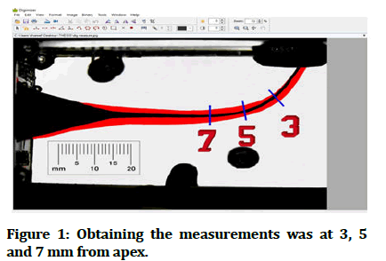
Superimposition of the pre and post images was formed via operating adobe photoshop software (adobe photoshop 2019, adobe systems incorporated and San Jose, CA, USA). The difference in width of canal (transportation) were measured at three levels (3, 5 and 7 mm) from canal apex, which correspond the curvature with high vulnerability to mishaps exist [10,11]. Obtaining the measurements was done through special software named digimizer image analysis software (MedCalc software, Ostend Belgium), by magnifying up to 300 X and using the photographed ruler in order to ease the calibration of the software via converting pixels measurement to mm measurements. So by this method it became feasible to assess the width of the resin eliminated from the outer and inner sides of simulated canal (Figure 1).
Figure 1: Obtaining the measurements was at 3, 5 and 7 mm from apex.
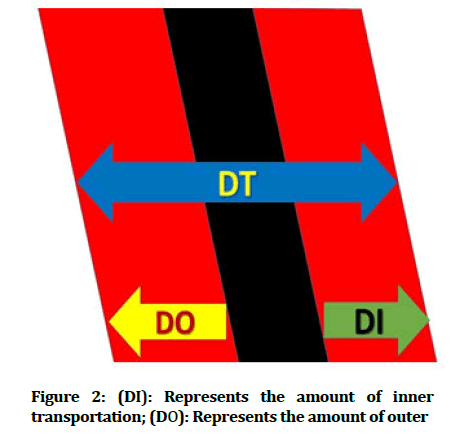
The measurements of transportation were attained by the following formula (T=DI-DO) (Figure 2).
Figure 2: (DI): Represents the amount of inner transportation; (DO): Represents the amount of outer
transportation; DT: Represents the total diameter of the final canal preparation.
The ideal result should be natural without shifting in direction of instrumented canal towards either positive or negative [11-13].
Furthermore, the centric ratio of the instrumented groups was done by applying the following formula: Centering ratio=(DI-DO)/DT × 100, Where, DT=(X2+Y2) symbolize the total width of the canal after preparation, the ideal ratio for file should be zero.
Results
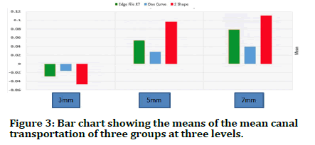
The results of the descriptive statistics which included the minimum, maximum, mean, standard deviation values of the mean canal transportation after instrumentation at the three measured levels in (mm) for the three groups were presented (Table 1).
| Levels | Groups | N | Mean | Std. deviation | Minimum | Maximum |
|---|---|---|---|---|---|---|
| 3 mm | Edge file X7 | 15 | -0.02878 | 0.001912 | -0.0331 | -0.0243 |
| One curve | 15 | -0.01563 | 0.001894 | -0.0191 | -0.0125 | |
| 2 Shape | 15 | -0.04669 | 0.004523 | -0.055 | -0.0379 | |
| Total | 45 | 0.030371 | 0.01321 | -0.055 | -0.0125 | |
| 5 mm | Edge file X7 | 15 | 0.053726 | 0.005062 | 0.0453 | 0.0627 |
| One curve | 15 | 0.027641 | 0.003467 | 0.0221 | 0.033 | |
| 2 Shape | 15 | 0.096544 | 0.016482 | 0.0707 | 0.1247 | |
| Total | 45 | 0.059304 | 0.030391 | 0.0221 | 0.1247 | |
| 7 mm | Edge file X7 | 15 | 0.07902 | 0.006123 | 0.0681 | 0.0905 |
| One curve | 15 | 0.039209 | 0.003349 | 0.0312 | 0.0428 | |
| 2 Shape | 15 | 0.110599 | 0.011975 | 0.0918 | 0.1296 | |
| Total | 45 | 0.076276 | 0.030557 | 0.0312 | 0.1296 |
Table 1: Descriptive statistical results of the mean transportation.
The following bar chart shows the means of transportation between three groups at three different (3 mm, 5 mm, 7 mm) levels (Figure 3).
Figure 3: Bar chart showing the means of the mean canal transportation of three groups at three levels.
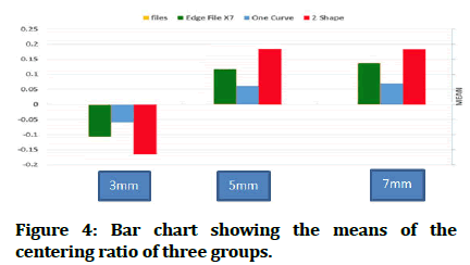
The consequences of the descriptive statistics which includes the minimum, maximum, mean, standard deviation values of the canal centering ratio after instrumentation at three measured levels for the three groups were provided (Table 2).
| Levels | Groups | N | Mean | Std. deviation | Minimum | Maximum |
|---|---|---|---|---|---|---|
| 3 mm | Edge file X7 | 15 | -10.7986 | 0.676339 | -11.5976 | -9.4705 |
| One curve | 15 | -5.896 | 0.67841 | -6.95 | -4.71 | |
| 2 Shape | 15 | -16.4064 | 1.495584 | -19.4961 | -14.2215 | |
| Total | 45 | -11.0337 | 4.45665 | -19.4961 | -4.71 | |
| 5 mm | Edge File X7 | 15 | 11.59009 | 0.928996 | 9.6423 | 12.7246 |
| One curve | 15 | 6.11786 | 0.540989 | 5.336 | 6.9188 | |
| 2 Shape | 15 | 18.45335 | 1.585007 | 15.3353 | 19.8901 | |
| Total | 45 | 12.05377 | 5.216714 | 5.336 | 19.8901 | |
| 7 mm | Edge file X7 | 15 | 13.75436 | 1.227698 | 12.0573 | 15.9816 |
| One curve | 15 | 6.94796 | 0.60854 | 6.1201 | 7.9012 | |
| 2 Shape | 15 | 18.36794 | 1.154231 | 16.0556 | 19.878 | |
| Total | 45 | 13.02342 | 4.850206 | 6.1201 | 19.878 |
Table 2: Descriptive statistical results of the canal centering ratio.
The following bar chart involves ratios of centering between the three groups at three different levels for direct comparison between them (Figure 4).
Figure 4: Bar chart showing the means of the centering ratio of three groups.
Both data of transportations and centric ratios were assessed by Shapiro-Wilk test. Which revealed the distribution of data was proper for parametric test. One way Analysis of Variance (ANOVA) test was achieved to decide occurrence of any statistically significant differences among noted means (ANOVA) test results. The test exhibit incidence of significant differences between groups, therefore, tukey's Honest Significant Difference (HSD) test was executed with the determination of examining the differences among the resultant groups and there were significant differences between the three groups. One curve groups showed the least amount of transportation and the best centric ability.
Discussion
Without a doubt the advent of Ni-Ti file and particularly the heat treated files have made significant influence in refining root canal instrumentation due to their superior ability in negotiating curved root canals and decreasing the risk of, zipping, stripping or ledging the root canal [14,15].
As consequence, studying the shaping abilities of recent heat treated file and also their features is critical since it brings the clinician with respected awareness about the latest endodontic systems obtainable therefore they can choose a proper system that has most safety and maximum efficacy [16].
Furthermore, the null hypothesis was rejected as there are differences in canal transportation and centering ability between the three heats treated rotary systems. Resin training blocks with Simulated curved canals were executed in this study and the motive behind that the measurements are highly reproducible and reliable [17].
Nevertheless, extracted human teeth also could be used in the study of canal transportation. Since it resemble the clinical environments. However, standardization of several variables such as length and width measurement of the root canal, stiffness of the dentine, root canal curvatures, as well as the calcification and pulp stones might influence on measuring the canal transportation, meanwhile, the resin block permits accurate standardization-n the degree curvature of canal, diameter and location of root canal curvature [18]. The device that has been selected in the digital photographic superimposition technique was digital stereomicroscope.
Throughout inspection of the statistics of this study leads to conclusion that no rotary systems have maintain the anatomy of root canal. All of the tested three systems have created canal straitening in varying degree. One curve rotary system has exhibited the highest competency to maintain the original outline of the root canal.
Furthermore, dissimilar heat treatment procedure of the files might produce different alloys with diverse mechanical properties causing in increased phase transformation temperature and increased flexibility might be the reason of alteration in canal transportation values among the tested groups. This finding come in agreement with Capar, et al. who found that different engineering process might produce different canal transportation values [19-22].
One curve, in this study have the lowest means of canal transportation and best centering ratios founded with one curve group followed by edge X7 and 2 Shape respectively. The excellent results obtained by one curve group could be attributed to the following: The file was manufactured from C wire alloy which have control memory feature produce a file with super elastic properties and highly flexible instruments which preserve the canal integrity aiding in less transportation [23-26]. The file have inconstant cross section alongside the cutting edge for better centering ability in the apical third and an excellent debris removal up to the medium and coronal portions [27]. One curve is a single file system which are claimed to be able to completely prepare and clean root canals with only one instrument [28-30].
Furthermore, the edge X7 file shown less satisfactory centering ratios and higher transportation standards when linked with the one curve, this findings were in agree with Al-Abady, et al. and disagree with Versiani, et al. These outcomes of being less than one curve could be correlated to the following reasons: These results could be related to the proprietary heat treatment process called fire wire which used by the manufacturer. This heat treatment process has created fire wire instruments with higher flexibility due to the unusual three dimensional part of the crystalline ground of the fire wire alloy, so this property allows files to better follow the canal; they have parabolic asymmetric triangular cross section [33]. Therefore shaping the root canal wall with 3 cutting points affects the centric ability of the file [34].
Meanwhile, the 2 shape recorded the highest marks of transportation and the minimum satisfactory centering ratios among the verified groups in this study which agree with Hussien, et al. and disagree with Singh, et al. [35,36]. These results could be attributed to the following: 2 Shape instruments were made of T wire alloy which is lack of control memory ability, according to previous studies that revealed that files with control memory are more flexible so resulted in adequate canal shape throughout the length of the canal with no major shaping errors.
Conclusion
Within the limitations of this study, it can be concluded that: The study showed that all the tested systems in this study produced variable degrees of canal transportation of the artificial canals during the instrumentation. The results showed there were significant differences at the three levels which represent the curved part of the canal. Rotary files which have control memory feature showed the least degree of transportation and well centric ability as compare it with other groups.
References
- Weine FS, Kelly RF, Lio PJ. The effect of preparation procedures on original canal shape and on apical foramen shape. J Endod 1975; 1:255-262. [Crossref][Googlescholar][Indexed]
- Nagaraja S, Murthy BSJ, Jocd J. CT evaluation of canal preparation using rotary and hand NI-TI instruments: An in vitro study. J Conserv Dent 2010; 13:16. [Crossref][Googlescholar][Indexed]
- Shenoi PR, Luniya DA, Badole GP, et al. Comparative evaluation of shaping ability of V-Taper 2H, ProTaper next, and HyFlex CM in curved canals using cone beam computed tomography: An in vitro study. Indian J Dent Res 2017; 28:181-186. [Crossref][Googlescholar][Indexed]
- Gambarini G, Galli M, Di Nardo D, et al. Differences in cyclic fatigue lifespan between two different heat treated NiTi endodontic rotary instruments: Wave one gold vs. edgeone fire. J Clin Exp Dent 2019; 11:e609-e613. [Crossref][Googlescholar][Indexed]
- MicroMega One Curve, A range of single files, in continuous rotation C. wire heat treatment. The Endo DNA. France, 2018.
- Micro-Mega SA. 2Shape Two files to shape. COLTENE. 2017.
- Pruett JP, Clement DJ, Carnes Jr, et al. Cyclic fatigue testing of nickel titanium endodontic instruments. J Endod 1997; 23:77-85. [Crossref][Googlescholar][Indexed]
- Forghani M, Hezarjaribi M, Teimouri H, et al. Comparison of the shaping characteristics of neolix and protaper universal systems in preparation of severely curved simulated canals. J Clin Exp Dent 2017; 9:e556-e559. [Crossref][Googlescholar][Indexed]
- Andrade Junior CV, Neto ND, Antunes S, et al. Transportation assessment in simulated curved canals after preparation with twisted file adaptive and BT race instruments. J Clin Exp Dent 2017; 9:e1136. [Crossref][Googlescholar][Indexed]
- de Oliveira Alves V, da Silveira Bueno CE, Cunha RS, et al. Comparison among manual instruments and path file and M two rotary instruments to create a glide path in the root canal preparation of curved canals. J Endod 2012; 38:117-120. [Crossref][Googlescholar][Indexed]
- Al-Asadi AI, Al-Hashimi R. In vitro assessing the shaping ability of three nickel titanium rotary single file systems by cone beam computed tomography. Int J Med Res Health Sci 2018; 7:69-74.
- Saleh A, Rashid AA. Canal central ability of four different endodontic single file systems in simulated L-shaped resin canals. Int J Dent Oral Health 2016; 2. [Crossref]
- Jasim AA, Al-Gharrawi HA. Evaluation of the canal transportation and centering ratio at different levels of simulated curved canals prepared by one shape, protaper next, protaper gold and two shape nickel titanium rotary files. Int J Med Res Health Sci 2019; 8:91-97.
- Fernandes FMB, Oliveira JP, Machado AM, et al. Effect of heat treatment on K3, K3XF and M two endodontic files. 2015; 33:03016. [Crossref][Googlescholar][Indexed]
- Tabassum S, Zafar K, Umer F. Nickel titanium rotary file systems: What's new? Eur Endod J 2019; 4:111-117. [Crossref][Googlescholar][Indexed]
- Campbell F, Cunliffe J, Darcey J. Current technology in endodontic instrumentation: Advances in metallurgy and manufacture. Br Dent J 2021; 231:49-57. [Crossref][Googlescholar][Indexed]
- Moawad E. Shaping and cleaning in endodontics. The university of liverpool, United Kingdom, 2017. [Crossref][Googlescholar][Indexed]
- Hulsmann M, Peters OA, Dummer PM. Mechanical preparation of root canals: Shaping goals, techniques and means. Endod Topics 2005; 10:30-76. [Crossref][Googlescholar][Indexed]
- Capar ID, Ertas H, Ok E, et al. Comparative study of different novel nickel titanium rotary systems for root canal preparation in severely curved root canals. J Endod 2014; 40:852-856. [Crossref][Googlescholar][Indexed]
- Pagliosa A, Sousa Neto MD, Versiani MA, et al. Computed tomography evaluation of rotary systems on the root canal transportation and centering ability. Braz Oral Res 2015; 29:1-7. [Crossref][Googlescholar][Indexed]
- Ozyurek T, Yılmaz K, Uslu GJ. Shaping ability of reciproc, wave one GOLD, and HyFlex EDM single file systems in simulated s-shaped canals. J Endod 2017; 43:805-809. [Crossref][Googlescholar][Indexed]
- Venino PM, Citterio CL, Pellegatta A, et al. A micro computed tomography evaluation of the shaping ability of two nickel titanium instruments, HyFlex EDM and protaper next. J Endod 2017; 43:628-632. [Crossref][Googlescholar][Indexed]
- Kishore A, Gurtu A, Bansal R, et al. Comparison of canal transportation and centering ability of twisted files, HyFlex controlled memory and wave one using computed tomography scan: An in vitro study. J Conserv Dent 2017; 20:161. [Crossref][Googlescholar][Indexed]
- Mohammadian F, Sadeghi A, Dibaji F, et al. Comparison of apical transportation with the use of rotary system and reciprocating hand piece with precurved hand files: An in vitro study. Iran Endod J 2017; 12:462-467. [Crossref][Googlescholar][Indexed]
- Kocak S, Sahin FF, Ozdemir O, et al. A Comparative investigation between protaper next, Hyflex CM, 2 shape, and TF adaptive file systems on cyclic fatigue resistance. J Dent Res Dent Clin Dent Prospects 2021; 15:172-177. [Crossref][Googlescholar][Indexed]
- Staffoli S, Grande NM, Plotino G, et al. Influence of environmental temperature, heat treatment and design on the cyclic fatigue resistance of three generations of a single file nickel titanium rotary instrument. Odontology 2019; 107:301-307. [Crossref][Googlescholar][Indexed]
- Yalniz H, Koohnavard M, Oncu A, et al. Comparative evaluation of dentin volume removal and centralization of the root canal after shaping with the protaper universal, protaper gold and one curve instruments using micro CT. J Dent Res Dent Clin Dent Prospects 2021; 15:47-52. [Crossref][Googlescholar][Indexed]
- Burklein S, Benten S, Schafer EJ. Quantitative evaluation of apically extruded debris with different single file systems: Reciproc, F360 and one shape versus M two. Int Endod J 2014; 47:405-409. [Crossref][Googlescholar][Indexed]
- Selivany BJ, Ahmed HA. Analysis of canal transportation, centering ability and remaining dentin thickness of different single file rotary systems in primary teeth: A CBCT assessment. J Duhok University 2019:1-10. [Crossref][Googlescholar]
- Arıcan Ozturk B, Atav Ates A, Fisekcioglu E. Cone beam computed tomographic analysis of shaping ability of XP endo shaper and protaper next in large root canals. J Endod 2020; 46:437-443. [Crossref][Googlescholar][Indexed]
- Al-Abady AM, Al-Zaka IM. Evaluation of the canal transportation and centering ability of different rotary Ni-Ti systems in simulated curved canals. Indian J Forensic Med Toxicol 2021; 15.
- Versiani MA, Carvalho KKT, Mazzi-Chaves JF, et al. Micro computed tomographic evaluation of the shaping ability of XP endo shaper, iRaCe and edge file systems in long oval shaped canals. J Endod 2018; 44:489-495. [Crossref][Googlescholar][Indexed]
- Khalil WAB, Merdad K, Abu-Haimed TS, et al. Cyclic fatigue resistance of protaper gold, edge file, one shape and protaper universal. Egypt Dent J 2018; 64:589-596. [Crossref][Googlescholar][Indexed]
- Burklein S, Fluch S, Schafer EJO. Shaping ability of reciprocating single file systems in severely curved canals: Wave one and reciproc versus wave one gold and reciproc blue. Odontology 2019; 107:96-102. [Crossref][Googlescholar][Indexed]
- Hussien AA, El-Gendy AAH, Yehia T. Shaping ability of different rotary nickel titanium systems (an in vitro study). Ain Shams Dent J 2020; 19:8-12. [Crossref]
- Singh S, Mirdha N, Shilpa PH, et al. Shaping ability of 2 shape and wave one gold files using cone beam computed tomography. J Int Soc Prev Community Dent 2019; 9:245-249. [Crossref][Googlescholar][Indexed]
Author Info
Shareef Radhi Jawad* and Shatha Abdul Kareem
Department of Restorative and Esthetic Dentistry, College of Dentistry, University of Baghdad, Baghdad, IraqCitation: Shareef Radhi Jawad, Shatha Abdul Kareem, Comparative Assessment of Canal Transportation and Centering Ratio of Three Heat Treated Rotary Systems in Severely Curved Simulated Resin Block (an in vitro study), J Res Med Dent Sci, 2023,11 (01): 183-188.
Received: 25-Oct-2022, Manuscript No. JRMDS-22-65204; , Pre QC No. JRMDS-22-65204 (PQ); Editor assigned: 28-Oct-2022, Pre QC No. JRMDS-22-65204 (PQ); Reviewed: 11-Nov-2022, QC No. JRMDS-22-65204; Revised: 27-Dec-2022, Manuscript No. JRMDS-22-65204 (R); Published: 13-Jan-2023


