Research - (2019) Volume 7, Issue 6
Comparative Evaluation of Acid Etching and Laser Etching Following the Application of Chlorhexidine on the Aesthetic Bracket
Nidhal H Ghaib* and Bayan Ghdhban
*Correspondence: Nidhal H Ghaib, College of Dentistry, University of Baghdad, Iraq, Email:
Abstract
Aim: The objective of this study was to identify the Er:CYSGG laser etching with and without sealant chlorhexidine is an alternative to acid etching by bonded ceramic bracket to enamel surface.
Materials and Methods: 42 upper premolars were assigned into four groups; Acid etching +ceramic brackets (AC), Laser etching+ ceramic brackets (LC), LC-CHX (laser etching+chlorhexidine sealant) and AC-CHX (acid etching + chlorhexidine sealant). An Universal testing machine (H50KT machine) were tested SBSs (shear bond strength). Scores adhesive remnant index (ARI) was assessed by a stereomicroscope. Two other teeth were not bonded and were examined with a SEM (scanning electron microscopic).
Results: Statistical analyses presented that SBSs of laser radiation were highly significantly difference than that of acid etching, also the values of SBS between the AC-CHX and LC-CHX is high significant difference. SEM examination shown different etching pattern.
Conclusion: Low power Er: CYSGG laser etching agreements clinically acceptable SBS and alternative for bonding of ceramic bracket. CHX sealant not more effect in SBS of acid etching, while it’s weaker in the laser etching.
Keywords
ErCr: YSGG laser, Acid etching, Chlorhexidine varnish
Introduction
Pretreatment conditioning to enamel surface is necessary to obtain good bond strength, therefore, it is consider the acid etching has been standard procedure for bonding orthodontic brackets because dissolves hydroxyapatitecrystals of tooth surface causing a rough and uneven surface, results easy penetrate adhesive resins to this rough surface, thus providing good micromechanical retention [1]. But the decalcified tooth surface when acid-etching has a prospective weakness of making the enamel more liable to a caries attack [2]. Therefore should be a required to make simpler clinical techniques and reduce enamel loss whilst keeping clinically good bond strength. Lately, It has attracted to using laser radiation will give an irregular surface pattern which is similar pattern in an acid etching [3]. Its can remove the smear layer and it does not include temperature or vibration, It can be modifies crystalline and chemical structure of the tooth surface and more resistant to caries also painless method [4]. Often the most type of laser used for this objective are Er, Cr:YSGG (erbium, chromium yttriumscandium- galliumgarnet) laser. This laser action causing in micro porosities on the surface of enamel that can possibly increase the resin flow and create micromechanical retention to the enamel, by stimulated emission of photons from excited atoms or molecules, and coherent light at a special wavelength, this wavelength matches the absorption peaks of water [5]. This laser is the most effective for hard and soft tissue ablation with least thermal effects on the pulp [6,7]. The Laser irradiation with adequate parameter was respected as an substitute to acid etch method for orthodontic treatment [8-10]. Fixed appliances causing easily accumulate dental plaque also difficulties in maintains sufficient oral hygiene, therefore increase prevalence of plaque-induced diseases [11]. Prevention of dental caries plays a vital role in orthodontic treatment, strategies chemotherapeutic treatment regimens have received much attention and have presented satisfactory results. Chlorhexidine (CHX) as chemical antimicrobial substance is proficient of inhibiting bacterial adhesion, metabolic activity, and colonization by reducing the bacterial growth [12]. Chlorhexidine combined with thymol in a varnish have excellent adsorption to the tooth surface for long period; prevent its immediate loss, thus acting as slow-releasing reservoirs [13]. These study has been established to evaluate and comparable the Er,Cr:YSGG laser etching with conventional acid-etching techniques, and to determine the suitability of these with chlorhexidine varnish as the sealant.
Material and Methods
Sample preparation
Recent 42 upper premolars had been collected from human and kept in a 0.1% thymol solution was changed weekly to prevent bacterial growth. The criteria to select is no caries, intact buccal enamel, no pretreatment chemical, these are determined by using 10X magnifying glass. The crown was exposed after every tooth was mounted vertically in a self-cure acrylic, the force of knife-edge, its parallel to the axis of the tooth during testing. Before etching procedure were pumiced/ washed-dried for (10) seconds. The divided for 4 groups A,B were etched using etching Gel rate 37% phosphoric acid was done for (30) seconds (depending to manufacturer's instructions), then rinsed/dried. The two another groups C,D were etched by laser Er,Cr:YSGG laser (Waterlase, I plus from Biolase USA, a wavelength of A2780 n ), the power output can be varied from 0-6W depending on the tissue to be cut. The power settings for laser groups had been selected on the basis of various studies on adequate SBS and pilot study, the using 2 W power, pulse duration is the 140 microseconds, 20 pulses (20H) for per second are pulse repetition rate [6,9,10] . The air and water levels were 90% and 80%, respectively. To standardize the distance, the laser beam was brought into line perpendicular to the enamel at a distance of 1 mm [14]. It was moved around in a sweeping fashion via hand over an approximately 4*4 an acrylic resin with a hole, was placed on the tooth surface during an exposure time of 15 seconds (Figure 1), which was enough to scan this area. The irradiated sample was dried with an air for 15 seconds.
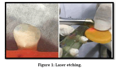
Figure 1: Laser etching.
Bonding procedure
The primer of the groups A,C was left without any additives chlorhexidine while the primer of the groups B,D, were incorporated, with CHX varnish contains 1:1 thymol +chlorhexidine (1 mg of each/gm). Two drops of CHX were added to every one drop of primer 2:1 and then mixed [15]. Thin uniform coat of mixture Transbond XT primer was smeared to the etched tooth surface and then light cure for 15 second all groups. ceramic brackets (Ortho Technologic.2019, U.S.A the base bracket base is 10.57 mm2) were bonded by using resin/primer (3M Unitek.,Monrovia, U.S.A), to standardization by (fixing 200 gm load on the upper part of the vertical arm of the surveyor), any excess adhesive material around the bracket base was gently removed by using dental prob. The light cured 5s on mesial and 5s on the distal; the intensity of light was 1200 mW/cm2, later exposed to thermo cycling 500 intervals in distilled water, between 5uC and 55uC, through a dwell time in every bath of 30 seconds and a move time of 15 seconds.
Testing shear bond
By using a chisel edge at a. crosshead speed of 1 mm/min, the force was expressed in newton’s (N), then converted to megapascals (Mpa), was calculated by dividing the force on the bracket base area.
Examine site failure (ARI)
After deboning, examined enamel surfaces and base of bracket under 10X magnifications with a stereomicroscope to conclude the quantity remaining the residual adhesive on the all of the tooth. The scores of ARI were defined by Artun, et al. [16] with the next scale utilize
1. The tooth no adhesive remained
2. On the enamel the adhesive was covered less than half.
3. The adhesive was covered more than half on the tooth.
4. All adhesive was covered on the enamel, with noticeable impression of the button mesh.
Scanning Electron Microscopy (SEM) Photographs representative morphological changes for two tooth in enamel surface. SEM samples were assessed according to the criteria of Silverstone et al. [17].
Statistical analysis
Data were collected and analyzed using SPSS program version 25. The descriptive statistics included means, standard deviations, frequency distribution and percentage, while the inferential statistics included independent sample t-test and Pearson's Chi-square test.
Results
MPa is expressed the SBS. ANOVA showed the presence of significant differences (p-value ≤0.000)
Comparing the effect of etching type on the she bond strength
The highest value conventional acid etching in shear bond strength than laser etching, Anova test revealed CB group in acid etching a highly significant difference than laser group(P-value P≤0.000) are shown in (Table 1 and Figure 2).
| Groups | Descriptive statistics | Comparison (d.f.=18) | |||||
|---|---|---|---|---|---|---|---|
| Phosphoric acid | LASER | ||||||
| Mean | S.D. | Mean | S.D. | Mean difference | t-test | p-value | |
| CB | 19.542 | 3.994 | 9.462 | 1.16 | 10.08 | 7.665 | 0.000 (HS) |
| CB CHX | 12.551 | 3.297 | 2.943 | 1.026 | 9.908 | 11.13 | 0.000 (HS) |
Table 1: Descriptive statistics and comparison effect of etching type on the SBS.
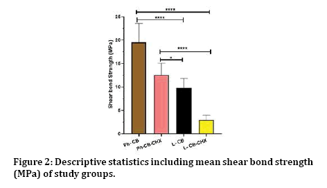
Figure 2: Descriptive statistics including mean shear bond strength (MPa) of study groups.
Comparing the effect of chlorhexidine on the shear bond strength
To distinguish any alteration in the mean rates for the shear-bond strength amongst the difference etching technique with and without CHX, a highly-significant difference (P≤0.000) between the mean rates of these groups has been found, (Table 2 and Figure 3).
| Etching types | Brackets' types | Descriptive statistics | Comparison (d.f.=18) | |||||
|---|---|---|---|---|---|---|---|---|
| Without CHX | With CHX | |||||||
| Mean | S.D. | Mean | S.D. | Mean difference | t-test | p-value | ||
| Phosphoric Acid | CB | 19.542 | 3.994 | 12.551 | 3.297 | 6.992 | 4.676 | 0.000 (HS) |
| LASER | CB | 9.462 | 1.16 | 2.943 | 1.026 | 6.519 | 13.315 | 0.000 (HS) |
Table 2: Descriptive statistics and comparison the effect of chlorhexidine on the shear bond strength of different etching.
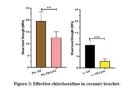
Figure 3: Effective chlorhexidine in ceramic bracket.
Adhesive Remnant Index (ARI)
The locations of bond failure After debonding the brackets, for each tooth was assessed (ARI), according to Artun andBergland (1984).
Frequency distribution of adhesive remnant index among the groups
Chi-square test showed that there are significantdifferences in the quantity of the adhesive covering on the enamel surface involving groups laser and acid etching. There were highly significant differences between groups, there are minimum amount of adhesive remnant was establish in laser groups as presented in (Tables 3 and 4)
| Groups | Score 0 | Score 1 | Score 2 | Score 3 | Total |
|---|---|---|---|---|---|
| A | 0 (0%) | 2 (20%) | 5 (50%) | 3 (30%) | 10 (100%) |
| B | 8 (80%) | 1 (10%) | 1 (10%) | 0 (0%) | 10 (100%) |
| C | 7 (70%) | 3 (30%) | 0 (0%) | 0 (0%) | 10 (100%) |
| D | 10 (100%) | 0 (0%) | 0 (0%) | 0 (0%) | 10 (100%) |
χ2= 32.747, d.f.= 9, p-value= 0.000 (HS)
Table 3: Frequency distribution and percentage of ARI of different groups (Ceramic brackets).
| Groups | χ2 | d.f. | p-value |
|---|---|---|---|
| A vs. C | 15.2 | 3 | 0.002 (HS) |
| B vs. D | 2.222 | 2 | 0.329 (NS) |
| A vs. B | 14 | 3 | 0.003(HS) |
| C vs. D | 1.569 | 1 | 0.210 (NS) |
Table 4: Comparison of ARI between different groups.
Scanning Electron Microscopy (SEM)
Photographs representative one tooth only from every group were chosen to observe alterations in enamel pattern surfaces. Its revealed the different etching patterns achieved for two groups, the acid etched on the enamel displays cracks in some areas, and produced enamel surface resembled type 1 pattern of etching according to Silverstone et al. While evaluation of the laser etching groups an increased the rough surface with dissolved alongside the prisms was detected, also showing micro cracks. Predominant type III etching pattern detected after laser etching (Figures 4 and 5).
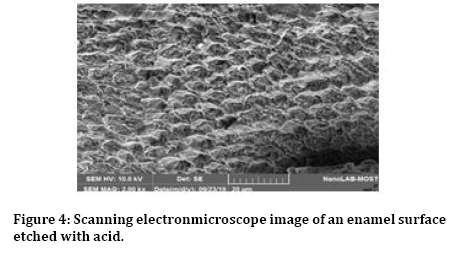
Figure 4: Scanning electronmicroscope image of an enamel surface etched with acid.
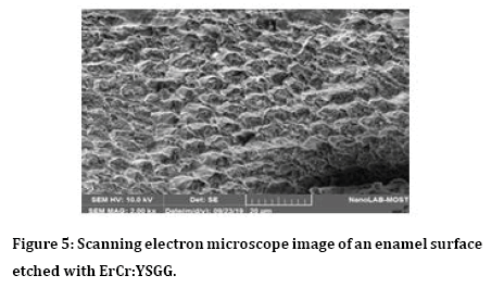
Figure 5: Scanning electron microscope image of an enamel surface etched with ErCr:YSGG.
Discussion
There is an increasing tendency to decrease in chair time both for the practitioner and patient, besides to reach a sufficient marginal seal, good shear bond strength and less bonding material around the bracket for avoid demineralization or white spot lesions in periphery and under the bracket [18]. Most of bacteria were identified on the adhesive more “than on the bracket material itself, this fact encouraged the development of antibacterial adhesives designed to reduce bacterial colonization without compromising the “adhesive’s mechanical properties [19].
The main advantage of using an Erbium laser is that it is adequate absorbed by water in “dental hard tissue”. It’s causing etching through a process of continuous micro explosions, which occur due to the vaporization of the water trapped within the hydroxyapatite matrix [20]. The surface created by laser irradiation is more acid resistant as result laser causing enamel modifies the calcium-phosphate ratio and leads to the formation of more stable and less acid soluble compounds, thus reducing the susceptibility to caries attack [21]. In present study, the laser parameters determine based on a previous studies [22] and pilot study, laser at 2 W for 15 s was gained SBS similar when using acid etching. Some researchers identified was not capable laser irradiation of etching enamel [23]. This may be was associated to sub-surface cracks detected in (SEM) images. Tarηin et al. [24] mentioned that the SBS the Er, Cr:YSGG laser -etched enamel was significantly less than from the acid etched . Another reported the laser using 2 W and 20 Hz provided adequate bond strength with consistency in the tag lengths [9]. The results of this study show that bond strengths done with the Er,Cr:YSGG hydrokinetic laser system at 2-W power setting were statistically different from those of acid etching. The bond strength had been reached in present study was 9.462 ± 1.160 MPa for Laser etching, however acid etching was higher 19.542 ± 3.994 MPa. Such variability may be because unobserved weaknesses at the tissue surface or craters, micro pores in enamel also melted bubbles formed in laser etching, or air bubble trapping within the resin. In orthodontic treatment SBS according Maijer, et al. [25] the 8 MPa to be adequate for orthodontic brackets. But Reynolds [26] reported bond strength from range 6 to 8 MPa to be adequate. Therefore bond strength values remained of the laser etching within these values; laser etching at these settings seems acceptable for clinical use. These results reach agreement with the results the findings of Verma et al. [27], Vijayan et al. [9] and Nakamura et al. [28], while our results are disagreement to those of Hoshing et al., Martinez-Insua et al. and Fraunhofer et al., [6,23,29] probably this Contradictory the findings relating they utilized more different power outputs of laser technique and varies nature curvature tooth structures, as result of the using sweeping motion by hand controlled of the laser beam during the etching may be reason a inadequately standardized etching pattern during the irradiated area. Further might be aid to solve this problem, optimal a standardized, etching technique by the Er, Cr:YSGG laser system.
The adding chlorhexidine varnish to the primary agent as the sealant. These particularly important more effects because tend to be reduce plaque accumulate on periphery around the brackets. CHX varnish as in Cervitec plus (1%thymol +1% Chlorhexidine). Its requires minimal application time and does not rely on patient compliance. CHX releases slowly into oral environment allowing for a prolonged effect of antimicrobial and connects to the glycoproteins via reversal electrostatic binding, however retained on the oral surfaces [30].
The present result SBS in acid etching with CHX sealant is 12.551 ± 3.297 lower than without CHX 19.542 ± 3.994 give emphasis to that an interaction may occur between the enamelbonding surface adhesion and CHX was not achieved or inadequate photo-polymerized when mixed with the CHX content, but practically is acceptable SBS according to Maijer, et al. While CHX with laser etching is more weakness in ceramic bracket 2.943 ± 3.297 because is used a silane coupling agent as chemical mediator between adhesive resin and the bracket base not provided a free end to reacts molecules with every of the acrylic bonding materials therefor fewer bond strength Finally, the bases of ceramic bracket have three dove tail therefore considerably less mechanical undercuts comparable are found in metal bracket are mesh base designs.
Highest ARI score was found for acid etching for group 2 and group 3. This means good bond strength because when the brackets debonded had no remaining of adhesive on the bracket base while wholly remaining of adhesive on tooth surface resulting in less risk in an enamel fracture. These results were in agreement with studies directed by Samruajbenjakul et al. [31]. Lower ARI score (0) was found for sealant with CHX and laser etching this is favorable for time saving purposes. This means more remaining of adhesive on the bracket base while a minor quantity remaining of adhesive on enamel surface resulting in relatively inferior bond strength. These results were comparable with studies showed by Sibi et al. [32].
Conclusion
The results of the study indicate that etching of enamel surface with an Er,Cr:YSGG less predictable bond strengths than did acid etching with 37% orthophosphoric acid for 30 seconds. But can be consider acceptable bond strength more practical and faster than conventional acid etching. CHX sealant did not influence bond strength in acid etching but not significant in laser etching mode of bond failure in laser group which is considered as the most preferable, in ceramic bracket because reducing fractures within the enamel itself during debonding.
References
- Zhu JJ, Tang AT, Matinlinna JP, et al. Acid etching of human enamel inclinical applications: a systematic review. J Prosthet Dent 2014; 112:122-35.
- Øgaard B, Rølla G, Arends J. Orthodontic appliances and enamel demineralization. Part 1. Lesion development. Am J Orthod Dentofacial Orthop 1988; 94:68–73.
- Shafiei F, Jowkar Z, Fekrazad R, et al. Micromorphology analysis and bond strength of two adhesives to Er,Cr:YSGG laser-prepared vs. bur-prepared fluorosed enamel. Microsc Res Tech 2014; 77:779-784.
- Ghaffari H, Mirhashemi A, Baherimoghadam T, et al. Effect of surface action on enamel cracks after orthodontic bracket debonding: Er,Cr:YSGG laser-etching vs. acid-etching. J Dent 2017; 14:259-266.
- Yassaei S, Fekrazad R, Shahraki N, et al. AComparison of shear bond strengths of metal and ceramic brackets using conventional acid etching technique and Er:YAG laser etching. J Dent Res Dent Clin Dent Prospects 2014; 8:27-34.
- Hoshing UA, Patil S, Medha A, et al. Comparison of shear bond strength of composite resin to enamel surface with laser etching versus acid etching: An in vitro evaluation. J Conserv Dent 2014; 17:320-324.
- Seifi M, Matini NS. Laser surgery of soft tissue in orthodontics: Review of the clinical trials. J Lasers Med Sci 2017; 8:1-6.
- Sağır S, Usumez A, Ademci E et al. Effect of enamel laser irradiation at different pulse settings on shear bond strength of orthodontic brackets. Angle Orthod 2013; 83:973-980.
- Vijayan V, Rajasigamani K, Karthik K, et al. Influence of erbium, chromium-doped: Yttrium scandium-gallium-garnet laser etching and traditional etching systems on depth of resin penetration in enamel: A confocal laser scanning electron microscope study. J Pharm Bioallied Sci 2015; 7:616-622.
- Gulec L, Koshi F, Karakaya İ, et al. Micro-shear bond strength of resin cements to Er, Cr: YSGG laser and/or acid etched enamel. Laser Physics 2018; 28:105601.
- Avriliyanti F, Suparwitri S, Alhasyimi AA. Rinsing effect of 60% bay leaf (Syzygium polyanthum wight) aqueous decoction in inhibiting the accumulation of dental plaque during fixed orthodontic treatment. Dent J 2017; 50:1-9.
- Kulkarni VV, Damle SG. Comparative evaluation of efficacy of sodium fluoride, (4) chlorhexidine and triclosan mouth rinse in reducing the mutans streptococci count in saliva. An in-vivo study. J Indian Soc Pedo Prev Dent 2003; 21:98-104.
- Patel PM, Hugar SM, Halikerimath S, et al. Comparison of the effect of fluoride varnish, chlorhexidine varnish and casein phosphopeptide-amorphous calcium phosphate (CPP-ACP) varnish on salivary Streptococcus mutans level: A six month clinical study. J Clin Diagnostic Res 2017; 11:53-59.
- Yilanci H, Usumez S, Usumez A. Can we improve the laser etching with the digitally controlled laser handpiece—xrunner? Photomed Laser Surg 2017; 35:324-331.
- Frey-Hugentobler CLM. Effects of different chlorhexidine pretreatments on adhesion of metal brackets in vitro. Doctoral dissertation, University of Zurich 2013.
- Årtun J, Bergland S. Clinical trials with crystal growth conditioning as an alternative to etch enamel pretreatment. Am J Orthod 1984; 85:333-340.
- Silverstone LM, Saxton CA, Dogan IL, et al. Variation in the pattern of acid etching on human dental enamel examined by scanning electron microscope. Caries Res 1975; 9:373–387.
- Mayfield BJ, Hamdan AM, Tufekci, et al. Development of white spot lesions during orthodontic treatment: Perceptions of patients, parents, orthodontists, and general dentists. Am J Orthod Dentofacial Orthop 2012; 14:337–344.
- Lim BS, Lee SJ, Lee JW, et al. Quantitative analysis of adhesion of cariogenic streptococci to orthodontic raw materials. Am J Orthod Dentofacial Orthop 2008; 133:882-888.
- Karaman E, Yazici AR, Baseren M, et al. Comparison of acid versus laser etching on the clinical performance of a fissure sealant: 24-month results. Operative Dent 2013; 38:151-158.
- Klein AL, Rodrigues LK, Eduardo CP, et al. Caries inhibition around composite restorations by pulsed carbon dioxide laser application. Eur J Oral Sci 2005; 113:239-244.
- Baygin O, Korkmaz FM, Tuzuner T, et al. The effect of different techniques of enamel etching on the shear bond strengths of fissure sealants. Dentistry 2011; 1:109.
- Martínez-Insua A, Da Silva Dominguez L, Rivera FG, et al. Differences in bonding to acid-etched or Er:YAG-laser-treated enamel and dentin surfaces. J Prosthet Dent 2000; 84:280-288.
- Tarçin B, Günday M, Oveçoglu HS, et al. Tensile bond strength of dentin adhesives on acid- and laser-etched dentin surfaces. Quintessence Int 2009; 40:865-874.
- Maijer R, Smith DC. Crystal growth on the outer enamel surface: An alternative to acid etching. Am J Orthod 1986; 89:183-193.
- Reynolds IR. A review direct orthodontic bonding. Br J Orthod 1975; 2:171–178.
- Verma M, Kumari P, Gupta R, et al. Comparative evaluation of surface topography of tooth prepared using erbium, chromium: Yttrium, scandium,gallium, garnet laser and bur and its clinical implications. J Indian Prosthod Soc 2015; 15: 23-28.
- Nakamura T, Wakabayashi K, Kinuta S, et al. Mechanical properties of new self-adhesive resin-based cement. J Prosthodont Res 2010; 54:59–64.
- Fraunhofer JA, Allen DJ, Orbell GM. Laser etching of enamel for direct bonding. Angle Orthod 1993; 1:73-76.
- DePaola LG, Spolarich AE. Safety and efficacy of antimicrobial mouthrinses in clinical practice. J Dent Hyg Fall Suppl 2007; 81: 13–25.
- Samruajbenjakul B, Kukiattrakoon B. Shear bond strength of ceramic brackets with different base designs to feldspathic porcelains. Angle Orthod 2009; 79: 571–576.
- Sibi AS, Kumar S, Sundareswaran S, et al. An in vitro evaluation of shear bond strength of adhesive precoated brackets. J Ind Orthod Soc 2014; 48:97-103.
Author Info
Nidhal H Ghaib* and Bayan Ghdhban
College of Dentistry, University of Baghdad, IraqCitation: Nidhal H Ghaib, Bayan Ghdhban, Comparative Evaluation of Acid Etching and Laser Etching Following the Application of Chlorhexidine on the Aesthetic Bracket, J Res Med Dent Sci, 2019, 7(6): 101-106.
Received: 28-Oct-2019 Accepted: 04-Dec-2019
