Research - (2021) Volume 9, Issue 11
Comparative Evaluation of Push out Bond Strength of Restorations with Bulk Fill, Flowable, and Conventional Resin Composite Restoration: An in vitro Study
Praktan B Gire, Neha Alone*, Ulhas Dudhekar, Abhilash Dudhekar and Minal Ganvir
*Correspondence: Neha Alone, Department of Prosthodontics, Swargiya Dadasaheb Kalmegh Smruti Dental College and Hospital, Wanadondri, Hingna Road, Nagpur, India, Email:
Abstract
Aim: The aim of this study was to evaluate the bond strengths of composite restorations made with different filler amounts and resin composites that were photo activated using a light-emitting diode (LED). Methodology: Thirty bovine incisors were selected, and a conical cavity was prepared in the facial surface of each tooth. All preparations were etched with 37% phosphoric acid, Te-Econom bond universal dental adhesive system was applied followed by photo activation, and the cavities were filled with a single increment of Tertic n cerem bulk, Smart dentin replacement (SDR) and Tetric N ceram nanohybrid, followed by photo activation. A push-out test to determine bond strength was conducted using a universal testing machine. Result: Data (MPa) were submitted to Student’s t-test at a 5% significance level. Among the three groups Conventional (5.86± 1.96) resin composite had a lower bond strength than the Bulk fill (6.53 ± 1.063) and Flowable (6.28± 1.085) resin composites ( p=0.014). After the test, the fractured specimens were examined using an optical microscope under magnification (10x). All the three composites demonstrated a high prevalence of adhesive failures (Conventional resin composite 90%; Bulk fill, 80%; and Flowable, 80%) Conclusion: Although all three composites demonstrated a high prevalence of adhesive failures, the bond strength values of the different resin composites photo activated by LED showed that the Tetric N Ceram nanohybrid had lower bond strength than the Tertic n cerem bulk fill and Smart dentin replacement flowable resin composites.
Keywords
Bulk Fill, Flowable, Dental composite, Push out, Adhesive failure
Introduction
The contraction of dental composites is stated to be about 1–5% of their volume. There is development of mechanical stresses inside these contracting materials when they are inserted into the bonded preparations. Rapid conversion in light-cured composites induces a correspondingly rapid increase in composite stiffness, producing high shrinkage stresses at the restoration-tooth interface [1]. Such stresses may disrupt the bonding between the composite and the cavity walls or may even cause cohesive failure of the restorative material or the adjacent tooth tissue. Polymerization shrinkage can strain the bond between tooth structure and the restoration, resulting in stress generation at the bonding interface which can cause marginal gap formation, post-operative sensitivity, and pulp irritation. Stress generation is often related with the:
• Geometry and size of the cavity/C-factor. • Composition of the materials. • Restorative technique [2].
Since the use of dental resin composites, reduction of the polymerization shrinkage has become an important issue. Much progress has been done to tackle this problem such as the development of Non-shrinking resins and modified filler particles, but did not lasted clinically [3]. Various Factors that can affect the shrinkage are inorganic filler content, the molecular weight of monomer system, and the degree of conversion of the monomer system [4].
During setting of the resin composites the polymerization shrinkage resulted in contraction stress. This contraction stress has been found to be dependent on the cavity configuration (C-factor), which is the ratio of the bonded to unbounded surface area of the restoration, the nature of the matrix material, the filler load, and the viscous–elastic properties [5]. Application of low elastic modulus liners between the tooth structure and layering techniques have been proposed to reduce the internal stress and deformation of the tooth structure [6,7]. Another important factor for both the shrinkage and the contraction stress is the elastic modulus of the resin composite [8]. Increase in the Elastic modulus of the resin composite is seen with the volume fraction of the inorganic filler content [9]. All shrinkage is not converted to contraction during curing because the polymer can rearrange and relieve stress. This flow is in mainly composed of a macroscopic and microscopic component. During the polymerization reaction macroscopic flow occurs at the free [10]. Polymer rearrangement within the resin composite resulted in this Microscopic flow. Other factors which result in this flow include Structure of the molecules, crosslink density of the network, the interaction of the matrix and filler particles and reaction kinetics [5].
Incremental build-up of composite resin is required to reduce polymerisation shrinkage of composite resin but the Layering techniques and multiple curing regimens of resin composites is time consuming. Nowadays there is a demand of composite material having faster and easier procedures and less curing time. New composite restoration such as bulk fill enables restoration build-up in thick layers, up to 4 mm and has become increasingly popular among dentist [11]. Thus, the purpose of this study was to evaluate the bond strengths of composite restorations that were made with different filler amounts and resin composites photo activated using a lightemitting diode (LED). The null hypothesis tested was that there is no difference in bond strength among the composite restorations that were made with different filler amounts and resin composites photo activated using LED (bulk-fill, flow, and conventional resin composite.
Material and Methods
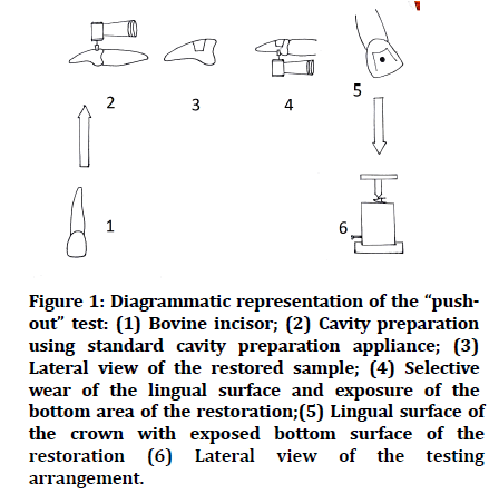
Thirty human Maxillary noncarious, crack free incisors were selected (Figure 1.1), and the crowns were cut off at the cement- enamel junction with a diamond disk (Mani, Utsunomiya, Japan). A conical cavity were with (top diameter of 4.5 mm, bottom diameter of 4.0 mm, and height of 3 mm) was prepared in the labial surface of each tooth using straight diamond bur (Mani, Utsunomiya, Japan) in a high-speed handpiece and copious air-water spray (Figure 1.2), (Figure 1.3). All preparations were etched with Ivoclar vivodent N-Etch gelTM (Schaan, Liechtenstein) containing 35% phosphoric acid for 30 seconds and washed with water for 30 s; the excess moisture was removed with absorbent paper. Bonding agent (Te-Econom BondTM, Schaan, Liechtenstein) was applied to the preparations according to the manufacturer’s instructions and photo activated using a LED curing unit (WoodpeckerTM, Guilin, Guangxi, China) with exposure for 20 sec. Teeth were divided into three groups, Group 1 Tertic n cerem bulk fill (Ivoclar Vivadent, Schaan, Liechtenstein.), Group 2 Smart dentin replacement, Group 3 Tetric N ceram nano hybrid (Ivoclar Vivadent, Schaan, Liechtenstein.) Then round diamond bur (ManiTM, Utsunomiya, Japan) was used to grind the lingual face of the crown in order to expose the bottom surface of the restoration. The mesial and distal areas of the crown in the lingual surface were conserved as a mode of strengthening the specimen for the push-out test (Figure 1.4).
Figure 1: Diagrammatic representation of the “pushout” test: (1) Bovine incisor; (2) Cavity preparation using standard cavity preparation appliance; (3) Lateral view of the restored sample; (4) Selective wear of the lingual surface and exposure of the bottom area of the restoration;(5) Lingual surface of the crown with exposed bottom surface of the restoration (6) Lateral view of the testing arrangement.
The push-out test was performed to evaluate the bond strength by using universal testing machine. An acrylic device was used to position the specimen such that the hole of specimen was bottom side up. The tip of the universal testing machine was applied on the bottom surface of restoration and compressive force was generated to provoke the rupture of the tooth-composite bond along the lateral wall (Figure 1.5). The speed used was 0.5 mm/min. The values were recorded (kgf) and converted into pressure values (Mpa). Statistical analysis was performed using MS Excel 2007 and statistical package for the social sciences (SPSS) version 20.0 (SPSS Inc, Chicago, USA). Descriptive statistical data was presented in the form of mean and standard deviation. One way analysis of variance (ANOVA) was performed to assess the mean significant difference between the different groups and LSD post hoc tests where use for multiple group comparison’s. The p value less than 0.05 was considered to be statistically significant. The fractured specimens were examined under stereo microscope (Wuzhou New Found Instrument Co. Ltd., China) with magnification (10x), and the modes of failure were classified as follows: adhesive failure, cohesive failure within the composite, or mixed failure involving adhesive, dentin, and composite (Figure 1.6).
Results
The bond strength results are shown in the Table 1. The bond strength values of the different resin composites showed that the Conventional (36.08 ± 1.96) resin composite had a lower bond strength than the Bulk fill (46.18 ± 1.063) and Flowable ( 38.02 ± 1.085) resin composites (p=0.014).
| Composite | Bond strength (Mpa) |
|---|---|
| Flowable resin composite | 6.28 |
| Bulk fill resin composite | 6.53 |
| Conventional resin composite | 5.86 |
Table 1: Mean of the push-out bond strength (Mpa).
Classification of the modes of failure for the three composite restorations with different filler amounts and resin composites is shown in Table 2.
| Composite | Cohesive | Failure mode of adhesive | Mixed |
|---|---|---|---|
| Flowable resin composite | 0 (0) | 80 (8) | 20 (2) |
| Bulk fill resin composite | 0(0) | 80 (8) | 20 (2) |
| Conventional resin composite | 0(0) | 90(9) | 10(1) |
Table 2: Percentage (%) of failure mode.
The three composites demonstrated a high prevalence of adhesive failures (Conventional resin composite 90%; Bulk fill, 80%; and Flowable, 80%).
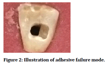
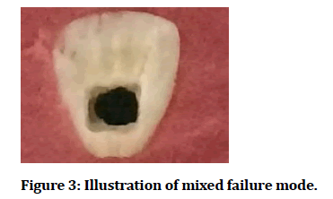
The modes of failure were classified as follows: adhesive failure, cohesive failure within the composite or mixed failure including adhesive, dentin, and composite. The number of the specimens is given in parentheses (Figures 2 and 3).
Figure 2:Illustration of adhesive failure mode.
Figure 3:Illustration of mixed failure mode.
Discussion
Frequently, evaluation of bond strength of endodontic cement is done by push-out test [2]. However, in the current study, push out test was used to evaluate bond strength of class V composite resin in a replicated class V cavity [2]. Bond strength of resin composite is also evaluated by other bond strength tests such as shear, tensile, microshear, and micro tensile evaluations [2]. However, flat surface is required to perform this test [2]. The C-factor (the cavity configuration factor is the ratio of the bonded surface area to unbounded or free surface area) in such a situation is very low and the development of shrinkage stress is not directed toward the bonding interface [2]. Thus push out test was used in the present study due to its ability to evaluate bond strength in a high C-factor cavity (3.0) with high stress generation directed toward the bonding area [2]. Thus, in the current study, the entire bonding area was submitted to the compressive force at the same time, allowing the shear bond strength to be evaluated in a cavity. In addition, low data variability and low standard deviations was used to confirm the confidence of the push out test. Besides, a high prevalence of adhesive failure was seen in the analysis of the mode of failure for all the composites tested, Flowable resin composite (80%), Bulk fill resin composite (80%), and Conventional resin composite (90%).
Presently, usage of bulk fill materials is trending among practitioners because of their more simplified procedures [11]. However, due to lack of available literature on their clinical performance, bulk-fill materials rank their properties relative to the conventional flow and paste composite types already on the market. In the present study, the restorations made with the Bulk fill resin composite showed higher mean bond strength values (6.53Mpa) than those achieved with the Flowable resin composites (6.28Mpa).This may be due to the difference in the filler loading (60 and 58% by volume for bulk fill), mechanical properties and rheological properties of the two groups of materials.
Bulk fill resin composite also showed higher mean bond strength values (6.53Mpa) than those achieved with conventional resin composites (5.86Mpa).
The difference can be explained by difference in depth of cure of the two groups of materials conventional and bulk fill resin composite, Bulk fill shows higher depth of cure than conventional resin composite. This is because of addition of a new initiator Ivocerin in addition to the camphoroquinone /amine initiator system which results in the increase in the depth of cure [12].
Our findings are in accordance with those of Colak et al. [12] and Garcia et al. [13], in their study bulk fill showed better result than flowable and conventional resin composite restoration. Flowable resin composite showed higher mean bond strength values (6.28 Mpa) than those achieved with the conventional resin composites (5.86 Mpa). This may be due to the Flowable composites are subjected to lower shrinkage than conventional resin composite during polymerization (2.96%). The main contributing factors to the reduced shrinkage of this material are their low flexural modulus and low filler loading. Flexural modulus is a function of many factors such as filler content, monomer chemistry, monomer structure, filler/matrix interactions and additives, Flowable resin composite have monomer structure based on UDMA which is characterized by a significant reduction in polymerisation shrinkage and higher molecular weight that contributes to reduced shrinkage stress [12].
Our findings are in accordance with those of Sagsoz et al. [14] and Damanhoury et al. [15], in their study flowable showed better result than conventional resin composite restoration.
Resin composites reveal viscoelastic behaviour and are altered during polymerization from a viscous plastic to a rigid elastic structure [16,17]. The polymerization shrinkage of the matrix, united with a limited adhesion force of adhesive systems to dental tissue, challenges the stability of a restoration. In addition, adhesive bonding of composites to teeth results in contraction stresses, the magnitude of which is dependent upon several factors [17,18]. Thus, the progress of contraction stress in dental composites is subjected upon the material composition of the material, including the type of monomer; the type and amount of filler; filler/matrix interactions; polymerization parameters such as the degree and rate of polymerization; placement; and curing technique [18]. In this study, the two composite resins Bulk fill composite resin and Flowable composite resin had nearly similar bond strengths, possibly because these factors were balanced. Hypothesis must be rejected because there was a difference in bond strength among the composite restorations photo activated using LED that were made with different filler amounts and resin composites (bulkfill, flow, and conventional resin composites).
Conclusion
Based on the results of this study, the Tetric N cerem Bulk fill and SDR resin composite restoration system resulted in push-out bond strength higher than those of Tetric N Ceram composite restoration. Thus, Tetric N cerem Bulk fill and SDR resin composite restoration showed higher bond strength values in a Class V cavity when compared to Tetric N Ceram composite restoration used in this study.
References
- Singh P, Kumar N, Singh R, et al. Overview and recent advances in composite resin: A review. Int J Sci Stud 2015; 3:169-172.
- Segreto D, Brandt WC, Correr-Sobrinho L, et al. Influence of irradiance on the push-out bond strength of composite restorations photoactivated by LED. J Contemp Dent Pract 2008; 2:089-096.
- Guggenberger R, Weinmann W. Exploring beyond methacrylates. Am J Dent 2000; 13:82–84.
- Peutzfeldt A. Resin composites in dentistry: The monomer systems. Eur J Oral Sci 1997; 105:97–116.
- Kleverlaan CJ, Feilzer AJ. Polymerization shrinkage and contraction stress of dental resin composites. Dent Materials 2005; 21:1150-1157.
- Choi KK, Condon JR, Ferracane JL. The effects of adhesive thickness on polymerization contraction stress of composite. J Dent Res 2000; 79:812–7.
- Kemp-Scholte CM, Davidson CL. Marginal integrity related to bond strength and strain capacity of composite resin restorative systems. J Prosthet Dent 1990; 64:658–664.
- Labella R, Lambrechts P, Van Meerbeek B, et al. Polymerization shrinkage and elasticity of flowable composites and filled adhesives. Dent Mater 1999; 15:128–37.
- Braem M, Lambrechts P, Van Doren V, et al. The impact of composite structure on its elastic response. J Dent Res 1986; 65:648–653.
- Asmussen E, Peutzfeldt A. Direction of shrinkage of light curing resin composites. Acta Odontol Scand 1999; 57: 310–315.
- Leprince JG, Palin WM, Vanacker J, et al. Physico-mechanical characteristics of commercially available bulk-fill composites. J Dent 2014; 42:993-1000.
- Colak H, Ercan E, Hamidi MM. Shear bond strength of bulk-fill and nano-restorative materials to dentin. Eur J Dent 2016; 10:040-5.
- Garcia D, Yaman P, Dennison J, et al. Polymerization shrinkage and depth of cure of bulk fill flowable composite resins. Operative Dent 2014; 39:441-448.
- Sagsoz O, Ilday NO, Karatas O, et al. The bond strength of highly filled flowable composites placed in two different configuration factors. J Conservative Dent 2016; 19:21.
- El-Damanhoury HM, Platt JA. Polymerization shrinkage stress kinetics and related properties of bulk-fill resin composites. Operative Dent 2014; 39:374-82.
- Papadogiannis D, Kakaboura A, Palaghias G, et al. Setting characteristics and cavity adaptation of low-shrinking resin composites. Dent Materials 2009; 25:1509-1516.
- Aleixo AR, Guiraldo RD, Fugolin AP, et al. Evaluation of contraction stress, conversion degree, and cross-link density in low-shrinkage composites. Photomed Laser Surg 2014; 32:267-73.
- Bucuta S, Ilie N. Light transmittance and micro-mechanical properties of bulk fill vs. conventional resin based composites. Clin Oral Investigations 2014; 18:1991-2000.
Author Info
Praktan B Gire, Neha Alone*, Ulhas Dudhekar, Abhilash Dudhekar and Minal Ganvir
1Practising Endodontist, Mumbai, Maharashtra, India2Department of Prosthodontics, Swargiya Dadasaheb Kalmegh Smruti Dental College and Hospital, Wanadondri, Hingna Road, Nagpur, India
3Department of Orthopaedics, Jawaharlal Nehru Medical College, DMIMS (DU), Sawangi (Meghe),Wardha, India
4School of Epidemiology and Public Health, Department of Community Medicine, Jawaharlal Nehru Medical College, DMIMS (DU),Sawangi(Meghe), Wardha, India
5Department of Periodontics, V.Y.W.S Dental College and Hospital, Amrawati, Maharashtra, India
Citation: Praktan B Gire, Neha Alone, Ulhas Dudhekar, Abhilash Dudhekar, Minal Ganvir, Comparative Evaluation of Push out Bond Strength of Restorations with Bulk Fill, Flowable, and Conventional Resin Composite Restoration: An in vitro Study, J Res Med Dent Sci, 2021, 9(11): 95-99
Received: 14-Sep-2021 Accepted: 27-Oct-2021


