Research - (2020) Volume 8, Issue 3
Comparative Evaluation of the External Gonial Angle in Adults Patients with Class I Malocclusion from Panoramic Radiographs and Lateral Cephalograms
Akhilesh Singh Parate1, Heeralal chokotiya2*, Divashree Sharma3, Siddharth Sonwane4, Pratibha Sharma5 and Sandeep Shrivastava5
*Correspondence: Heeralal chokotiya, Department of Dentistry, N.S.C.B Medical College Jabalpur, India, Email:
Abstract
Introduction: One of the most important measurements required for Orthodontic treatment and Orthognathic surgery is gonial angle. It is difficult to determine the accurate measurement of each gonial angle on cephalometric radiographs because of superimposition of the left and right angles.
Objectives: the aim of the present study was to evaluate external gonial angle using orthopantomogram and comparing it with lateral cephalogram in skeletal class I malocclusion.
Materials and Methods: From 14 to 29-year adult patients, a total of 100 panoramic and cephalometric radiographs were obtained and the gonial angle was determined by the tangent of the inferior border of the mandible and the most distal aspect of the ascending ramus and the condyleon both panoramic and cephalometric radiographs. To mark and measure the angles and other parameters, cephalometric protractor and calipers were used.
Results: The mean gonial angle was 123.30 ± 6.04o and 120.13o ± 5.770 degrees on cephalometric and panoramic and radiographs, respectively. There was no significant difference between the values of gonial angles determined by lateral cephalogram and panoramic radiography (P=0.995).
Conclusion: The value of the gonial angle measured on panoramic radiography was the same as that measured on the routinely used cephalometric radiography.
Keywords
Radiography, Panoramic, Cephalometry, Gonial angle
Introduction
Orthodontic diagnosis and treatment planning require essential and supplementary diagnostic aids for detailed study of dental occlusion, soft tissue proportions and hard tissue relationships [1]. However, an essential diagnostic aid includes panoramic radiography, study models, and photography.
Panoramic radiography was introduced in 1961 [2]. The information about the teeth maturation periods, their inclinations and surrounding tissues [2,3] are frequently used in orthodontic practice which are obtained by Panoramic radiographs. Both right and left angles without any superimposition4 are also visualized by panoramic radiographs. In diagnosis and treatment planning for orthodontic and orthognathic surgery, Lateral cephalogram is considered as supplementary diagnostic aid. It ascertains the dimensions of lines, angles and planes between anthropometric landmarks established by physical anthropologists and point by including measurements, description and appraisal of the morphologic configuration and growth changes in the skull. Because of superimposition of left and right angles [4] determination of gonial angle is difficult.
With the same degree of accuracy as from lateral cephalogram [5] the size of gonial angle can be determined from the panoramic radiograph also, as reported by various studies. The gonial angle on lateral cephalograms is important in forecasting growth [6-9] which represents mandibular morphology with respect to the mandibular ramus and mandibular body. For diagnosing panoptic jaw lesions and changes in anatomical landmarks, such as the condition of the mandibular ramus width and angle, width of cortical bone in the inferior border of the mandible, and the development of calcification in the oral cavity or cervical region [10] Panoramic radiographs are used.
No previous report described how to produce accurate, detailed images of the gonial angle [11- 19], though various studies evaluated the gonial angle on panoramic radiographs. A detailed method for drawing the inferior border of the mandible in which only one line is made on the inferior border of the mandible, so that there is no change in the condition of the gonial angle is essential. Additionally, if the gonial angle on panoramic radiographs is anatomically similar to the gonial angle on lateral cephalograms or has a characteristic structure, panoramic radiographs may prove to be more useful and could become a high-value-added dental examination. Thus, using a novel method, we examined the utility of measuring the gonial angle on panoramic radiographs and the correlation between the gonial angle on panoramic radiographs and lateral cephalograms. The aims of the present study were to evaluate external gonial angle using orthopantomogram and comparing it with lateral cephalogram in skeletal class I malocclusion and to evaluate the difference in sex dimorphism.
Materials and Methods
This cross-sectional study was conducted at Department of Orthodontics in Mansarovar Dental College and Hospital Bhopal (MP) India, the preoperative Orthodontic record of 100 cephalograms and orthopantomogram obtained from patients visiting in college, and department of orthodontics for treatment. A total of 50 male and 50 female patients were included in the study with age ranging from 14 to 29 years. This age range are selected because no teeth missing (other than third molar) and who had not received orthodontic treatment. They were selected based on the following inclusion criteria:
(1) Skeletal class I malocclusion.
(2) All the lateral cephalometric radiographs and orthopantographs were taken with same apparatus with standard exposure conditions and in the Natural Head Position (NHP).
(3) The radiographs are high quality and sharpness.
(4) Fully erupted permanent incisors and first molars.
The subjects with previous history of trauma or craniofacial malformation affecting the facial symmetry, pathological jaw lesion and Orthodontic treatment were not included in the study. Before the commencement of the study, all participants were informed about the study and written consents were obtained from them and Ethical approval was obtained from the hospital research committee for the study.
The radiographs were taken with XTROPAN 2000 Panoramic system in natural head position. The film distance to the X-Ray tube was fixed at 152 cm. The distance from the film to the mid sagittal plane of the patient head was also fixed at 15 cm. Films were exposed at 80KV, 10mAs and a filter of 2.5mm aluminum equivalent was used.
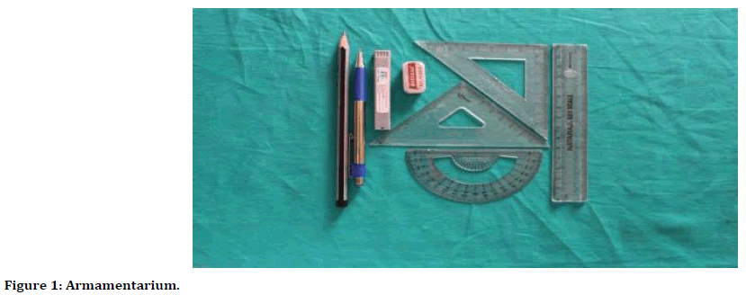
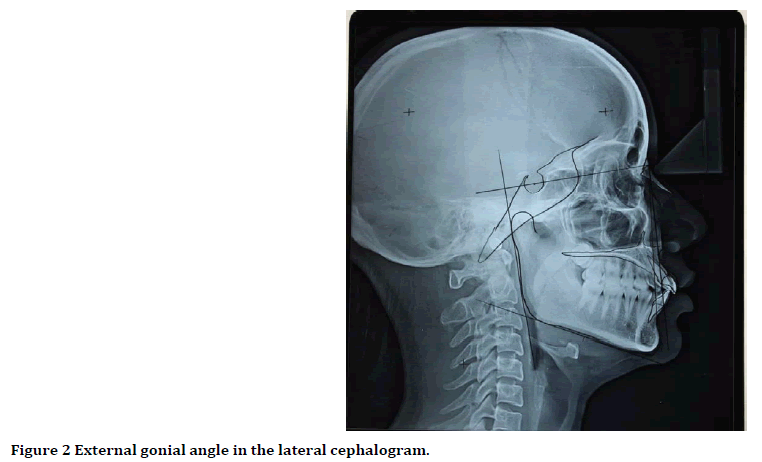
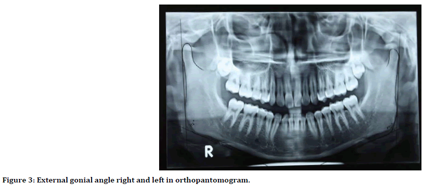
The selected radiographs have been traced onto a sheet of cellulose acetate by using a 2H pencil (Figure 1). The Cephalometric landmarks were located, identified and marked as per Steiner’s analyses (Figure 2). The measured angle was widely accepted as a skeletal analysis’s parameter, hence in our study we have taken SNA, SNB, ANB; angles or parameter to prove skeletal class I malocclusion. The lines and angles were drawn and measured using a cephalometric protractor and calipers. In the lateral cephalograms, the gonial angle was measured at the point of intersection of the plane tangential to the lower border of the mandible and that tangential to the distal border of the ascending ramus and the condyle (Figure 2). Mandibular plane was drawn on basis of tweeds analysis. In the panoramic radiographs, the gonial angle was measured by drawing a line tangent to the lower border of the mandible and another line tangent to the distal border of the ascending ramus and the condyle on both sides (Figure 3). After 15 days, both the lateral cephalogram and orthopantomograms were traced and angles were measured. The obtained values were compared with previous values and mean values were taken for analysis.

Figure 1. Armamentarium.

Figure 2. External gonial angle in the lateral cephalogram.

Figure 3. External gonial angle right and left in orthopantomogram.
Statistical analysis
The recorded values were analyzed using SPSS software version 16.0, one-way ANOVA test was applied. The mean gonial angle on lateral cephalogram and on OPG (right and left sides) with standard deviations according to gender was calculated. Pearson product-moment correlation was used to measure the degree of correlation between right and left gonial angles on OPG and on lateral cephalogram.
One-way analysis of variance (ANOVA) was applied for differences in gonial angle between the groups. For inter-observer reliability, measurements were repeated after two weeks with randomly selected radiographs and paired t-test was performed. The statistical significance was set at ≤ 0.05 (p-value) and a level of P<0.01 considered to be highly significant.
Results
The mean value of the gonial angle in lateral cephalograms was 123.3° ±6.04° (Table 1) and the gonial angle in females (Table 2) was 123.24o and that in males (Table 3) 123.36o with no statistically significant difference between the two genders.
| Method | Sex | Min | Max | Mean | SD | F | P value |
|---|---|---|---|---|---|---|---|
| Lat. Ceph. | All patients (n=100) | 109 | 133 | 123.3 | 6.04 | 0.005 | 0.995 |
| Male (N=50) | 113 | 132 | 121.36 | 5.92 | |||
| Female (N=50) | 109 | 133 | 123.24 | 6.22 | |||
| OPG-Avg | All patients (n=100) | 105 | 132 | 120.13 | 5.77 | 0.228 | 0.796 |
| Male (N=50) | 105 | 129.5 | 119.74 | 5.6 | |||
| Female (N=50) | 109 | 132 | 120.52 | 5.96 |
Table 1: Mean, standard deviation and range of gonial angle in lateral cephalogram and OPG.
| Variables | Mean | Standard deviation | Range |
|---|---|---|---|
| Gonial angle in cephalogram in females | 123.24 | 6.21 | 109-133 |
| Gonial angle in OPG in females | 125.52 | 5.96 | 109-132 |
| Right gonial angle in OPG in females | 120.28 | 7 | 108-133 |
| Left gonial angle in OPG in females | 120.76 | 5.94 | 109-133 |
Table 2: Mean, standard deviation and range of gonial angle in lateral cephalogram and OPG in females.
| Variables | Mean | Standard deviation | Range |
|---|---|---|---|
| Gonial angle in cephalogram in males | 123.36 | 5.91 | 113-132 |
| Gonial angle in OPG in males | 119.74 | 5.59 | 105-129 |
| Right gonial angle in OPG in males | 119.52 | 5.69 | 106-128 |
| Left gonial angle in OPG in males | 119.96 | 5.94 | 104-131 |
Table 3: Mean, standard deviation, and range of gonial angle in lateral cephalogram and OPG in males.
The mean value of the gonial angle in panoramic radiographs was 120.13° ± 5.77° (Table 1) and the gonial angle in females was 125.52° and that in males 119.74° with no statistically significant difference between the two genders. There was no significant difference between the values of gonial angles determined by lateral cephalogram and panoramic radiography (P=0.995). Also, in panoramic radiography, there was no significant difference between the right and left gonial angles (P=0.796).
Discussion
Our study result illustrates that, there was no statistically significant difference found between orthopantomograms and lateral cephalograms with mean age group of 19.76 years in both male and female adult patients. Similar study was carried out by Shahabi, et al. with mean age group of 18.24 years and study was concluded no evidence of statistically significant between them [2]. In comparison with both above study, our studies report lesser external gonial angle values, but statistically insignificant.
Larheim, et al. carried out similar study with aim to evaluate values of external gonial angle on right and left on orthopantomograms. However, values obtained were similar and the study was concluded reporting that there was no significant difference found in value of right and left gonial angles [14].
The gonial angle obtained by panoramic radiography was 2.2-3.6 degrees less than that of lateral cephalogram as said by Fisher-Brandies. In contrast to the results reported in the present study they observed that there are significant differences in the gonial angle obtained by the two different radiographs. As the type of malocclusion and age of the samples was not specified in the study of Fisher-Brandies there was disparity in the results and there was no significant difference found in value of right and left gonial angles [20] because present study was performed in adults with class I malocclusion
It was concluded that the size of gonial angle can be determined from the orthopantomogram with the same degree of accuracy as from the commonly used lateral cephalogram, the gonial angle being formed by the tangent of the lower border of the mandible and distal border of the ascending ramus and the condyle on each side by Mattila, et al. They avoided the disturbing influences of the superimposed images found on lateral cephalogram by showed that the right and left gonial angles can be easily determined individually from the orthopantomogram. So, it can be proved that the orthopantomogram is the more obvious choice for determination of the gonial angle. There is no significant difference in the values of gonial angle in both the panoramic and lateral cephalograms as shown in the present study.
No statistical difference between the right and left gonial angles in the panoramic radiograph was mentioned in this study. Mattila, et al. reported that, there was not any statistically significant gender differences in the gonial angle determined from the two different types of radiographs [5].
The size of the gonial angle which angle was not statistically significant in any of the three tooth retention categories was not much affected by gender as said by Ohm, et al. [21]. There were no statistically significant differences between the two genders with age ranges of 14-29 years in present study. Clinicians should be vigilant when predicting skeletal cephalometric parameters from panoramic radiographs, because of their lower predictability percentages though panoramic radiographs provide information on the vertical dimensions of craniofacial structures which was concluded by Akcam, et al. [22]. There were no significant differences between the mean values of the external gonial angle in the panoramic radiograph and lateral cephalogram and the mean values of the right and left gonial angles in panoramic radiographs as shown in the result of present study. There will be no considerable effect of gender on the gonial angle in both radiographs so both the radiographs can be used to determine the gonial angle as accurately. The right and left gonial angles can be measured easily without superimposition of anatomic landmarks in panoramic radiography, which occurs frequently in a lateral cephalogram.
Conclusion
It can be concluded that between the two gender there was no statistically significant difference found in gonial angles obtained from lateral cephalogram and orthopantomogram. In the accuracy of measurement of gonial angle between lateral cephalogram and orthopantomogram there is no statistical significance. The gonial angle can be determined as accurately as a lateral cephalogram by panoramic radiography. Without superimposed images of anatomical structures, the panoramic radiography can be used to determine the gonial angle accurately. Therefore, the Orthopantomogram can be a better choice than a lateral cephalogram.
References
- Bhullar MK, Uppal AS, Kochhar GK, et al. Comparison of gonial angle determination from cephalograms and orthopantomogram. Indian J Dent 2014; 5:123-126.
- Shahabi M, Ramazanzadeh BA, Mokhber N. Comparison between the external gonial angle in panoramic radiographs and lateral cephalograms of adult patients with Class I malocclusion. J Oral Sci 2009; 51:425-429.
- Zangouei-Booshehri M, Aghili HA, Abasi M, et al. Agreement between panoramic and lateral cephalometric radiographs for measuring the gonial angle. Iran J Radiol 2012; 9:178-182.
- Zach GA, Langland OE, Sippy FH. The use of the orthopantomograph in longitudinal studies. Angle Orthod 1969; 39:42-50.
- Mattila K, Altonen M, Haavikko K. Determination of the gonial angle from the orthopantomogram. Angle Orthod 1977; 47:107-110.
- Slagsvold O, Pedersen K. Gonial angle distortion in lateral head films: a methodologic study. Am J Orthod1977; 71:554-564.
- Haskell B, Day M, Tetz J. Computer-aided modeling in the assessment of the biomechanical determinants of diverse skeletal patterns. Am J Orthod1986; 89:363-382.
- Kasai K, Richards LC, Kanazawa E, et al. Relationship between attachment of the superficial masseter muscle and craniofacial morphology in dentate and edentulous humans. J Dent Res 1994; 73:1142-1149.
- Akcam MO, Altiok T, Ozdiler E. Panoramic radiographs: A tool for investigating skeletal pattern. Am J Orthod Dentofacial Orthop 2003; 123:175-181.
- Almog DM, Tsimidis K, Moss ME, et al. Evaluation of a training program for detection of carotid artery calcifications on panoramic radiographs. Oral Surg Oral Med Oral Pathol Oral Radiol Endod 2000; 90:111-117.
- Xie QF, Ainamo A. Correlation of gonial angle size with cortical thickness, height of the mandibular residual body, and duration of edentulism. J Prosthet Dent 2004; 91:477-482.
- Yanikoğlu N, Yilmaz B. Radiological evaluation of changes in the gonial angle after teeth extraction and wearing of dentures: A 3-year longitudinal study. Oral Surg Oral Med Oral Pathol Oral Radiol Endod2008; 105:55-60.
- Huumonen S, Sipilä K, Haikola B, et al. Influence of edentulousness on gonial angle, ramus and condylar height. J Oral Rehabil 2010; 37:34-38.
- Larheim TA, Svanaes DB. Reproducibility of rotational panoramic radiography: mandibular linear dimensions and angles. Am J Orthod 1986; 90:45-51.
- Ceylan G, Yaníkoglu N, Yílmaz AB, et al. Changes in the mandibular angle in the dentulous and edentulous states. J Prosthet Dent 1998; 80:680-684.
- Dutra V, Devlin H, Susin C, et al. Mandibular morphological changes in low bone mass edentulous females: evaluation of panoramic radiographs. Oral Surg Oral Med Oral Pathol Oral Radiol Endod 2006; 102:663-668.
- Okşayan R, Aktan AM, Sökücü O, et al. Does the panoramic radiography have the power to identify the gonial angle in orthodontics? Scientific World J 2012; 2012:219708.
- Joo JK, Lim YJ, Kwon HB, et al. Panoramic radiographic evaluation of the mandibular morphological changes in elderly dentate and edentulous subjects. Acta Odontol Scand 2013; 71:357-362.
- Cho IG, Chung JY, Lee JW, et al. Anatomical study of the mandibular angle and body in wide mandibular angle cases. Aesthetic Plast Surg 2014; 38:933-940.
- Fischer-Brandies H, Fischer-Brandies E, Dielert E. The mandibular angle in the orthopantomogram. Radiologe 1984; 24:547-549.
- Ohm E, Silness J. Size of the mandibular jaw angle related to age, tooth retention and gender. J Oral Rehabil 1999; 26:883-891.
- Akcam MO, Altikok T, Ozdiler E. Panoramic radiographs: A tool for investigating skeletal pattern. Am J Orthod 2003; 123:175-181.
Author Info
Akhilesh Singh Parate1, Heeralal chokotiya2*, Divashree Sharma3, Siddharth Sonwane4, Pratibha Sharma5 and Sandeep Shrivastava5
1Department of Dentistry, Government Medical College Shahdol, MP, India2Department of Dentistry, N.S.C.B Medical College Jabalpur, MP, India
3Department of Dentistry, SS Medical College Rewa, MP, India
4Department of Orthodontics, Government. Dental College Nagpur, MH, India
5Department of Orthodontics, Mansarovar Dental College, Bhopal, MP, India
Citation: Akhilesh Singh Parate, Heeralal chokotiya, Divashree Sharma, Siddharth Sonwane, Pratibha Sharma, Sandeep Shrivastava, Comparative Evaluation of the External Gonial Angle in Adults Patients with Class I Malocclusion from Panoramic Radiographs and Lateral Cephalograms, J Res Med Dent Sci, 2020, 8 (3):157-162.
Received: 23-Apr-2020 Accepted: 18-May-2020
