Research - (2021) Volume 9, Issue 7
Comparative Histological Study of the Oesophagus of Mammals
*Correspondence: Y Durga Devi, Department of Anatomy, Sree Balaji Medical College & Hospital Affiliated to Bharath Institute of Higher Education and Research, India, Email:
Abstract
The mucosal layer of all four animals in the present study consisted of three layers. This layer probably lines all organs that communicate outside the body and may have protective and to some extent secretory in function. In this study the thickness of the mucosa increased gradually in goat and cow. The muscularis extrema comprised of striated muscle fibres in the upper end and smooth muscle fibres in the lower end in human. In dog and cow skeletal muscle fibres were present throughout the length of the oesophagus. The serosal layer consisted of connective tissue fibres in all four mammals.
Keywords
Oesophagus, Digestive system, Serosal layerIntroduction
The oesophagus, as the gullet is a straight tube that extends from the pharynx to the stomach and serves as a food pipe. The architecture is that of a typical hollow organ with four layers mucosa, submucosa, muscularis extrema, and serosa/adventitia.
The basic structure of the mammalian oesophagus is well established, many years ago. Much research is performed to carry out the study the origin of adult and highly differentiated structures from an already existing simple types [1-4].
The study of internal structure of animals will enlightens about the establishment of broad historic relations also about the path of the development of the race. The basis of mammalian digestive tube is same from oesophagus till anus.
The species variation known to occur involves mainly the lining epithelium, glands that secretes mucus. The presence, number and distribution of the mucus secreting oesophageal glands in the submucosa are said to vary considerably in different species.
Hence the present comparative study on the histological features of oesophagus of mammals such as human, goat and dog were undertaken.
Materials and Methods
Study design
Collection of specimens
Oesophagus of human was collected from cadavers, from the Department of Anatomy of Sree Balaji Medical College and Hospital Chrompet, Chennai. Oesophagus of goat and cow was collected from slaughterhouse. Oesophagus specimens of dog was obtained from, dogs died of road accidents. Six animals of each species were utilised for the study. Tissues, from upper and lower end of the oesophagus were taken.
Fixation and processing of tissues
The collected oesophagus tissues were fixed in 10% formalin solution for one week. Processing of the specimens into paraffin blocks were prepared as per the standard technique of dehydration in ascending grades of alcohol, clearing in xylene (3 changes) and finally impregnation and embedding in molten paraffin wax.
Cutting of sections
5 micron serial sections were floated in water bath and taken onto pre cleaned slides. These were then incubated in 58°C in oven for five minutes, before staining.
Staining with haematoxylin and eosin
The slides with thin sections of tissue was again preheated, then descended using xylene (3 changes) , grades of alcohol , rinsed in tap water for 5 minutes until water runs off evenly , stained using Harris haematoxylin for 15 minutes , gently washed in running tap water to wash the excess haematoxylin stain . Then eosin stain is used for 2 minutes, washed in tap water for 5minutes then ascended with acetone (2 changes) and xylene (3 changes), finally mounted using DPX.
Van Gieson's method for collagen fibres solution
- Haematoxylin.
- Solution A
- Haematoxylin - 1g Alcohol 95% - 100ml.
- Solution B.
- Ferric chloride 29% aqueous-4ml Hydrochloric acid concentrated-1ml Distilled water-95 ml.
Working solution
Equal parts of solution A & B.
Vangieson's solution
Acid fuchsin 1% aqueous - 2.5ml
Picric acid-saturated aqueous - 97.5ml
Oesophagus of human, dog, and goat were collected for the study. Six animals are used for the study. The collected oesophageal tissues were fixed and stained for histological studies. Thickness of different layers was measured using ocular and stage micrometre.
Results
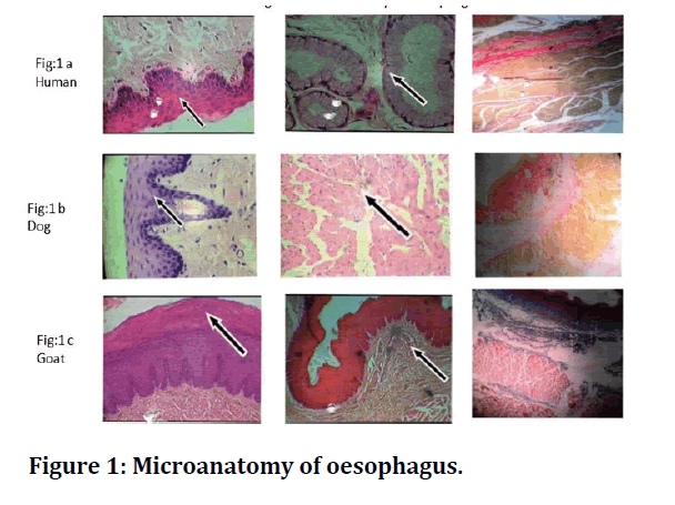
The histological examination of human oesophagus shows that the thickness and stratification of the epithelium was observed to be more in the upper end of the oesophagus when compared to that of the lower end (Figure 1A). Many peg-like protrusions of the lamina propria indented the epithelium. In the oesophagus of dog, the height of the longitudinal folds appeared to be uniform in upper end as well as the lower end. The lamina layer was composed of evenly distributed connective tissue fibres. In the submucosal layer, the mucous alveoli were formed of cuboidal cells with distinct cell boundaries (Figure 1b). The epithelial layer of goat samples was comprised of keratinised stratified squamous epithelium which rested on a basement membrane and the submucosal glands were absent throughout the length of the oesophagus (Figure 1c).

Figure 1: Microanatomy of oesophagus.
The arrangement of structural components in the oesophagus of mammals is depicted in Table 1.
Table 1: Structural components of oesophagus.
| Epithelium | Muscularis mucosa | Submucosal glands | Muscularis externa | |
|---|---|---|---|---|
| Human | Non keratinized | Continuous and thick | Few and scattered | Striated in upper end and smooth muscle in the lower end |
| Dog | Non keratinized | Complete and distinct | Numerous | Striated muscle fibers throughout the length |
| Goat | Keratinized | Discontinuous in some areas | absent | Striated in the upper half and smooth muscle near the cardia |
Discussion
Oesophagus at two levels upper and lower end of all 3 mammals was compared under light microscope [3-6]. The muscularis mucosa was composed of smooth muscle fibres but its arrangement and distribution varied in all three animals. In the submucosa the submucosal glands were absent throughout the length of the oesophagus of goat. The glands were numerous in dog and decreased in human [6-10]. The serosal layer consisted of connective tissue fibres in mammals. The present study at light microscopic level provides scope to study it under ultrastructural level. By understanding the varies layers of the oesophagus and correlating it with its function and physiology can form basis for comprehending any pathological variation in each individual layer.
Conclusion
Although studies have been made on the oesophagus in all species. Histological structure of different regions of the oesophagus varies widely among species, depending upon their physiological activities and the type of ingesta and this form base for the present study. The present study at light microscopic level provides scope to study it under ultra-structural level. By understanding the varies layers of the oesophagus and corelating it with its function and physiology can form basis for comprehending any pathological variation in each individual layer Thus the present study may be useful for clinicians especially in understanding the etiology of tumours in the oesophagus with respect to food habits. Comparative histological study in mammals, like the present study can be supportive and useful for surgeons for xenografting.
Funding
No funding sources.
Ethical Approval
The study was approved by the Institutional Ethics Committee.
Conflict of Interest
The authors declare no conflict of interest.
Acknowledgements
The encouragement and support from Bharath Institute of Higher Education and Research, Chennai, is gratefully acknowledged. For provided the laboratory facilities to carry out the research work.
References
- Culling CFA. The Microscope of histopathology and histochemical techniques. 3rd Edn. Butterworths, London 1974; 593-594.
- Di Fiore MSH. Atlas of human histology. 4th Edn. 1977; 125-127.
- Busch C. The structure of oesophagus of the dog. Acta Anat 1980; 107:339-360.
- Byrnes CK, Pisko-Dubeienski ZA. An anatomical sphincter of the oesophageal-gastric junction. Bull Soc Int Chir 1963; 22:62-68.
- https://anth.la.psu.edu/research/research-labs/weiss-lab/documents/CQ44_GraysAnatomy.pdf
- Grahame T. Structure of the Oesophagus of domestic animals .Vet Res 1926; 38:308-311.
- Henk WG, Hoskins JD, Abdelbaki YZ. Comparative morphology of esophageal mucosa and submucosa in dogs from 1 to 337 days of age. Am J Veterinary Res 1986; 47:2658-65.
- Ian Whitmore. Oesophageal striated muscle arrangement and histochemical fibretypes in guinea-pig, marmoset, macaque and man. J. Anat 1982; 134: 684 -695
- Jamdar MN, Ema AN. The submucosal glands and the orientation of the musculature in the oesophagus of the camel. J Anat 1982; 135:165-171.
- John BAE. Developmental changes in the oesophageal epithelium in man. J Anat 1952; 86:431-441.
Author Info
Department of Anatomy, Sree Balaji Medical College & Hospital Affiliated to Bharath Institute of Higher Education and Research, Chennai, Tamil Nadu, IndiaCitation: Y Durga Devi, Comparative Histological Study of the Oesophagus of Mammals, J Res Med Dent Sci, 2021, 9(7): 397-399
Received: 07-Jul-2021 Accepted: 22-Jul-2021
