Research Article - (2022) Volume 10, Issue 5
Comparing the Antimicrobial Activity of Catgut Suture Coated with Neem, Ginger and Mint Infused In Silver Nanoparticles-An in-Vitro Study
Guy Patrick Sandou1*, Subhashree R2, S Rajeshkumar3 and Thiyaneswaran Nesappan4
*Correspondence: Guy Patrick Sandou, Department of Implantology, Saveetha Academy of Implant Dentistry, Saveetha Dental College and Research Institute, Chennai, India, Email:
Abstract
Introduction: Plants play an important part in drug production, and the pharmaceutical industry relies extensively on
natural ingredients to produce new drugs. According to a WHO survey, folk medicine is used by 80% of the world's
population for primary health care. Nanoparticles have attracted a lot of attention due to their special properties and
potential applications due to their large surface area and surface plasmon resonance effect.
Aim: The aim of this study was to evaluate the antimicrobial effect of green synthesis of neem, ginger and mint infused in
silver nanoparticles.
Methodology: Neem, ginger and mint were washed thoroughly and shade dried. The dried plant material was finely
powdered and grounded using a slender blender. An aqua extract of neem, ginger and mint was prepared separately. 50 ml
of silver nanoparticles were mixed with a 50 ml solution of aqua extract. Kept in a shaker and UV-vis Spectrophotometer
readings are taken at 12,24,48 hrs to check for the formation of nanoparticles. UV-Visible spectrophotometry was used to
classify the synthesized silver nanoparticles, revealing peak absorption at 440 nm and a composite at 380 nm. The uniformly
scattered spherical shaped silver nano-composite with a size of about 14-20 nm was clearly visible in the TEM pictures. The
biosynthesized nanocomposite was coated on catgut suture material to test its anti-peri-implantitis effect. In the in-vitro
study, the antimicrobial activity of the three plants was analysed by agar well method using organisms Lactobacillus,
Streptococcus mutans and Candida albicans.
Results: Preventing any post-surgical infection is an important parameter for obtaining the desired outcome. This study
showed that neem, ginger, mint Ag coated sutures showed an effective antimicrobial action against L. bacilli, S. mutans and
C. albicans, which are the most commonly found oral microbiota.
Conclusion: Ginger Ag+chitosan nanocomposite showed a significant potent effect against E. fecalis, S. mutans,
Pseudomonas, C. albicans.
Keywords
Neem, Ginger, Mint, Silver Nanoparticles, In-Vitro Study
Introduction
Plants play an essential part in drug production, and the pharmaceutical industry relies extensively on natural ingredients to produce new drugs. According to a WHO survey, folk medicine is used by 80% of the world's population for primary health care. Nanoparticles have attracted much attention due to their unique properties and potential applications due to their large surface area and surface plasmon resonance effect. Nanotechnology is a branch of applied science and technology that seeks to create devices and drug forms that are 1 to 100 nanometers in size. Nano-medicine is a term that has recently been used to describe the use of nanotechnology in the treatment, detection, surveillance, and regulation of biological systems. Secure materials, such as synthetic biodegradable polymers, lipids, and polysaccharides, were used to create nanocarriers [1].
Phytotherapeutics require a technical approach to administer the components in to improve patient compliance and prevent repetitive delivery. Designing nano-medicine for herbal constituents will help with this nanomedicine. Nano-medicine not only helps to resolve non-compliance by decreasing toxicity and improving bioavailability, but they also help to improve medicinal value by reducing toxicity and increasing bioavailability. Nanotechnology is one such novel solution. Herbal drug delivery systems with nano-sized drug delivery systems have a promising future for improving operation and solving issues associated with plant medicines [2].
Peri-implantitis is an inflammatory reaction in the tissues around an implant that results in the removal of supporting bone. For chosen embed systems, the general recurrence of peri-implantitis was estimated to be 5% to 8%. The gingival tissue's intersection with dental implants' neck can help prevent microorganism colonisation and peri-implantitis [3].
The majority of people have used herbal plants because of their pharmacological properties. Scientists have recently become more interested in herbal medicine as a result of the rise of antibiotic-resistant bacteria. Ginger, neem, mint is a medicinal plant that is used as a flavouring agent in food in both fresh and dried forms all over the world. Ginger, neem and mint also have anti-tumorigenic, anti-apoptotic, and antioxidant properties, which can be due to its active polyphenol compounds gingerols and shogaols. Herbal nanoparticles were recently isolated from the ginger extract and used in a drug delivery device without causing any adverse side effects [4].
The aim of this study was to evaluate the antimicrobial effect of green synthesis of neem, ginger and mint infused in silver nanoparticles.
Materials And Methods
Neem ginger and mint plants were washed thoroughly and shade dried. The dried plant material was finely powdered and grounded using a slender blender. One gram of neem powder, ginger powder, and mint powder was mixed with 100 ml of distilled water, stirred, boiled, and filtered separately. 50 ml of silver nanoparticle was mixed with 50 ml solution of neem, ginger and mint aqua extract separately (0.574 grams). Silver nitrate (AgNO3) and chitosan were obtained from Sigma Aldrich, and microbial media such as Mueller Hinton Agar and Rose Bengal Agar were obtained from Hi media laboratories Pvt. Ltd. Kept in a shaker, and UV-vis Spectrophotometer readings are taken at 12 hrs, 24 hrs, 48 hrs to check for the formation of nanoparticles. The practical solution is centrifuged, and the pellets are collected. Supernatants dried in a hot air oven, and the scrapings collected. The herbal extract was then transferred to the container for further analysis (Figure 1).
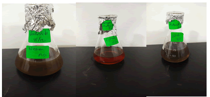
Figure 1: Picture representing neem Ag+, Ginger Ag+, Mint Ag+ herbal extract preparation respectively.
Synthesis of dried herbal extract mediated silver nanoparticles
In 90 mL of distilled water, 1 mM silver nitrate (AgNO3) was dissolved. 10 mL filtered, dried herbal extract was added to that. For 48 hours, the reaction mixture was stirred at 600-700 rpm in a magnetic stirrer. The synthesis was verified using a UV-Visible double beam spectrophotometer after the colour change was detected regularly. The solution of synthesised silver nanoparticles was centrifuged for 10 minutes at 8000 rpm. The supernatant was discarded after centrifugation, and the pellet was collected. For characterisation and other tests, the collected pellet was placed in an Eppendorf tube at 4°C.
Synthesis of dried herbal extract mediated AgNPs based nanocomposite.0.5 g of chitosan was weighed and applied to 49.5 mL of distilled water to make the nanocomposite. 0.5 mL glacial acetic acid was added to that. The chitosan mixture was stirred continuously for around 2-3 hours at 800 rpm using a magnetic stirrer. To 50 mL of chitosan solution, 50 mL of pre-centrifuged dried herbal extract AgNPs solution was added. This nanocomposite solution was stirred using a magnetic stirrer at 800 rpm for three days.
Coating dried herbal extract mediated AgNPs based nanocomposite on Cat-gut suture materialA sterile catgut suture material was added to the biosynthesised nanocomposite solution, and the coating process was carried out using a magnetic stirrer while stirring at 800-950 rpm and heating at 50ºC. The catgut suture material and the biosynthesised AgNPs-based nanocomposite solution were dried and coated in a sterile glass Petri plate and held in a hot air oven at 60ºC-70ºC for 6 hours. The coated surface of the dried and fully coated suture material was compared to the control catgut suture material's coated surface under a digital microscope. For further studies, the coated catgut suture material was placed in a sterile Eppendorf Tube.
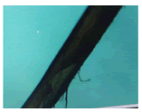
Figure 2: Picture represents TEM of Ginger Ag+ coated catgut suture.
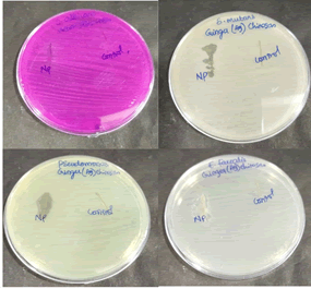
Figure 3: Agar well method antimicrobial activity of Ginger Ag+.
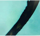
Figure 4: Picture represents TEM of Neem Ag+ coated catgut suture.
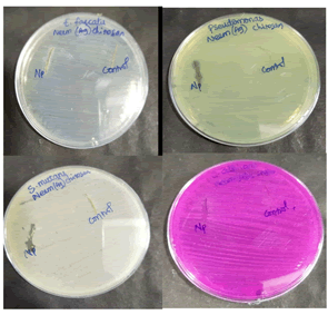
Figure 5: Agar well method antimicrobial activity of Neem Ag+.
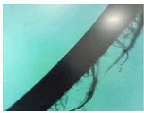
Figure 6: Picture represents TEM of Mint Ag+ coated catgut suture.
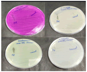
Figure 7: Agar well method antimicrobial activity of Mint Ag+.
Characterisation studiesTransmission Electron Microscopy (TEM) was used to examine the size and shape of the green synthesised silver nanoparticles, and a UV-Visible double beam spectrophotometer in the wavelength range of 250-750 nm was used to confirm the synthesis of silver nanoparticles. (Figure 2, Figure 4 and Figure 6)
Antimicrobial activityAgar well diffusion method: Pseudomonas aeruginosa, Streptococcus mutans, Enterococcus faecalis, and Candida albicans were isolated from patients at Saveetha Dental College and Hospitals in Poonamallee, Chennai.
Antibacterial activityAgar-well diffusion method: Antibacterial activity of the herbal formulation against Lactobacillus, Streptococcus mutans and strains. To determine the zone of inhibition, this operation was carried out on MHA agar. Muller Hinton agar was made and sterilised for 45 minutes at 120 pounds. The media was poured onto the sterilised plates and left to set. After the wells were cut with a good cutter, the test organisms were swabbed. The plates were packed with varying amounts of nanoparticles and incubated at 37°C for 24 hours. After the incubation time, the zone of inhibition was evaluated. (Figure 3, Figure 5 and Figure 7)
Antifungal activityAgar-well diffusion method: Candida albicans is the test pathogen in the agar well diffusion assay. Sabouraud's Dextrose Agar was used to make the medium. Nanoparticles of varying amounts were added to the prepared and sterilised medium after the wells were swabbed with test species. At 28°C, the plates were incubated for 48-72 hours. After the incubation time, the region of inhibition was evaluated.
Result
Preventing any post-surgical infection is an essential parameter for obtaining the desired outcome. This study showed that neem, ginger, mint Ag coated sutures showed an effective antimicrobial action against L. bacilli, S. mutans and C. albicans, which are the most commonly found oral microbiota (Figure 8, Figure 9 and Figure 10).
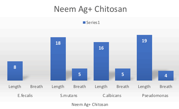
Figure 8: bar graph represents Neem Ag+ Chitosan antimicrobial effect on E. fecalis, S. mutans, C. albicans, Pseudomonas.
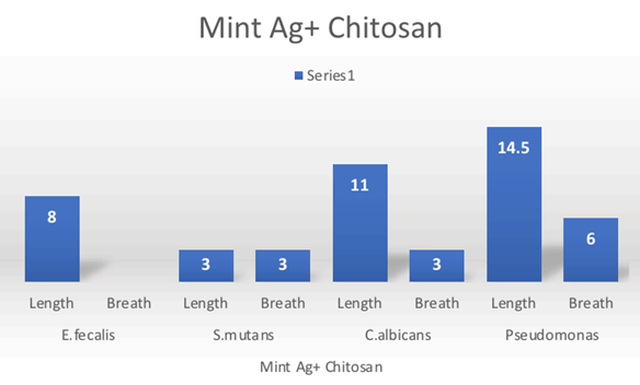
Figure 9: Bar graph represents Mint Ag+ Chitosan antimicrobial effect on E. fecalis, S. mutans, C. albicans, Pseudomonas.
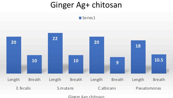
Figure 10: Bar graph represents Ginger Ag+ Chitosan antimicrobial effect on E. fecalis, S. mutans, C. albicans, Pseudomonas.
Antimicrobial herbal traditional therapies or plants range from the rapidly growing flora of woods to those grown in farm fields. They have been studied, examined, proven to have antimicrobial properties, or are being checked now. All around the world, these herbs grow spontaneously or are cultivated primarily for this purpose. As a result of advances in herbal medicine, new and popular herbs have arisen. Incorporating herbal extracts into nanomedicine has several additional benefits, including bulk dosing and the ability to counteract absorption, which is a significant issue, attracting the interest of major pharmaceutical companies.
If left untreated, pathogens can cause a wide range of human diseases, from self-limiting illnesses to potentially lethal medical conditions. Nonetheless, due to widespread use, antibiotic resistance is on the rise, leaving previously treatable infections untreatable. Globalisation also facilitated the proliferation of (resistant) strains of other infectious agents. Recently research in the field of newer antibiotic therapies is best suitable in antibiotic resistance. In our study, 3 plants, namely neem, ginger and mint silver nanoparticle antimicrobial activity was analysed using the agar well diffusion method and outcomes were analysed by a zone of inhibition.
Chandrasekaran studied Neem nanoemulsion formulated using neem oil; the formulated neem NE showed antibacterial activity against the bacterial pathogen Vibrio vulnificus by disrupting the integrity of the bacterial cell membrane [5].Verma et al. study showed at higher temperatures and alkaline pH, the formation of AgNPs increased over time. The AgNPs developed had improved antimicrobial properties and demonstrated a zone of inhibition against bacteria (Escherichia coli) isolated from a garden soil sample [6].
El-Refai studied the antibacterial properties of ginger nanoparticles were tested against Staphylococcus aureus, Klebsiella pneumonia, Candida albicans, Bacillus subtilis, Erwinia carotovora, and Proteus Vulgaris, as well as antifungal properties against Candida albicans. Antimicrobial analyses show that these nanoparticles have solid antimicrobial activity compared to antibiotic norm performance [7]. Yang et al. experimented with silver nanoparticles using ginger extract against aquatic microorganisms, and results showed significant antimicrobial effects against typical aquatic pathogenic strains of Vibrio anguillarum, Vibrio alginolyticus, Aeromonas punctata, Vibrio parahaemolyticus, Vibrio splendidus and Vibrio harveyi [8].
Discussion
Sarkar study suggested that Silver nanoparticles made from mint plant leaf extracts demonstrated antibacterial activity against Escherichia coli, Pseudomonas aeruginosa, Bacillus subtilis (Gram Negative), and Staphylococcus aureus microorganisms (Gram-Positive). The antimicrobial influence, however, differed depending on the salt concentration. Furthermore, the silver nanoparticles produced by mint plant leaves extracts inhibited Escherichia coli and Pseudomonas aeruginos [9]. Gabriela, in the study, revealed that silver nanoparticles synthesised had antibacterial activity against pathogenic bacteria such as E. coli and Staphylococcus aureus. For treatments of 150, 250, and 350 l of extract, the minimum inhibitory concentration needed for S. aureus was 11.1, 26.3, and 51.5 g, respectively [10-12].
While nanopharmaceuticals may appear to provide limitless possibilities in the field of drug delivery for the diagnosis and treatment of a variety of diseases, their safety should not be overlooked. Engineered nano-sized materials' physicochemical and structural properties can change as their size decreases, resulting in various material interactions that could lead to toxicological effects. Scientists must agree that the toxicological assessment of nanomaterials and nanomedicines is still in its early stages, and there are few reports on their safety and toxicity [13].
Conclusion
Ginger Ag+ chitosan nanocomposite showed a significant potent effect against E. fecalis, S. mutans, Pseudomonas, C. albicans. Sutures made of catgut have high tensile strength and are quickly absorbed by our bodies. It has been used in a variety of surgical procedures due to its unique characteristics. Dried ginger extract mediated silver nanocomposite has been developed to make suture material more resistant to peri-implantitis causing pathogens. In the TEM image, the nanocomposite had a uniformly distributed spherical shape and a size of about 14-20 nm. The established suture material had a strong antimicrobial effect against all pathogens tested, indicating that it will play an essential role in potential biomedical applications in the dental field.
References
- Azmi L, Shukla I. Perspectives of phytotherapeutics: Diagnosis and cure. Phytomedicine 2021; 1:225-250.
- Jerobin J, Makwana P, Kumar RS, et al. Antibacterial activity of neem nanoemulsion and its toxicity assessment on human lymphocytes in vitro. Inte J Nanomed 2015; 10:77.
[Crossref] [Google Scholar] [Pubmed]
- Chauhan P, Tyagi BK. Herbal novel drug delivery systems and transfersomes. J Drug Delivery Ther 2018; 8:162-168.
- El-Refai AA, Ghoniem GA, El-Khateeb AY, et al. Eco-friendly synthesis of metal nanoparticles using ginger and garlic extracts as biocompatible novel antioxidant and antimicrobial agents. J Nanostruct Chem 2018; 8:71-81.
- Fragkioudakis I, Tseleki G, Doufexi AE, et al. Current concepts on the pathogenesis of peri-implantitis: A narrative review. Euro J Dent 2021; 15:379-387.
[Crossref] [Google Scholar] [Pubmed]
- Gabriela ÁM, Gabriela MD, Luis AM, et al. Biosynthesis of silver nanoparticles using mint leaf extract (Mentha piperita) and their antibacterial activity. Adv Sci Engineer Med 2017; 9:914-923.
- Kaur A, Kaur L, Singh G, et al. Nanotechnology based herbal formulations: A survey of recent patents, advancements and transformative headways. Rec Patent Nanotech 2021; 16:90-96.
[Crossref] [Google Scholar] [Pubmed]
- Devi VK, Jain N, Valli KS. Importance of novel drug delivery systems in herbal medicines. Pharmacog Rev 2010; 4:27.
[Crossref] [Google Scholar] [Pubmed]
- Sosnowska M, Kutwin M, Adamiak AD, et al. Green synthesis of silver nanoparticles by using aqueous mint (Mentha piperita) and cabbage (Brassica oleracea var. capitata) extracts and their antibacterial activity. Annals of Warsaw University of Life Sciences-SGGW. Animal Sci 2017;
- Farokhzad OC, Langer R. Impact of nanotechnology on drug delivery. ACS nano 2009; 3:16-20.
[Crossref] [Google Scholar] [Pubmed]
- Singh B, Awasthi R, Ahmad A, et al. Phytosome: Most significant tool for herbal drug delivery to enhance the therapeutic benefits of phytoconstituents. J Drug Del Therapeut 2018; 8:98-102.
- Verma A, Mehata MS. Controllable synthesis of silver nanoparticles using Neem leaves and their antimicrobial activity. J Rad Res Appl Sci 2016; 9:109-115.
- Yang N, Li F, Jian T, et al. Biogenic synthesis of silver nanoparticles using ginger (Zingiber officinale) extract and their antibacterial properties against aquatic pathogens. Acta Oceanolog Sinic 2017; 36:95-100.
Author Info
Guy Patrick Sandou1*, Subhashree R2, S Rajeshkumar3 and Thiyaneswaran Nesappan4
1Department of Implantology, Saveetha Academy of Implant Dentistry, Saveetha Dental College and Research Institute, Chennai, India2Department of Maxillofacial Prosthodontics And oral Implantology, Saveetha Academy of Implant Dentistry, Saveetha Dental College and Research Institute, Chennai, India
3Department of Pharmacology, Nanobiomedicine Lab, Saveetha Dental College and Research Institute, Chennai, India
4Department of Maxillofacial Prosthodontics and Oral Implantology, Saveetha Dental College and Research Institute, Chennai, India
Citation: Guy Patrick Sandou, Subhashree R, S Rajeshkumar, Thiyaneswaran Nesappan, Comparing the Antimicrobial Activity of Catgut Suture Coated with Neem, Ginger and Mint Infused In Silver Nanoparticles-An in-Vitro Study , J Res Med Dent Sci, 2022, 10(5): 123-128.
Received: 23-Feb-2022, Manuscript No. 48598; , Pre QC No. 48598; Editor assigned: 25-Feb-2022, Pre QC No. 48598; Reviewed: 09-Mar-2022, QC No. 48598; Revised: 22-Apr-2022, Manuscript No. 48598; Published: 05-May-2022
