Research - (2021) Volume 9, Issue 6
Comparison of Different Obturation Techniques in Polymer-Based Prototyped Primary Incisors: An in vitro Study
*Correspondence: Manal A Almutairi, Division of Pediatric Dentistry, Department of Pediatric Dentistry and Orthodontics, Dental college, King saud university, Saudi Arabia, Email:
Abstract
Background: Several factors affect the success of pulp therapy of primary teeth, including cleaning and shaping of the root canals and the quality of obturation as the most important steps. Achieving a void-free root canal filling is a challenge in current clinical pulpectomy practice and successful pulpectomy in primary teeth depends on quality of obturation.
Aim: To compare the quality of different obturation techniques in polymer-based prototyped primary incisors using digital radiographic images.
Materials and Methods: Sixty polymer-based prototyped primary incisors were instrumented and randomly divided into six groups of 10 teeth each. Obturation technique was done as follows: Group 1- MetapexR by injection, Group 2- MetapexR by reamer, Group 3- MetapexR by motor driven lentulo spiral, Group 4- ZOE by injection, Group 5- ZOE by reamer and Group 6- ZOE by motor driven lentulo spiral. The quality of obturation was evaluated by using digital radiographic images. The obtained data were analysed using Chi-square test, Kruskal-Wallis, and Mann-Whitney test.
Results: There were significant differences between all groups in the quality of obturation. The best scores for length of obturation were ranked as followed: Group 4, Group 6, Group 1, and Group 2. While the best scores regarding density of obturation were ranked as followed: Group 4, Group 2, Group 1, and Group 3.
Conclusions: Based on results of this study Group 4- ZOE inserted by injection exhibit best scores in quality of obturation, while Group 5- ZOE inserted by reamer exhibit lowest scores.
Keywords
Primary teeth, Pulpectomy, Digital radiographic, Zinc oxide eugenol paste, Metapex, Obturation
Introduction
A pulpectomy is one of the treatment options used to maintain primary teeth with radicular pulpal tissue inflammation or become nonvital until regular exfoliation [1,2]. The treatment consists of removing the infected pulp tissue and debris from the canal and obturation with resolvable antibacterial paste [3]. Successful pulpectomy procedure in necrotic primary teeth depends upon aseptic root canal preparation and hermetic seal of the root canal system [4,5].
The ideal biomechanical preparation of primary root canals is challenging due to their unpredictable root canal anatomy [6,7]. Moreover, significant differences in the root canal anatomy among the used specimens (lack of paired samples) may be a source of bias in evaluating the effectiveness of a specific technique in endodontic studies [8,9]. For this reason, the use of prototypes with individualized root canal anatomy may bring advantages in this regard [10]. In addition, bioethical issues of risk cross-infection from infected specimens are now posing a challenge to this activity in some organizations.
To improve the success of the endodontic treatment, the properties of filling materials and the appropriate insertion into the root canal are critical. The most prevalent root canal filling materials for primary teeth are zinc oxide eugenol paste (ZOE), calcium hydroxide (Ca (OH)2), and iodoform paste, or a combination [11,12]. ZOE is probably the most used filling material for primary teeth in the United States [13]. However, it is a slowly resorbing material and is difficult to remove if extruded beyond the root apex into periapical areas. On the contrary, Ca (OH)2- based materials are to resorb simultaneously with physiological root resorption [14]. The ability of these pastes to completely adapt to canal walls or root apical sealing without overfilling and not to create voids in the paste is limited and can depend upon the delivery system used to carry obturating material to the canals [15].
Several techniques have been used to fill the material into the root canals of primary teeth. An ideal filling technique should assure complete filling of the root canal without overfill and minimal or no voids [16]. The most common obturation technique used with ZOE paste in clinical practice are lentulo spiral/paste carriers, either handheld or rotary, while Ca (OH)2/iodoform combination pastes such as VitapexR, MetapexR are available in premixed syringes, and the material is carried to the root canal directly through disposable tips [17]. Other techniques may be used to deliver the obturation material to the roots of primary teeth canal, including an endodontic pressure syringe, a disposable tuberculin syringe, an Injection Technique, a Navi Tip syringe and reamer [18]. However, there is at present no agreements as to which technique provides the best sealing of root canal filling material in primary teeth. Therefore, this in vitro study aimed to compare the quality of different obturation techniques in polymer-based prototyped primary incisors using digital radiographic images. The tested null hypothesis was there is no difference in the combination of filling pastes and method of obturation.
Material and Methods
The Ethics Committee approved this study protocol of the College of Dentistry Research Center at King Saud University, Riyadh, Saudi Arabia, under the research number FR-0597. In this study, the sample size was calculated at the level of significance σ =0.05 with e size 0.5 through power=0.95 to be at least 60 teeth/10 per group.
Sixty artificial resin primary maxillary central incisors with standardized internal anatomy (Denarte, Sao Paulo, Brazil) were used in this study. All resin-based primary maxillary central incisors were accessed from the palatal surface with a 1012 diamond spherical drill (KG Sorensen, Cotia, Sao Paulo, Brazil). Root canal length (17 mm) was established by introducing a #15 Kerr file into the root canal until it was visible at the apex, and the working length was set 1 mm short. The ProFileR (DENTSPLY, Maillefer, Ballaigues, Switzerland) reciprocating instrument was enlarged sequentially to size 30-35/0.04 in a rotary handpiece (Bien-Air Dental, Langgasse 60, Biel, Switzerland) at 300 rpm with the minimum torque was used to clean and shape the root canal with three pecking movements, followed by irrigation until reaching the working length. The root canals were irrigated with 10 ml normal saline after each filing step. Next, the canals were dried with absorbent paper points (Spident, Incheon, Korea). In each session, to overcome biases resulting from operator fatigue, only five root canals were prepared. All root canals were prepared by a single trained operator.
After the preparations, the apexes of the prepared roots were covered with red modelling wax (Golden Modelling Wax, Telangana, India) to produce a halo space to serve as a collection area for any extruded canal filling material. Finally, the roots were mounted in 2x2 cm cold cured acrylic resin (Major Ortho, Major Prodotti Dentari S.p.A., Moncalieri, Italy) blocks from 1 mm below the cementoenamel junction.
Obturation Technique: Sixty Teeth were randomly divided into six groups (n=10), according to filling paste: MetapexR (Meta biomed co., Ltd, Chungcheongbuk-do, korea) or ZOE paste (Pulpdent, Watertown, MA, USA) and application methods as follows:
Group 1: Prepared canals were filled with MetapexR by injection. Commercially available MetapexR paste in a syringe form (Meta biomed co., Ltd, Chungcheongbuk-do, korea). Initially, placed the disposable tip and was fitted it beforehand in the prepared canal. Once the syringe reached the apex, it was continuously pressed and slowly withdrawn gradually until the paste filled the canal orifice.
Group 2: Prepared canals were filled with MetapexR by using a hand reamer. A 21 mm reamer of size 30 was used to deliver filling paste into canal according to working length. The reamer was smeared with filling paste inserted into the canal and rotated in the counterclockwise direction. Then pumped it up and down with a wiping motion against the canal walls. It was then withdrawn it from the canal. This process was repeated until the canal orifice appeared to be filled with the cement dispensed.
Group 3: Prepared canals were filled with MetapexR by motor-driven lentulo spiral. A lentulo spiral size (#30) was mounted on slow speed handpiece according to working length. The lentulo spiral coated with the Metapex R, inserted into the canal, and withdrew gently while still rotating. This procedure was repeated 3–5 times until the canal orifices were filled visibly.
Group 4: Prepared canals were filled with ZOE by injection. Achieved a creamy consistency by mixing one volume unit of powder and a two-volume unit of liquid on a dry glass slab at room temperature for 45 seconds. The ZOE paste was inserted on 3 ml syringe (Crystal-ject, Bu Kwang Medical Inc, Korea) and fitted disposable tip (Meta biomed co., Ltd, Chungcheongbuk-do, korea) then filled canal like group1.
Group 5: Prepared canals were filled with ZOE by a hand reamer method like group 2.
Group 6: Prepared canals were filled with ZOE by motordriven lentulo spiral method like group 3.
To control paste delivery in all groups, a rubber stopper was placed around each instrument at a distance determined by preoperative measurements. When the canals were visibly filled in all groups, a wet cotton pellet was used to lightly tamp the material into the canal. Later, the access cavity was filled with a thick mixture of ZOE.
Radiographic technique. After completion of the pulpectomy procedure and to obtain accurate and consistent radiographs that would allow for good tooth structure visualization and reproducibility, teeth were exposed with digital radiographic at standardized kilovolts (70 kVp), milliamperes (6 mA) for exposure time (0.160s) with a target-film distance of 15 mm so that radiographic errors were minimized. Radiographic evaluation was done in both buccolingual and mesiodistal direction using paralleling cone technique with both receptor and tooth aligned in the same direction for each tooth in all six groups.
Radiographic evaluation. The quality of obturation was radiographically evaluated by trained and knowledgeable pediatric dentist blinded to filling technique for the length and density of obturation using a modification of the methods reported by Coll and Sadrian 1996 [19] and Sigurdsson et al. [20].
The obturation length of filled root canals was recorded, according to the distance of the filling past from the apex as follows:
Score-1 (under filling): canal filled more than 2 mm short of the apex.
Score-2 (optimal filling): canal having filled ending at the radiographic apex or up to 2 mm short of apex.
Score-3 (overfilling): any canal showing filled outside the root apex.
The density of the root canal filling was recorded as follows:
Score-1 (Unacceptable): presence of more than three voids in different areas of root canal. Score-2 (Acceptable): good filling, in which between one to three voids were observed. Score-3 (Good): optimal filling, with no voids.
Examination of radiographic criteria/scores were performed twice, 72 hours apart, by single investigator who was blind to groups. A data collection form was used to record the required data. The obtained data were statistical analyzed using the Chi-square test, Kruskal- Wallis and Mann-Whitney tests to detect differences and interactions among groups. A P-value of <0.05 was used to report the statistical significance of results. The statistical analysis was carried out with SPSS V21.0 (SPSS Inc., Chicago, Ill, USA).
Results
The Kappa value for intra-examiner reproducibility was interpreted as almost perfect. The intra-observer kappa value for the length of obturation 0.91, indicating almost perfect reliability; the intra-observer kappa value for the density of obturation was 0.98, indicating almost perfect reliability.
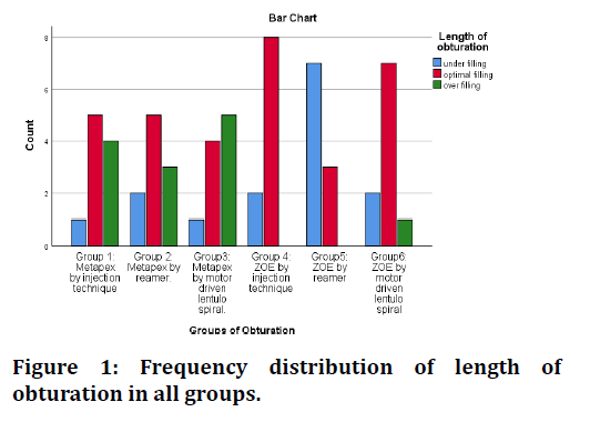
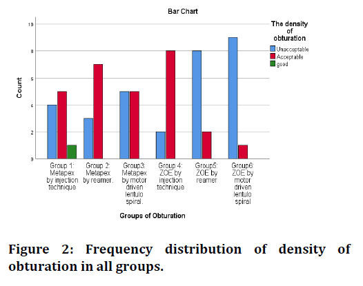
Frequency distribution of length of obturation and frequency distribution of density of obturation in all groups are shown in Figures 1 and 2. Table 1 shows the frequency distribution of the quality of obturation categorized by length and density of obturation in each group. The evaluation length of obturation within different obturation techniques showed that Group 4 (ZOE inserted by injection) exhibited the highest number of the optimally filled canal (80%), followed by Group 6 (ZOE inserted by Motor-Driven Lentulo Spiral, 70%), Group 1(MetapexR inserted by Injection, 50%), Group 2 (MetapexR inserted by Reamer, 50%), and Group3 (MetapexR inserted by Motor Driven Lentulo Spiral, 40%). However, Group 5 (ZOE inserted by Reamer, 70%) indicated the highest number of underfilled canals. The difference between all groups was statistically significant regarding the length of obturation (p<0.05).

Figure 1. Frequency distribution of length of obturation in all groups.

Figure 2. Frequency distribution of density of obturation in all groups.
| Group | Quality of obturation | |||||||||
|---|---|---|---|---|---|---|---|---|---|---|
| Length of obturation | Density of obturation | |||||||||
| Under-filling | Optimal filling | Over-filling | Total | P value* | Unacceptable | Acceptable | Good | Total | P value* | |
| Group 1: Metapex by Injection | 1(10.0) | 5(50.0) | 4(40.0) | 10(100.0) | 0.008 | 4(40.0) | 5(50.0) | 1(10.0) | 10(100.0) | 0.025 |
| Group 2: Metapex by Reamer | 2(20.0) | 5(50.0) | 3(30.0) | 10(100.0) | 3(30.0) | 7(70.0) | 0(0.0) | 10(100.0) | ||
| Group3: Metapex by Motor Driven Lentulo Spiral | 1(10.0) | 4(40.0) | 5(50.0) | 10(100.0) | 5(50.0) | 5(50.0) | 0(0.0) | 10(100.0) | ||
| Group 4: ZOE by Injection | 2(20.0) | 8(80.0) | 0(0.0) | 10(100.0) | 2(20.0) | 8(80.0) | 0(0.0) | 10(100.0) | ||
| Group 5: ZOE by Reamer | 7(70.0) | 3(30.0) | 0(0.0) | 10(100.0) | 8(80.0) | 2(20.0) | 0(0.0) | 10(100.0) | ||
| Group6: ZOE by Motor Driven Lentulo Spiral | 2(20.0) | 7(70.0) | 1(10.0) | 10(100.0) | 9(90.0) | 1(10.0) | 0(0.0) | 10(100.0) | ||
| *p<0.05 statistically significant; p> 0.05 statistically non-significant NS | ||||||||||
Table 1: Distribution of all experimental groups according to the quality of obturation.
Regarding the density of obturation, Group 4 (ZOE inserted by injection) showed the highest number of the acceptably filled canal (80%), followed by Group 2 (MetapexR inserted by Reamer, 70%), Group 1 (MetapexR inserted by injection technique, 50%) and Group 3 (MetapexR inserted by Motor-Driven Lentulo Spiral., 50%). On the other hand, the highest number of the unacceptable filled canal was observed in Group 6 (ZOE inserted by Motor-Driven Lentulo Spiral, 90%),followed by Group 5 (ZOE inserted by Reamer, 80%). The difference between all groups was statistically significant regarding the density of obturation (p<0.05). Analysis of data with the Kruskal-Wallis test showed a significant difference between experimental groups in quality of obturation (p<0.05) (Table 2).
| Groups of Obturation | Quality of Obturation | N | Mean Rank | Kruskal-Wallis H | p-value* |
|---|---|---|---|---|---|
| Group 1: Metapex by injection | Length of obturation | 10 | 38.15 | 16.751 | 0.005 |
| Group 2: Metapex by reamer | 10 | 33.55 | |||
| Group3: Metapex by motor driven lentulo spiral | 10 | 40.4 | |||
| Group 4: ZOE by injection | 10 | 26.8 | |||
| Group 5: ZOE by reamer | 10 | 15.05 | |||
| Group 6: ZOE by motor driven lentulo spiral | 10 | 29.05 | |||
| Group 1: Metapex by injection | The density of obturation | 10 | 35.15 | 15.072 | 0.01 |
| Group 2: Metapex by reamer | 10 | 36.65 | |||
| Group3: Metapex by motor driven lentulo spiral | 10 | 30.75 | |||
| Group 4: ZOE by injection | 10 | 39.6 | |||
| Group 5: ZOE by reamer | 10 | 21.9 | |||
| Group 6: ZOE by motor driven lentulo spiral | 10 | 18.95 | |||
| *p<0.05 statistically significant; p> 0.05 statistically non-significant NS | |||||
Table 2: Mean and Sum Rank scores of qualities of obturation in all groups.
Comparing the two techniques using Mann Whitney’s test, found no significant difference in the quality of obturation between the two filling pastes in both injection technique and motor-driven lentulo spiral technique (p>0.05). Conversely, the application technique using reamer showed a statistical difference between ZOE and MetapexR in quality of obturation (p<0.05) (Table 3).
| Application technique | Filling paste | N | Mean Rank | Sum of Rank | Mann-Whitney U | p-value* | |
|---|---|---|---|---|---|---|---|
| Injection | Length of obturation | Metapex | 10 | 12.6 | 126 | 29 | 0.061 |
| ZOE | 10 | 8.4 | 84 | ||||
| The density of obturation | Metapex | 10 | 9.9 | 99 | 44 | 0.588 | |
| ZOE | 10 | 11.1 | 111 | ||||
| Reamer | Length of obturation | Metapex | 10 | 13.45 | 134.5 | 20.5 | 0.015 |
| ZOE | 10 | 7.55 | 75.5 | ||||
| The density of obturation | Metapex | 10 | 13 | 130 | 25 | 0.028 | |
| ZOE | 10 | 8 | 80 | ||||
| Motor Driven Lentulo Spiral | Length of obturation | Metapex | 10 | 12.55 | 125.5 | 29 | 0.084 |
| ZOE | 10 | 8.45 | 84.5 | ||||
| The density of obturation | Metapex | 10 | 12.5 | 125 | 30 | 0.057 | |
| ZOE | 10 | 8.5 | 85 | ||||
| *p<0.05 statistically significant; p> 0.05 statistically non-significant NS | |||||||
Table 3: Mean and sum rank scores of qualities of obturation in two filling paste.
Discussion
The ultimate aims of root canal filling are to confirm that the paste is well adapted to the canal walls, that the root is fully filled over its length (apical sealing without overfilling), and that no voids or gaps are created in the paste [1,8]. One of the causes that contributes to deficiencies in the length and density of obturation is the procedure used to deliver the obturation material through the root canal [8,10,13-15].
Different obturation methods have been studied in vivo and in vitro in various studies, such as the penetration of dye, bacteria, or radioisotopes, clearing techniques following tooth sectioning, and radiographic assessment [5,8,9,11-13]. For radiographic evaluation both traditional radiographs and new digital radiographs have been used in the past for this reason [8,15,21-24]. Previous research has shown the advantages of digital radiograph for evaluating filled canals [8,10,24]. The present study used digital radiograph for evaluating filled canals. The majority of the evaluations, on the other hand, were limited to natural primary teeth and compared two or three methods. The present study aims to fill this gap, since the best technique of root canal obturation in primary teeth is still required. Differently from previous studies, in which the sample consisted of natural extracted teeth [8,14,15]; this study assessed the quality of root canal obturation using polymer-based prototyped primary maxillary central incisors. When evaluating the consistency of a root canal filling, it's important to understand the anatomic diversity of the root canals rather than a single tooth, since procedure problems are actually more common in variations in regular root canals than in standardization anatomy. According to previous studies, the use of these prototyped resin replicas is very promising and has the ability to be used for educational purposes, endodontic testing, and analysis due to sample standardization, which allows laboratory-based studies to test root canal instrumentation [10,22].
The present study used both filling pastes of obturation to compare quality of obturation because the evidence suggests that zinc oxide/iodoform/calcium hydroxide (ZO/iodoform/CH) and zinc oxide eugenol (ZOE) may be a better choice for pulpectomy [23]. The purpose of this research was to compare the efficiency of different obturation techniques in primary teeth. The costeffectiveness, ease of availability, and material manipulation of six different obturation methods were considered in this report.
In the present study, modifications were used for the criteria of obturation of Coll et al. Sigurdsson et al. [19,20]. Other criteria in the literature were reported by Memarpour et al. [15] The root canal filling was considered in this study as acceptable regarding density of obturation if observed one to three voids in the canal. The ideal filling technique should provide a minimum number of voids, since voids in the obturating material will provide channels for leakage [1,8,15].
Different studies reported that the lentulo spiral is an effective technique for obturation of primary teeth [5,22]. However, in the present study, the motor driven lentulo spiral technique demonstrated the worst results regarding density of obturation as 90% of root canal fillings with ZOE inserted by motor driven lentulo spiral technique and 50% of root canal fillings with MetapexR inserted by motor driven lentulo spiral technique were considered unacceptable in quality of obturation. Wherever, the obturations completed with ZOE by injection technique were considered optimal length and acceptable obturation technique in 80% of prepared canal. These results are inconsistence with previous studies which had reported poor results regarding obturation density when using ZOE by this technique [21,22,24]. However, this technique still exhibited the disadvantages reported by previous study [15], including the possibility of ZOE being locked in the needle during injection and the need for repeatedly changing the needle, and even some cartridges fractured. In the present study, in order to overcome such a problem, the needles used were plastic and disposable tip with disposable syringe.
For the obturations completed with MetapexR, though, were considered good or acceptable in all obturation techniques of prepared canal. Similar results were seen in a vitro study conducted by Aragao et al. [10]. They found VitapexR, and ZOE performed better effectiveness of canal filling with syringe [10]. These results are inconsistent with the study reported by Guelmann et al. [8] in which VitapexR syringe technique displayed poor results.
The results also demonstrated an influence of the application technique in quality of obturation. A statistically significant difference was noticed regarding quality of obturation when using reamer. Removing and reinserting the reamer on a regular basis is timeconsuming. There results which study indicated worse scores in quality of obturation by this technique that could be because use by hand, have a major tendency to be underfilled and created void. The hand reamer's design allows it to delivered paste in an unevenly manner into root canals and allowing the paste to underfilled and create unacceptable density in the root canals. The results of this study inconsistence with finding of Nagar et al., who observed a higher number of voids with an insulin syringe than with a conventional hand reamer [25]. The discrepancies in the results from other studies may be explained by differences in the methods used.
The results of this investigation should consider the limitations of the study, including the limited sample size in each group. Additionally, since the present study used two-dimensional radiographic to evaluate the quality, and since the voids are three-dimensional objects, it seems logical that the recorded may be different than the three-dimensional radiographic images. Despite the advantage of using cone bean computed tomography (CBCT) for evaluation quality of obturation has limitation for use in polymer-based prototyped primary maxillary central incisors. The major difference in composition of polymer-based prototyped compare to filling show greater distortion in the image. According to Akhil et al., digital radiograph performed better than CBCT in detected voids. In day practice, digital radiograph provides easy accessible, user friendly, use low radiation, and are less expensive compared to CBCT [25]. However, further studies should be performed with larger sample size and using Micro-CT for higher resolution tomographic technique to evaluate the obturation quality.
Conclusion
Based on the results of the current in vitro study, the following conclusions can be made:
• All tested filling pastes and techniques may be used to obturate primary teeth.
• Injection technique produced the best results in both length and quality of obturation.
• Group 4- ZOE inserted by injection exhibit best scores in quality of obturation, while Group 5- ZOE inserted by reamer exhibit lowest scores.
• Obturation technique by using reamer showed a statistical difference between ZOE and MetapexR in quality of obturation.
Acknowledegements
The author wishes to thanks the College of Dentistry, King Saud University, Riyadh, KSA for providing the facilities used to carry out this study. The authors also would like to thank Prof. Salama for his helpful advice and comments.
Financial Support and Sponsorship
Nil.
Conflicts of Interest
There are no conflicts of interest.
References
- Nowak A, Christensen J, Mabry T, et al. Pediatric dentistry: Infancy through adolescence. 6th Edn. Elsevier Inc., New York, NY, 2018.
- Rodd HD, Waterhouse PJ, Fuks AB, et al. British society of pediatric dentistry UK national clinical guidelines in paediatric dentistry. Pulp therapy for primary molars. IntJ Paediatr Dent 2006; 16:15-23.
- Pulp therapy for primary and immature permanent teeth. The reference manual of pediatric dentistry. Chicago, Ill.: American Academy of Pediatric Dentistry 2020; 384-92.
- Rood HD, Waterhouse PJ, Fuks AB, et al. UK clinical guidelines in paediatric dentistry: pulp therapy for primary molars. Int J Paediatr Dent 2006; 16: S15–23.
- Bawazir OA, Salama FS. Clinical evaluation of root canal obturation methods in primary teeth. Pediatr Dent 2006; 28:39-47.
- Salama FS, Anderson RW, McKnight-Hanes C, et al. Anatomy of primary incisor and molar root canals. Paediatr Dent 1992; 14:117–8.
- Barja-Fidalgo F, Moutinho-Ribeiro M, Oliveira MA, et al. A systematic review of root canal filling materials for deciduous teeth: Is there an alternative for zinc oxide-eugenol? ISRN Dent 2011; 2011:367318.
- Guelmann M, McEachern M, Turner C. Pulpectomies in primary incisors using three delivery systems: An in vitro study. J Clin Pediatr Dent 2004; 28:323-6.
- Versiani MA, Pécora JD, Sousa-Neto MD. Microcomputed tomography analysis of the root canal morphology of single-rooted mandibular canines. Int Endod J 2013; 46:800-807.
- Aragao AC, Pintor AVB, Marceliano-Alves M, et al. Root canal obturation materials and filling techniques for primary teeth: In vitro evaluation in polymer-based prototyped incisors. Int J Paediatr Dent 2020; 30:381-389.
- Sadrian R, Coll JA. A long-term follow up on the retention rate of zinc oxide eugenol filler after primary tooth pulpectomy. Pediatr Dent 1993; 15:249-53.
- Ranly DM, Garcia-Godoy F. Current and potential pulp therapies for primary and young permanent teeth. J Dent 2000; 28:153-61.
- Fuks AB. Pulp therapy for the primary and young permanent dentitions. Dent Clin North Am 2000; 44:571-96.
- Mani SA, Chawla HS, Tewari A, et al. Evaluation of calcium hydroxide and zinc oxide eugenol as root canal filling materials in primary teeth. J Dent Child 2000; 67:142-7.
- Memarpour M, Shahidi S, Meshki R. Comparison of different obturation techniques for primary molars by digital radiography. Pediatr Dent 2013; 35:236-40.
- Mahajan N, Bansal A. Various obturation methods used in deciduous teeth. Int J Med Dent Sci 2015; 4:708–13.
- Walia T, Ghanbari AH, Mathew S, et al. An in vitro comparison of three delivery techniques for obturation of root canals in primary molars. Eur Arch Paediatr Dent 2017; 18:17-23.
- Ahsana Asif, Subramanian EMG. Obturation techniques in primary teeth. Int. J Res Pharm Sci. 2020; 11:5956-5959.
- Coll JA, Sadrian R. Predicting pulpectomy success and its relationship to exfoliation and succedaneous dentition. Pediatr Dent 1996; 18:57-63.
- Sigurdsson A, Stancill R, Madison S. Intracanal placement of Ca (OH)2: A comparison of techniques. J Endod 1992; 18:367-70.
- Akhil JEJ, Prashant B, Shashibushan KK. Comparative evaluation of three obturation techniques in primary incisors using digital intra-oral receptor and CBCT: An in vitro study. Clin Oral Investig 2019; 23:689-696.
- Almeida LHS, Krüger MM, Pilownic KJ, et al. Root canal filling techniques for primary molars: An in vitro evaluation. Giornale Italiano di Endodonzia 2019; 33:14-20.
- Coll JA, Dhar V, Vargas K, et al. Use of non-vital pulp therapies in primary teeth. Pediatr Dent 2020; 42:337-349.
- Jafarzadeh M, Saatchi M, Jafarnejadi P, et al. Digital radiographic evaluation of the quality of different root canal obturation techniques in deciduous mandibular molars after preparation with rotary technique. J Dent 2019; 20:152-158.
- Nagar P, Araali V, Ninawe N. An alternative obturation technique using insulin syringe delivery system to traditional reamer: An in-vivo study. J Dent Oral Biosci 2011; 2:7–9.
Author Info
Division of Pediatric Dentistry, Department of Pediatric Dentistry and Orthodontics, Dental college, King saud university, Riyadh, Saudi ArabiaCitation: Manal A Almutairi,Comparison of Different Obturation Techniques in Polymer-Based Prototyped Primary Incisors: An in vitro Study , J Res Med Dent Sci, 2021, 9(6): 198-204
Received: 15-May-2021 Accepted: 21-Jun-2021
