Research - (2022) Volume 10, Issue 11
Comparison of Flap Techniques for Crown Lengthening Procedures Using an All Tissue Laser (Waterlase Iplus®) and Conventional Surgical Flaps: A Cross Sectional Study
Iram Rafique Pawane* and Sankari Malaiappan
*Correspondence: Iram Rafique Pawane, Department of Periodontics, Saveetha Institute of Medical and Technical Sciences (SIMATS) Saveetha University, Chennai, India, Email:
Abstract
Aim: To determine which technique is suitable for crown lengthening procedures using an all tissue laser (WATERLASE IPLUS®) and scalpel with apically displaced flap based on patient satisfaction and wound healing. Background: Lasers have been used because of several potential benefits such as antibacterial effect and stimulation of wound healing. Lasers help in hemostasis and delay epithelial migration which may facilitate the outcome of flap surgery. The all tissue laser (WATERLASE IPLUS®); composed of erbium, chromium: yttrium- scandium-gallium-garnet (Er;Cr:YSGG) is a suitable tool for soft tissue surgery such as unerupted tooth exposures, maxillary and lingual frenectomy, gingivectomy, pyogenic granuloma and pulpotomy excision. The use of this laser in minimally invasive operations is highly effective and helps manage soft tissues during restoration procedures. Materials and Methods: Twenty one patients (7 males and 14 females) aged 19-45 years, systemically healthy, with adequate width of attached gingiva (>2mm) presenting; subgingival caries, inadequate tooth structure, lack of esthetics due to gummy smile etc. Medically compromised subjects, patients with periodontitis, chronic smokers, pregnant and lactating women and patients who have previously undergone surgical procedures in the same area were excluded. 12 patients underwent laser assisted crown lengthening with osseous reduction using WATERLASE IPLUS® and 8 patients who underwent apically displaced flap with osseous reduction. All data was assessed in the form of mean ± standard deviation, statistical significance was assessed using IBM SPSS version 23, independent t test used to assess intergroup statistical significance; (p < 0.05 was considered statistically significant) Results: Intergroup comparison of the VAS scores for discomfort observed at baseline after the procedure and 7th day of the study suggested that there was a significant difference of the VAS scores, with the patients in the laser group displaying significantly lower VAS scores for discomfort compared with the scalpel. On comparing the healing scores using Landry’s simplified healing index there was no statistical difference seen (p>0.05). Conclusion: It can be concluded that using an all tissue laser assisted crown lengthening with osseous reduction showed better healing and comparatively improved patient VAS scores and compliably to the apically displaced flap using a conventional scalpel.
Keywords
All tissue laser, Crown lengthening, Apically displaced flap, Scalpel, Osseous reduction, Laser crown lengthening
Introduction
The frequent challenge that exists when dentists encounter a case presented for crown lengthening is the perfect symphony of function with esthetics; especially in the anterior esthetic zone. Traditionally, the scalpel along with the use of a surgical hand piece is used to remove soft and hard tissue respectively; but newer aids such as lasers have evolved the practice to include efficiency, better patient compliance and satisfaction. First introduced into the field of dentistry in 1960 by Miaman, lasers have since seen multiple applications [1]. A variety of clinical situations exist in daily clinical practice; subgingival caries, fractured teeth, attrition, gummy smiles, etc. here is where interdisciplinary dentistry helps save these teeth by crown lengthening with one common objective: to avoid violating the biological width, in either an aesthetic or functional procedure. The biologic width preservation is the therapeutic endpoint; the dentogingival junction which is 2.04 mm long with two components; the connective tissue attachment that is 1.07 mm long and a 0.97 mm long epithelial attachment. Finally, a 3mm tooth structure over the osseous crest is considered optimal in order to prevent any loss of attachment (Gargiulo, Wentz, and Orban 1961) Here is where conventional crown lengthening can be replaced using lasers [2].
The all tissue laser (WATERLASE IPLUS®) composed of an erbium, chromium: yttrium-scandium-gallium- garnet (Er;Cr:YSGG) laser encompasses a plethora of applications in soft and hard tissue management and is a suitable tool for surgery such as unerupted tooth exposures, maxillary and lingual frenectomy, gingivectomy, pyogenic granuloma and pulpotomy excision. Some series of cases have also shown acceptable flapless Crown.
Lengthening.(Shah, Pradhan, and Bhattacharyya 2015; Tianmitrapap, Srisuwantha, and Laosrisin 2021) (Jacobson and Starr 2009)The use of All Tissue Laser in minimally invasive operations effectively helps manage healing in soft tissues during restoration procedures. Minimal discomfort, rapid hemostasis, shorter recovery time and immediate restoration placement, lasers have an additional advantage over the scalpel in functional crown lengthening procedures [3].
Therefore, our current clinical study was conceived to determine which technique is suitable for surgical crown lengthening procedures with osseous reduction using an all tissue laser (WATERLASE IPLUS®) or a scalpel with apically displaced flap based on patient satisfaction and wound healing.
Materials and Methods
Study Design and Population
This cross sectional study was conducted in a parallel design conducted in accordance with the ethical principles, including the World Medical Association Declaration of Helsinki ((wma) and World Medical Association (WMA) 2009) and was independently approved by the Ethical Committee of Saveetha Dental College [4]. All the subjects taken in this study were provided an informed consent mentioning detailed information regarding the type of study, advantages and potential side effects of participation. Twenty one patients (7 males and 14 females) aged 19-45 years were randomly selected according to the inclusion criteria from the outpatient department of the Department of Periodontics and Oral Implantology, Saveetha Dental College and Hospitals, Chennai.
Inclusion Criteria
Systemically healthy patients with adequate width of attached gingiva (>2mm) and with the presence of one of the following: subgingival caries, inadequate tooth structure, lack of esthetics due to gummy smile; were included in the study [5].
Exclusion Criteria
Medically compromised patients, patients with periodontitis, chronic smokers, pregnant and lactating women and patients who have previously undergone surgical procedures in the same area were excluded from the study [6].
Randomisation and Grouping
Crown lengthening procedure was explained in detail to all the subjects and a written as well as a video consent was taken from them. Patients were then divided into two groups by tossing a coin. The following groups were as follows:
Group I: composed of 12 patients who underwent all tissue laser assisted crown lengthening (WATERLASE IPLUS®) and,
Group II-comprising 8 patients who underwent conventional scalpel surgical procedure by apically displaced flap.
Prior to the crown lengthening procedure, patients received a thorough scaling and necessary oral hygiene instructions were given for proper maintenance of oral hygiene. Preoperative calibrating exercise was given to two operators (Operator A and reviewer B) to standardize the protocol and to minimize the bias while recording the VAS scores and healing index. Neither operator had any knowledge of the study they took part in [7].
Laser Assisted Soft Tissue Crown Lengthening Procedure
Topical anesthetic gel was applied with sterile cotton onto the surgical site area. The area was adequately anesthetized with 2% lignocaine and 1:80,000 adrenalines. Safety precautions such as safety glasses were put on by the clinician, assistant and the patient prior to the procedure. An All Tissue laser (WATERLASE IPLUS®, Biolase Technologies; CA, USA) with a wavelength of 2780nm was used for the procedure. After sufficient anesthesia was achieved, we proceed to perform a flapless crown lengthening using a laser tip (in contact mode for soft tissues) for contouring the soft tissue along the demarcated area done by a periodontal probe to trace symmetry with paint brush-like strokes and internal irrigation was provided by the console to progressively resect the bone after the changing the mode to osseous contouring (in non-contact mode for hard tissue and implants), the laser bandage provided clotting for the minimal bleeding that occurred and the patient VAS scores and satisfaction of treatment was gauged. The patient was recalled after one week for review [8].
Crown Lengthening Procedure By Apically Displaced Flap
After topical anesthesia was administered, Local anesthesia was delivered by infiltration. A number 15 blade was utilised to give the internal bevel incision and a full thickness flap was reflected [9]. The underlying bone was reduced using a diamond bur with ample of irrigation with saline so as to achieve a proper dimension of biological width and to expose the required tooth length in a scalloped fashion to follow the desired contour of the overlying gingiva and the flap was sutured back using 3-0 silk sutures, suture removal was done after 7 days of surgery.
VAS scores were recorded for pain intensity and Landry’s Healing Index was recorded for soft tissue appearance after one week. There was no fallout of subjects for the study [10].
Statistical Analysis
All data was taken from the study was assessed in the form of mean ± standard deviation statistical significance was assessed using IBM SPSS version 23 independent t test were used to assess intergroup statistical significance; (p < 0.05 was considered statistically significant) [11].
| Mean | P value | |
|---|---|---|
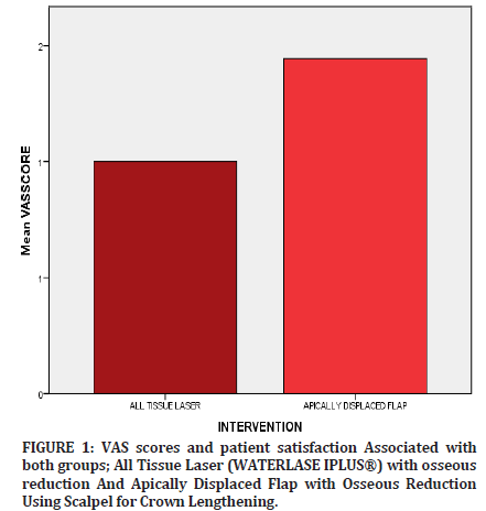
| All Tissue Laser | 1.00±0.739 | >0.05 (NS) |
| (Waterlase Iplus®) | ||
| Apically Displaced Flap | 1.44±0.527 | >0.05 (NS) |
Table 1: Vas Scores Done Using Independent Sample T Test (P<0.05).

Figure 1: VAS scores and patient satisfaction Associated with both groups; All Tissue Laser (WATERLASE IPLUS®) with osseous reduction And Apically Displaced Flap with Osseous Reduction Using Scalpel for Crown Lengthening.
| Mean | P value | |
|---|---|---|
| Between groups (ALL TISSUE LASER (WATERLASE IPLUS®) and APICALLY DISPLACED FLAP | 2.099 | >0.05 |
| ANOVA was used to compare both groups to assess which groups had better healing, p value was seen to be statistically insignificant p>0.05 | ||
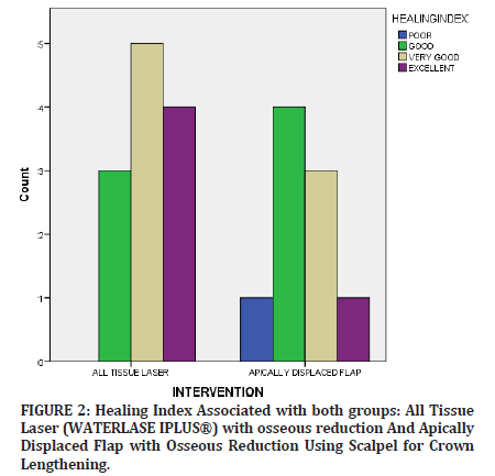
Table 2: Comparing both crown lengthening groups, All Tissue Laser (WATERLASE IPLUS®) with osseous reduction And Apically Displaced Flap with Osseous Reduction Using Scalpel for Crown Lengthening to assess healing scores.

Figure 2: Healing Index Associated with both groups: All Tissue Laser (WATERLASE IPLUS®) with osseous reduction And Apically Displaced Flap with Osseous Reduction Using Scalpel for Crown Lengthening.
Clinical Relevance
Crown lengthening procedures are common procedures in the dental practice to place restorations forfunction and esthetics. Lasers are widely used for various procedures to aid in better healing, minimal postoperative pain and now with flapless osseous contouring using all tissue erbium lasers [12]. With this study the authors have tried to determine which gives better outcomes with respect to healing and patient satisfaction crown lengthening procedures with osseous reduction using an all tissue laser (WATERLASE IPLUS®) or a scalpel with apically displaced flap [13].
Results
Independent sample t test values depict no statistical difference between the two groups: crown lengthening done using all tissue laser (WATERLASE IPLUS®) and apically displaced flap (NS) non-significant (p>0.05).
Bar graphs represent association of vas scores between both intervention groups; all tissue laser and apically displaced flap. X axis represents intervention groups and y axis represents vas scores. There was no statistically significant difference in the VAS scores (p<0.05)].
Bar graphs represent association of healing index between both intervention groups; all tissue laser and apically displaced flap. The X axis represents intervention groups and the Y axis represents the healing index. There was no statistically significant difference in the VAS scores (p>0.05)].
Discussion
Functional crown lengthening enables the clinician to deliver a prosthesis that does not violate the biological width and encompasses the adjustment of soft tissue as well as underlying bone in most cases [14].
The biologic width is imperative for periodontal health and the tooth preparation for restorations given in the anterior esthetic zone are usually placed subgingivally. (Ingber, Rose, and Coslet 1977) The 1mm needed from the base of the junctional epithelium to the tip of the alveolar bone is what prevents inflammation, resorption or the initiation of periodontitis. It has been proven that 3 mm (1 mm supracrestal connective tissue attachment, 1 mm junctional epithelium and 1 mm sulcus depth: adequate width even when margins are placed 0.5mm subgingivally) between the preparation and crest of alveolar bone maintains the health of the periodontium for 4- 6 months. If this width is violated, it leads to detrimental effects on the periodontium including recession or bone resorption [15-17].
The emergence of lasers in the field of periodontics and esthetics has been widely noted, the ease of use, patient acceptance, minimal pain and bleeding is an asset to the dental practice. The use of all tissue lasers can now pave the field for more applications in dentistry including hard tissue contouring, corticotomy and resection, applications in implantology and flapless procedures [18].
Aesthetic crown lengthening usually done for short clinical crowns, improves the gingival contour to achieve symmetry, and may include modifying tissue hyperplasia in some cases. Both indications can be performed using conventional gingivectomy or apically displaced flap techniques using a scalpel or to provide precision, bloodless field of surgery, predictable outcomes with minimal postoperative complications and no sutures, lasers are now preferred amongst dental practitioners [19].
The energy produced by laser onto a target tissue can have the following interactions:
Reflection
Transmission
Scattering and
Absorption.
When this energy is absorbed by the tissue, temperature within it is elevated and produces photochemical effects depending on the amount of water present within the target. (Rawlani 2017)When a temperature of 100°C is reached, vaporization of the water occurs in a process called ablation. At a temperature below 100°C and above 60°C, proteins are denatured without vaporizing the underlying tissue surface, alternatively, above temperatures of 200°C, tissue is dehydrated and burnt, resulting in an undesirable effect known as carbonization [20,21].
The absorption of this wavelength of light is done by absorbers called chromophores that have an affinity for specific wavelengths of energy. The primary chromophores present in dental hard tissues are water and hydroxyapatite and in soft tissue are hemoglobin and melanin. Different wavelengths of laser have different absorption coefficients corresponding to the primary tissue components, the all tissue laser works using both modes, a hard tissue and soft tissue mode and works according to the selection of the particular type of target tissue [22].
The all tissue laser (WATERLASE iPLUS) by BIOLASE is the only laser that provides an Er,CR:YSGG component, encompassing an operating voltage of 100 VAC +/- 10%, 50/60 Hz frequency, and a current rating of 5A/8A, 2780nm medium, 0.5-5 mm interaction zone from the target; this laser functions in contact (soft tissue) and non-contact (hard tissue/implants) mode, uses distilled water for irrigation that prevents necrosis of the bone during osseous recontouring and flushing during its application in cavity preparation (this laser is also found to make tooth enamel more resistant to carious lesions(Tudose et al. 2020) by increasing the tolerance against acids that demineralize enamel), and availability of multiple tips for these various applications on hard and soft tissue makes this laser system very versatile to use. The temperatures measured are higher for the Er,Cr:YSGG laser and are managed by the internal irrigation [23].
The Er,Cr:YSGG laser has shown commendable results in soft tissue management in pediatric dentistry too, with applications in not only crown lengthening but in exposure of unerupted teeth, labial and lingual frenectomy, excision of oral lesions and gingivectomy procedures
The All tissue laser shows promising results in the treatment protocol for peri implantitis. In a study with a 6 year follow up, bone levels were maintained in sites with peri implantitis treated by Er:YAG laser and DBBM. As the Erbium laser does not damage the titanium surface of the implant it is possible to debride granulation tissue and modify the surface of the implant, decorticate surrounding bone etc without disturbing the implant surface it is now possible to predict outcomes of such implants [24].
When compared, both procedures; laser and conventional crown lengthening, resulted in satisfactory removal of soft and hard tissue, it must be noted that, although precise and efficient, resection of had tissue by laser was time consuming compared to using a surgical hand piece, whose main drawback was in the instrument used that lacked internal irrigation and required external irrigant (cold saline) to prevent tissue necrosis. Internal irrigation is provided in the laser console that works efficiently and prevents necrosis. The use of the laser provided precision compared to the surgical hand piece. Patients exposed to the all-tissue laser had minimal to no bleeding, coupled with an inbuilt illumination on the hand piece aided in better visualization, whereas the scalpel caused bleeding and low visibility.
After 7 days the patients were analyses in groups, suture removal and irrigation of the patients in the conventional scalpel group was done. Landry's healing index is based on five criterias: redness or inflammation, presence of granulation tissue, bleeding, suppuration, and epithelialization. The score is ranged 1-5 with score 5 depicting excellent healing and score 1 denoted as very poor. The patients treated with crown lengthening using the all tissue laser demonstrated slightly better results compared to the patients in the conventional scalpel group. In a study by (Lingamaneni, Mandadi, and Pathakota 2019) this healing index was used to indicate gingival wound healing using Low Level Laser [25].
Therapy after Gingivectomy and Gingivoplasty Operations
The teeth in the esthetic zone i.e. maxillary and mandibular anterior and premolars were sites taken into consideration, as the authors wanted to assess the healing without keratinized tissue as a confounding factor, subjects with adequate width of attached gingiva were involved in the study to assess patient satisfaction and esthetic outcomes. It was observed that the crown lengthening done by the all tissue laser demonstrated better healing and preoperative compliance to the treatment. Patients in the conventional scalpel group demonstrated fear and hesitance to the treatment when the procedure was explained.
A VAS score also employed to assess discomfort and pain perception amongst the laser and scalpel group. The Visual Analogue Scale (VAS) is a 10 cm line drawn with end points defining extreme limits of pain and discomfort the patient is asked to rate their pain on a scale of 0-10, the distance between 0 and the mark defines the patient’s pain or level of discomfort. Patients in the laser group demonstrated lower VAS scores at 7 days compared to the scalpel group, this could be due to deposition of protein coagulum that seals the sensory nerves on application that also result in reduced inflammation in the laser group. Patient satisfaction was generally found to be better with the laser than the scalpel group.
Future research can include longer follow-up periods recording interdental alveolar bone height and formation, a larger sample size and subjects with varying width of attached gingiva and biotypes to further analyse healing outcomes [26].
Conclusion
It can be concluded that using an all tissue laser (WATERLASE IPLUS®) assisted crown lengthening with osseous reduction showed better healing and comparatively improved patient VAS scores, satisfaction and compliably to the apically displaced flap using a conventional scalpel.
Conflict of Interest
The authors declare no conflict of interest financial or otherwise.References
- AlMoharib HS, Steffensen B, Zoukhri D, et al. Efficacy of an Er: YAG laser in the decontamination of dental implant surfaces: An in vitro study. J Periodontol 2021; 92:1613-21.
- Mohanty R, Lenka B, Nayak R, et al. A New Pioneering Advancement: Waterlase. Indian J Forensic Med 2020; 14:8237.
- Capodiferro S, Tempesta A, Limongelli L, et al. Minimally invasive (flapless) crown lengthening by erbium: YAG laser in aesthetic zone. F1000Res 2020; 9.
- Chaudhary Z, Verma M, Tandon S. Treatment of oral submucous fibrosis with ErCr: YSGG laser. Indian J Dent Res 2011; 22:472.
- Chaudhary Z, Mohanty S, Sharma P, et al. Role of ErCr: YSGG Laser and PRF in Verrucous Carcinoma. J Maxillofac Surg 2018; 76:e66-7.
- Farista S, Kalakonda B, Koppolu P, et al. Comparing laser and scalpel for soft tissue crown lengthening: A clinical study. Glob J Health Sci 2016; 8:55795.
- Gargiulo AW, Wentz FM, Orban B. Dimensions and relations of the dentogingival junction in humans. J Periodontol 1961; 32:261-7.
- Ingber JS. The" biologic width"--a concept in periodontics and restorative dentistry. Alpha Omegan 1977; 70:62-5.
- Jacobson N, Starr C. Flapless implant placement with crown lengthening procedure to correct crown height: A case report. Implant Dent 2009; 18:387-92.
- Kumar G, Dhillon JK, Rehman F. A comparative evaluation of retention of pit and fissure sealants placed with conventional acid etching and Er, Cr: YSGG laser etching: A randomised controlled trial. Laser Ther 2016; 25:291-8.
- Kumar G, Rehman F, Chaturvedy V. Soft tissue applications of Er, Cr: YSGG laser in pediatric dentistry. Int J Clin Pediatr Dent 2017; 10:188.
- Landry GJ, Silverman DA, Liem TK, et al. Predictors of healing and functional outcome following transmetatarsal amputations. Arch Surg 2011; 146:1005-9.
- Lingamaneni S, Mandadi LR, Pathakota KR. Assessment of healing following low-level laser irradiation after gingivectomy operations using a novel soft tissue healing index: A randomized, double-blind, split-mouth clinical pilot study. J Indian Soc Periodontol 2019; 23:53.
- Matulic N, Bago I, Sušic M, et al. Comparison of Er: YAG and Er, Cr: YSGG Laser in the treatment of oral leukoplakia lesions refractory to the local retinoid therapy. Photobiomodul Photomed Laser Surg 2019; 37:362-8.
- Crichton N. Visual analogue scale (VAS). J Clin Nurs 2001; 10:706-6.
- Nugala B, Kumar BS, Sahitya S, et al. Biologic width and its importance in periodontal and restorative dentistry. J Conserv Dent 2012; 15:12.
- Ong M, Tseng SC, Wang HL. Crown lengthening revisited. Clin adv periodontics 2011; 1:233-9.
- Onur SG. Evaluation of pain perception and wound healing after laser-assisted frenectomy in pediatric patients: a retrospective comparative study. Photobiomodul Photomed Laser Surg 2021; 39:204-10.
- Verma SK, Maheshwari S, Singh RK, Chaudhari PK. Laser in dentistry: An innovative tool in modern dental practice. J Maxillofac Surg 2012; 3:124.
- Rubelman PA. The Apically Positioned Flap. Quintessence Publishing (IL); 1981.
- McGuire MK, Todd Scheyer E. Laser-assisted flapless crown lengthening: a case series. Int J Periodontics Restorative Dent 2011; 31:357.
- Tianmitrapap P, Srisuwantha R, Laosrisin N. Flapless Er, Cr: YSGG laser versus traditional flap in crown lengthening procedure. J Dent Sci 2022; 17:89-95.
- Tudose DA, Biclesanu C. Optimizing the treatment of dental caries with laser Er Cr: YSGG 2,790 nm (WaterLase Biolase USA). Rom J Stomatol 2020; 66:188.
- Weiner GP. Laser dentistry practice management. Dent Clin 2004; 48:1105-26.
- General Assembly of the World Medical Association. World Medical Association Declaration of Helsinki: ethical principles for medical research involving human subjects. J Am Coll Dent 2014; 81:14-8.
- Yamamoto A, Kubota T, Komatsu Y, et al. Efficacy of Erbium: YAG Laser for Regenerative Surgical Treatment of Peri-implantitis: Clinical, Microbiological, and Biomarker Analyses. Int J Periodontics Restorative Dent 2021; 41.
Indexed at, Google Scholar, Cross Ref
Indexed at, Google Scholar, Cross Ref
Indexed at, Google Scholar, Cross Ref
Indexed at, Google Scholar, Cross Ref
Indexed at, Google Scholar, Cross Ref
Indexed at, Google Scholar, Cross Ref
Indexed at, Google Scholar, Cross Ref
Indexed at, Google Scholar, Cross Ref
Indexed at, Google Scholar, Cross Ref
Indexed at, Google Scholar, Cross Ref
Indexed at, Google Scholar, Cross Ref
Indexed at, Google Scholar, Cross Ref
Indexed at, Google Scholar, Cross Ref
Indexed at, Google Scholar, Cross Ref
Indexed at, Google Scholar, Cross Ref
Indexed at, Google Scholar, Cross Ref
Indexed at, Google Scholar, Cross Ref
Indexed at, Google Scholar, Cross Ref
Indexed at, Google Scholar, Cross Ref
Indexed at, Google Scholar, Cross Ref
Indexed at, Google Scholar, Cross Ref
Author Info
Iram Rafique Pawane* and Sankari Malaiappan
Department of Periodontics, Saveetha Institute of Medical and Technical Sciences (SIMATS) Saveetha University, Chennai, IndiaCitation: Iram Rafique Pawane, Sankari Malaiappan, Comparison of Flap Techniques for Crown Lengthening Procedures Using an All Tissue Laser (Waterlase Iplus®) and Conventional Surgical Flaps: A Cross Sectional Study, J Res Med Dent Sci, 2022, 10(11):288
Received: 11-Oct-2022, Manuscript No. jrmds-23-94642; Accepted: 13-Oct-2022, Pre QC No. jrmds-23-94642; Editor assigned: 13-Oct-2022, Pre QC No. jrmds-23-94642; Reviewed: 27-Oct-2022, QC No. jrmds-23-94642; Revised: 31-Oct-2022, Manuscript No. jrmds-23-94642; Published: 07-Nov-2022
