Research - (2021) Volume 9, Issue 10
Comparison of Prevalence of Leukoplakia in Males and Females among Chennai Population
Akash N and Lakshminarayanan Arivarasu*
*Correspondence: Lakshminarayanan Arivarasu, Department of Pharmacology, Saveetha Dental College & Hospitals, Saveetha Institute of Medical and Technical Sciences, Saveetha University, India, Email:
Abstract
Aim: The aim of the study is to assess the prevalence of leukoplakia in males and females among the Chennai population. Introduction: Leukoplakia can be defined as a Thickened, white patch inside the mouth. Most leukoplakia patches are non-cancerous, but some show unique signs of cancer. They look differently in different areas of the mouth. Oral leukoplakia malignancy significantly differs with different tobacco habits. The most common cause of oral leukoplakia in makes is cigarettes and chewing tobacco. The most common cause of oral leukoplakia in women is betel leaves with calcium hydroxide and chewing tobacco. Methodology: This a descriptive study of prevalence of leukoplakia in males and females among the Chennai population. The collection of data of Saveetha dental college and hospitals called DIAS has been taken for the research study. Analysing the data, ratio is obtained, and SPSS software has been used to plot the graphs and the final data has been interpreted. Results and discussion: The total number of male and female patients has been obtained from the DIAS, they are then looked into the general examination to check the prevalence of oral leukoplakia. The data of patients with oral leukoplakia are segregated. Conclusion: From the study we can conclude that, the prevalence of leukoplakia in Chennai population is found to be 7.84%, and the prevalence is more among the males, which is about 5.68%.
Keywords
Leukoplakia, Tobacco smoking, Lesion, Oral mucositis
Introduction
Leukoplakia can be defined as a Thickened, white patch inside the mouth [1]. Most leukoplakia patches are noncancerous, but some show unique signs of cancer. They look differently in different areas of the mouth [1-6]. They form thickened, white patches on the gums, feathery or brizzle appearance on the insides of the cheeks, spots, or thickness over the bottom of the mouth and, sometimes hypertrophy of the tongue along with whitel lesions over papilla [1,2,7]. There ll be a diminished taste and sensation of heat and cold. These patches can't be scraped off, which is the significant feature differentiating them from oral submucous fibrosis. Leukoplakia is the most common premalignant or potentially malignant lesion of the oral mucosa [8-10]. It seems preferable to use the term leukoplakia as a clinical term only. When a biopsy is taken, the term leukoplakia should be replaced by the diagnosis obtained histologically [11]. There are two types of biopsies taken for oral leukoplakia, they are oral brush biopsy and excisional biopsy. Oral brush biopsy involves removing cells from the surface of the lesion with a small, spinning brush. This is a non-invasive procedure but does not always result in a definitive diagnosis and Excisional biopsy [12-14]. This involves surgically removing tissue from the leukoplakia patch or removing the entire patch if it's small. An excision biopsy is more comprehensive and usually results in a definitive diagnosis. Study says that the prevalence of oral leukoplakia is about 1.5 to 4.3% all around the world [15]. And there is 10.9 to 27.3% malignant transformation, which include oral cancers. The most involved type of cancer involved here is squamous cell carcinoma. The annual percentage of malignant transformation varies in different parts of the world, probably because of differences in tobacco and dietary habits [16,17]. Although epithelial dysplasia is an important predictive factor of malignant transformation, it should be realized that not all dysplastic lesions will become malignant. On the other hand non-dysplastic lesions may become malignant as well [18-20]. The tongue and the floor of the mouth can be high-risk sites about malignant transformation of leukoplakia [21]. Epithelial dysplasia is the malignant lesion, but not that serious like oral cancer, this is because of lack of metastasis of the lesion. It is found that the most important cause of the oral leukoplakia is the usage of tobacco [22,23]. Study says that the prolonged usage of tobacco has a significant increase in the malignancy of the oral leukoplakia lesion. It is found that the incidence of oral leukoplakia is maximum with the people more than 10 pack years [24]. And the increase in pack years increases the malignancy too. Untreated oral leukoplakia has a 83% chance of causing oral cancer, Our research team has worked on the nanoparticles over cancers [25-30].
Oral leukoplakia malignancy significantly differs with different tobacco habits. The most common cause of oral leukoplakia in makes is cigarettes and chewing tobacco. The most common cause of oral leukoplakia in women is betel leaves with calcium hydroxide and chewing tobacco [2,31]. The least malignant usage is the filtered cigarettes followed by bedi, cigars, betel leaves with calcium hydroxide and the most malignant leukoplakia is caused in chewing tobacco. The usage of chewing tobacco for a minimum of 3 years, has a 76% incidence of oral leukoplakia [8]. The consumption of alcohol doubles the risk of leukoplakia. The cessation of tobacco habits has been shown to be an effective measure regarding the incidence of leukoplakia and, thereby, the incidence of oral cancer as well [32,33]. Tobacco Cessation Counselling is usually given for these patients. Nicotine tablets are advised in case they prefer for replacements. Relaxation techniques are taught to the patients, they play a major role in tobacco cessation. Our team has extensive knowledge and research experience that has translate into high quality publications [34-53].
The oral leukoplakia is a severe oral lesion caused by various tobacco usage and the risk doubles in case of alcohol consumption. The prevalence of the oral leukoplakia differs in various places, this study compares the prevalence of oral leukoplakia in males and females in chennai population.
Materials and Methods
This is a descriptive study of prevalence of oral leukoplakia in males and females in the Chennai population. The collection of data of Saveetha dental college and hospitals called DIAS has been taken for the research study. The DIAS stands for Dental Information Archiving System and has a collection of data of all the patients who have undergone diagnosis or treatment in Saveetha Dental College and Hospitals. They have a completed case history of about 39.9 million. The full history of patients starts from PID 170604508 to PID 210510467, this gives us a count of 21,905,959 patients, which is the total number of patients visited. The males are segregated, and the count is 12,705,462, the female count is 9,200,497. The DIAS gives us the number of patients with oral leukoplakia. The obtained data has been analysed to get the ratio of number of patients to number of patients with oral leukoplakia. SPSS software has been used to plot the graphs and the final data has been interpreted.
Results and Discussion
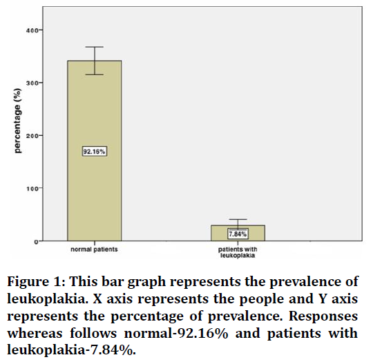
Oral leukoplakia is a malignant lesion seen in the oral cavity, they prolong to cause oral carcinoma. The total number of male and female patients has been obtained from the DIAS, they are then looked into the general examination to check the prevalence of oral leukoplakia. The data of patients with oral leukoplakia are segregated. Those cases have been examined with a preference of chief complaints followed by secondary complaints. As Figure 1 represents the prevalence of oral leukoplakia. From the graph, we find the ratio of the prevalence of patients with oral leukoplakia is found to be 7.84%, which is about 1717427 patients who have oral leukoplakia. The most common cause of the oral leukoplakia over Chennai were smoking and drinking, rarely chewing tobacco. The combination of smoking and drinking is the most common cause, this is obtained from the history of the patient.

Figure 1. This bar graph represents the prevalence of leukoplakia. X axis represents the people and Y axis represents the percentage of prevalence. Responses whereas follows normal-92.16% and patients with leukoplakia-7.84%.
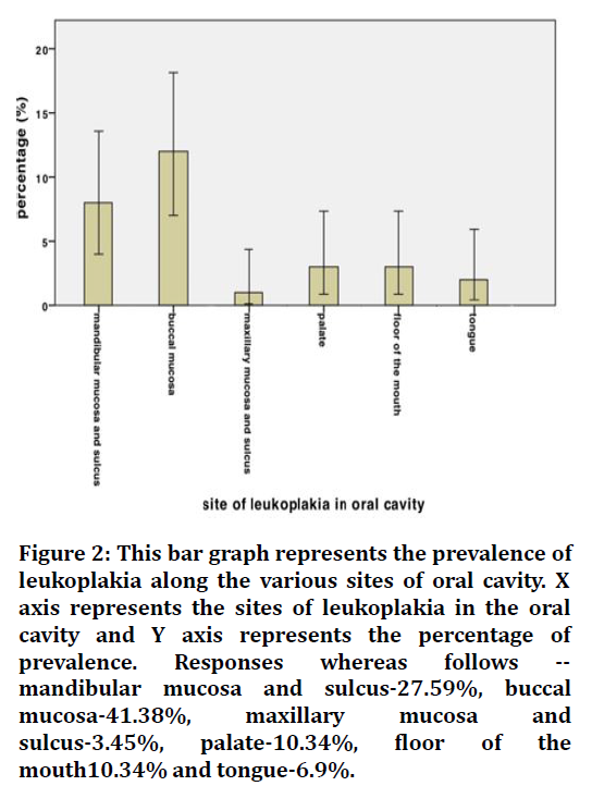
Figure 2 presents the prevalence of leukoplakia along the sites of oral leukoplakia. The oral leukoplakia are the ones which are seen all around the oral cavity. The leukoplakia lesions differ along the sites of the oral cavity, they appear as spots in tongue, buccal mucosa and floor of the mouth [1]. They appear as small white patches on the sulcus of both maxilla and mandible and floor of the mouth. They appear as fibrous or hairy structures along the buccal mucosa. These lesions are unscrappable, this differentiates them from oral submucous fibrosis. From the graph we can see that the leukoplakia is mostly seen along buccal mucosa which is of 41.38% followed by mandibular sulcus which is of 27.59%. The buccal mucosa is mostly involved because of smoking and chewing tobacco. The buccal mucosa of the cheeks is responsible for the suction of nicotine over the cigarettes. The mandibular sulcus is mostly involved because people keep the chewable tobacco over the sulcus for a long period.

Figure 2. This bar graph represents the prevalence of leukoplakia along the various sites of oral cavity. X axis represents the sites of leukoplakia in the oral cavity and Y axis represents the percentage of prevalence. Responses whereas follows -- mandibular mucosa and sulcus-27.59%, buccal mucosa-41.38%, maxillary mucosa and sulcus-3.45%, palate-10.34%, floor of the mouth10.34% and tongue-6.9%.
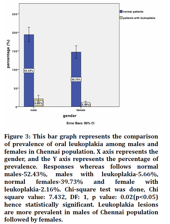
Figure 3 represents the comparison of prevalence of leukoplakia lesions among the males and females in Chennai population.

Figure 3. This bar graph represents the comparison of prevalence of oral leukoplakia among males and females in Chennai population. X axis represents the gender, and the Y axis represents the percentage of prevalence. Responses whereas follows normal males-52.43%, males with leukoplakia-5.66%, normal females-39.73% and female with leukoplakia-2.16%. Chi-square test was done, Chi square value: 7.432, DF: 1, p value: 0.02(p<0.05) hence statistically significant. Leukoplakia lesions are more prevalent in males of Chennai population followed by females.
From the graph we can see that the prevalence of leukoplakia is more prevalent in males, which is about 5.68%. From the data, it is observed that oral leukoplakia is seen in 9,754,986 males. The prevalence of leukoplakia among females of Chennai is about 2.16% which is about 3,709,643 females.
The most common cause among the males are seen to be smoking followed by the combination of smoking and drinking whereas the most common cause among the females are observed to be chewing tobacco and betel leaves with calcium hydroxide.
Biopsies are taken for these patients and the diagnosis are obtained and medication is provided for the patients.
These people are presented to the tobacco cessation council, and they advise the people to quit the habit, in extreme cases nicotine replacement is given. Patient was followed up to obtain the results of Tobacco cessation counselling.
The inference of the study is the oral leukoplakia lesion is mostly seen in males of Chennai population, because of the various tobacco habits like smoking and chewing tobacco. The most involved site for these oral leukoplakia is found to be buccal mucosa. The prevalence of the oral leukoplakia in Chennai population is found to be 7.84%. The limitation of this study is the limited demographic data. The increase in demographic data would give a prominent result.
Conclusion
From the study we can conclude that, the prevalence of leukoplakia in Chennai population is found to be 7.84%, and the prevalence is more among the males, which is about 5.68%. The most involved site for the leukoplakic lesion among Chennai population is found to be buccal mucosa and mandibular sulcus.
Acknowledgement
The authors would like to acknowledge the help and support rendered by the Department of Pharmacology, Saveetha Dental College and Hospitals, Saveetha Institute of Medical and Technical Sciences, Saveetha University, Chennai.
Funding
The present study is funded by Sri Vijay Furniture, Puducherry.
Conflict of Interest
The authors declare no potential conflict of interest.
References
- Ramos-GarcÃa P, González-Moles MÃ, Mello FW, et al. Malignant transformation of oral proliferative verrucous leukoplakia: A systematic review and meta-analysis. Oral Diseas 2021.
- Rubert A, Bagán L, Bagán JV. Oral leukoplakia, a clinical-histopathological study in 412 patients. J Clin Exp Dent 2020; 12:e540.
- Jaisankar AI, Arivarasu L. Free radical scavenging and anti-inflammatory activity of chlorogenic acid mediated silver nanoparticle. J Pharma Res Int 2020; 106-12.
- Karthik V, Arivarasu L, Rajeshkumar S. Hyaluronic acid mediated zinc nanoparticles against oral pathogens and its cytotoxic potential. J Pharm Res Int 2020; 113-7.
- Shankar SB, Arivarasu L, Rajeshkumar S. Biosynthesis of hydroxy citric acid mediated zinc nanoparticles and its antioxidant and cytotoxic activity. J Pharm Res Int 2020; 108-12.
- Shree MK, Arivarasu L, Rajeshkumar S. Cytotoxicity and antimicrobial activity of chromium picolinate mediated zinc oxide nanoparticle. J Pharma Res Int 2020; 28-32.
- Das D, Maitra A, Panda CK, et al. Genes and pathways monotonically dysregulated during progression from normal through leukoplakia to gingivo-buccal oral cancer. NPJ Genomic Med 2021; 6:1-9.
- Liu W, Yao Y, Shi L, et al. A novel lncRNA LOLA1 may predict malignant progression and promote migration, invasion, and EMT of oral leukoplakia via the AKT/GSK-3Ã? pathway. J Cell Biochem 2021.
- Wu S, Rajeshkumar S, Madasamy M, et al. Green synthesis of copper nanoparticles using Cissus vitiginea and its antioxidant and antibacterial activity against urinary tract infection pathogens. Artificial Cells Nanomed Biotechnol 2020; 48:1153-1158.
- Varshini A, Arivarasu L. Herbal sources used by the public against infections. Int J Pharma Res 2020; 84-92.
- https://www.euro.who.int/__data/assets/pdf_file/0009/429939/Tobacco-Mental-Health-Policy-Brief.pdf
- Pogoda K, Ciesluk M, Deptula P, et al. Inhomogeneity of stiffness and density of the extracellular matrix within the leukoplakia of human oral mucosa as potential physicochemical factors leading to carcinogenesis. Translational Oncol 2021; 14:101105.
- Abijeth B, Ezhilarasan D. Syringic acid induces apoptosis in human oral squamous carcinoma cells through mitochondrial pathway. J Oral Maxillofac Pathol 2020; 24:40.
- Sohaib M, Ezhilarasan D. Carbamazepine, a histone deacetylase inhibitor induces apoptosis in human colon adenocarcinoma cell line HT-29. J Gastrointestinal Cancer 2020; 51:564-70.
- Fawzy MM, Nofal A, El-Hawary EE. Proliferative verrucous leukoplakia. Indian J Dermatol Venereol Leprol 1997; 87:455.
- Bánóczy J. The therapy of oral leukoplakia. Oral Leukoplakia 1982; 182â??189.
- Shathviha PC, Ezhilarasan D, Rajeshkumar S, et al. Ã?-sitosterol mediated silver nanoparticles induce cytotoxicity in human colon cancer HT-29 cells. Avicenna J Med Biotechnol 2021; 13:42.
- Bánóczy J. Oral â??White lesionsâ? other than leukoplakia. Oral Leukoplakia 1982; 146â??181.
- Kaumheimer GJ. Kerosene as a remedy and its variability. J Am Med Assoc 1989; 132.
- Solai Prakash AK, Devaraj E. Cytotoxic potentials of S. cumini methanolic seed kernel extract in human hepatoma HepG2 cells. Environ Toxicol 2019; 34:1313-1319.
- Bánóczy J. Results of clinical follow-up studies in oral leukoplakia. Oral Leukoplakia 1982; 15-27.
- Ezhilarasan D, Apoorva VS, Ashok Vardhan N. Syzygium cumini extract induced reactive oxygen species-mediated apoptosis in human oral squamous carcinoma cells. J Oral Pathol Med 2019; 48:115-21.
- Ghazali N, Bakri MM, Zain RB. Aggressive, multifocal oral verrucous leukoplakia: proliferative verrucous leukoplakia or not?. J Oral Pathol Med 2003; 32:383-92.
- Akash N, Arivarasu L, Rajeshkumar S. Anti-inflammatory and antioxidant potential of hyaluronic acid mediated zinc nanoparticles. J Pharma Res Int 2020; :33-37.
- Demirbas A. Global renewable energy projections. Energy Sources 2009; 4:212-24.
- Karthik V, Arivarasu L. Viruses and their treatment-A review. J Complementary Med Res 2020; 11:121-126.
- Stephen B. Herbal formulation: Review of efficacy, safety, and regulations. Int J Res Pharm Sci 2008; 23:1506â??1510.
- Suleria RH. Plant-based functional foods and phytochemicals: From traditional knowledge to present innovation.
- Shue YJ, Chen PC, Wang MC, et al. Tyrosinase inhibitory effect and antioxidant activity of formosan phyla nodiflora for cosmetic use. Planta Med 2008; 74:PD7.
- Cunningham JA, Leatherdale ST, Chaiton M, et al. Offering nicotine patches to all households in a high community with smoking rates: Pilot test of a population-based approach to promote tobacco cessation. Int J Population Data Sci 2021; 6.
- Liau M, Cheong BE, Teoh PL. Antioxidant and anticancer properties of solvent partitioned extracts of Phyla nodiflora L. J Microbiol Biotechnol Food Sci 2021; 2021:42-6.
- Rajeshkumar S, Kumar SV, Ramaiah A, et al. Biosynthesis of zinc oxide nanoparticles using Mangifera indica leaves and evaluation of their antioxidant and cytotoxic properties in lung cancer (A549) cells. Enzyme Microbial Technol 2018; 117:91-95.
- . Nandhini NT, Rajeshkumar S, Mythili S. The possible mechanism of eco-friendly synthesized nanoparticles on hazardous dyes degradation. Biocatalysis Agricultural Biotechnol 2019; 19:101138
- Rajkumar PV, Prakasam A, Rajeshkumar S, et al. Green synthesis of silver nanoparticles using Gymnema sylvestre leaf extract and evaluation of its antibacterial activity. South African J Chem Eng 2020; 32:1-4.
- Rajasekaran S, Damodharan D, Gopal K, et al. Collective influence of 1-decanol addition, injection pressure and EGR on diesel engine characteristics fueled with diesel/LDPE oil blends. Fuel 2020; 277:118166.
- Vairavel M, Devaraj E, Shanmugam R. An eco-friendly synthesis of Enterococcus sp.â??mediated gold nanoparticle induces cytotoxicity in human colorectal cancer cells. Environ Sci Pollution Res 2020; 27:8166-75.
- Santhoshkumar J, Sowmya B, Kumar SV, et al. Toxicology evaluation and antidermatophytic activity of silver nanoparticles synthesized using leaf extract of Passiflora caerulea. South African J Chem Eng 2019; 29:17-23.
- Raj R K. Ã?-sitosterol-assisted silver nanoparticles activates Nrf2 and triggers mitochondrial apoptosis via oxidative stress in human hepatocellular cancer cell line. J Biomed Materials Res 2020; 108:1899-908.
- Saravanan M, Arokiyaraj S, Lakshmi T, et al. Synthesis of silver nanoparticles from Phenerochaete chrysosporium (MTCC-787) and their antibacterial activity against human pathogenic bacteria. Microb Pathogene 2018; 117:68-72.
- Gheena S, Ezhilarasan D. Syringic acid triggers reactive oxygen speciesâ??mediated cytotoxicity in HepG2 cells. Human Exp Toxicol 2019; 38:694-702.
- Ezhilarasan D, Sokal E, Najimi M. Hepatic fibrosis: It is time to go with hepatic stellate cell-specific therapeutic targets. Hepatobiliary Pancreatic Diseas Int 2018; 17:192-197.
- Ezhilarasan D. Oxidative stress is bane in chronic liver diseases: Clinical and experimental perspective. Arab J Gastroenterol 2018; 19:56-64.
- Dua K, Wadhwa R, Singhvi G, et al. The potential of siRNA based drug delivery in respiratory disorders: Recent advances and progress. Drug Develop Res 2019; 80:714-730.
- Gomathi AC, Rajarathinam SX, Sadiq AM, et al. Anticancer activity of silver nanoparticles synthesized using aqueous fruit shell extract of Tamarindus indica on MCF-7 human breast cancer cell line. J Drug Delivery Sci Technol 2020; 55:101376.
- Ramesh A, Varghese S, Jayakumar ND, et al. Comparative estimation of sulfiredoxin levels between chronic periodontitis and healthy patientsâ??A case-control study. J Periodontol 2018; 89:1241-1248.
- Duraisamy R, Krishnan CS, Ramasubramanian H, et al. Compatibility of nonoriginal abutments with implants: Evaluation of microgap at the implantâ??abutment interface, with original and nonoriginal abutments. Implant Dent 2019; 28:289-95.
- Ezhilarasan D, Apoorva VS, Ashok Vardhan N. Syzygium cumini extract induced reactive oxygen species-mediated apoptosis in human oral squamous carcinoma cells. J Oral Pathol Med 2019; 48:115-121.
- Arumugam P, George R, Jayaseelan VP. Aberrations of m6A regulators are associated with tumorigenesis and metastasis in head and neck squamous cell carcinoma. Arch Oral Biol 2021; 122:105030.
- Joseph B, Prasanth CS. Is photodynamic therapy a viable antiviral weapon against COVID-19 in dentistry? Oral Surg Oral Med Oral Pathol Oral Radiol 2021..
- Gnanavel V, Roopan SM, Rajeshkumar S. Aquaculture: An overview of chemical ecology of seaweeds (food species) in natural products. Aquaculture 2019; 507:1-6.
- Markov A, Thangavelu L, Aravindhan S, et al. Mesenchymal stem/stromal cells as a valuable source for the treatment of immune-mediated disorders. Stem Cell Res Therapy 2021; 12:1-30.
- Mathew B, Rohisha IK, Kumar H. Video assisted awareness programme on ill effects of tobacco and pan-chewing among migrant labourers. Indian J Applied Res 2020; 1â??4.
- Apollonio DE, Malone RE. Marketing to the marginalised: Tobacco industry targeting of the homeless and mentally ill. Tobacco Control 2005; 14:409-415.
Author Info
Akash N and Lakshminarayanan Arivarasu*
Department of Pharmacology, Saveetha Dental College & Hospitals, Saveetha Institute of Medical and Technical Sciences, Saveetha University, Chennai, IndiaCitation: Akash N, Lakshminarayanan Arivarasu,Comparison of Prevalence of Leukoplakia in Males and Females among Chennai Population , J Res Med Dent Sci, 2021, 9(10): 87-91
Received: 09-Sep-2021 Accepted: 29-Sep-2021
