Research - (2020) Volume 8, Issue 4
Comparison of Shaping Ability of XP-Endo Shaper in Simulated J-Shaped Canals with Various Sizes
Ayfer Atav Ates*, Burçin Arıcan and Vasfiye Işık
*Correspondence: Ayfer Atav Ates, Department of Endodontics, Istanbul Okan University, Turkey, Email:
Abstract
Objective: To evaluate the shaping efficiency of the XP-endo Shaper® (XPS; FKG Dentaire, La Chaux-de-Fonds, Switzerland) in simulated canals with various sizes.
Materials and Methods: Sixty-four simulated J shaped root canals, which had various apical diameters and 2% taper were used. Apical diameters of group 1, group 2, group 3, and group 4 were 0.15 mm, 0.30 mm, 0.40 mm, 0.50 mm, respectively. Simulated resin canals were prepared with the XP-endo Shaper at 37°C inside a cabinet. The transportation value (TV), centering ability (CA), and the amount of resin removed (RR) were calculated using the superimposition of initial and final images. These parameters were calculated with the AutoCAD software program (Autodesk, San Rafael, CA) based on 18 reference points with 1-mm intervals of the canal. The data were analyzed with Kruskal-Wallis and then Dunn’s multiple comparison tests. The significance level was set at p ≤ 0.05.
Results: Shaping activity of XPS continued in group 4, but the amount of resin removed was statistically less than group 1. Group 2 and group 3 removed a statistically higher resin amount than group 1 and group 4 (p>0.05). Within groups, transportation value statistically increased in the middle region (points 4-6). The centering ratio of the groups was different at all points, being highest at point 8 and point 9.
Conclusion: Under the limitations of this study, XPS respected the root canal anatomy and adapted to the canal walls, even in large simulated canals.
Keywords
Large Canals, MaxWire, Resin blocks, Root canal preparation, XP-endo shaper
Introduction
Disinfection of the root canal system is one of the main objectives of root canal treatment. To achieve this purpose, root canals must be shaped and enlarged enough to eliminate microorganisms, infected dentine, and pulp remnants [1]. Besides all these expected features, the instrumentation system should minimize the untouched canal wall areas with increasing three-dimensional adaptation, while respecting the main shape of the canal without removing dentine excessively [2,3]. Because of this approach, the big sized of NiTi instruments have been used for the preparation of teeth with resorption, trauma, and open apices which may have large root canals and apical diameters. These instruments have increased taper, less flexibility and less centering ability [4]. To eliminate these disadvantages, new NiTi single-file systems are being developed to handle these tough cases.
XP-endo Shaper® (XPS; FKG Dentaire, La Chaux-de-Fonds, Switzerland), which is made with MaxWire alloy (Martensite-Austenite Electropolishing-Flex, FKG Dentaire) has been launched with its superior facilities. It has a tip size of ISO #30 with .01 taper in the martensitic phase at room temperature. Thanks to its unique technology, the file transforms from martensite to austenite phase when exposed to body temperature and expands at least 30/.04 [5]. According to the manufacturer claims, MaxWire alloy gives the instrument perfect flexibility. Due to its small taper and core diameter, XPS can easily adapt to the three-dimensional anatomy of the root canal [6].
According to our knowledge, there is not any study that investigated the shaping ability of this novel instrument in anatomically challenging canals with various apical diameters since it was produced. For this purpose, this study aims to evaluate and compare the shaping ability of XPS in canals with various sizes and to observe the maximum limits.
Materials and Methods
Endo-training resin blocks (Endo Training Blocks, Dentsply Maillefer) with different apical diameters were designed and produced for the present study. All canals were J shaped and had 2% taper, 10-mm radius of curvature, 70º angle of curvature, and 16 mm working length. The apical diameters of groups were designed as follows; 0.15 mm, 0.30 mm, 0.40 mm, 0.50 mm for group 1-4, respectively. Each group consisted of 18 samples.
All samples were irrigated with 5 ml distilled water and then were stained using black ink (Winsor & Newton, Colart Tianjin Art Materials, Tianjin, China). Then, a photo camera (Canon EOS 700 D, Canon Incorporated, Tokyo, Japan) adapted to standard setup was used to take initial images. These images were transferred to Adobe software program (Adobe Systems, Inc., San Jose, CA) to check if the black ink could reach all through the root canals. To stabilize the resin blocks, a metal holder was used during the instrumentation procedures. Next, the negotiation of the canals was controlled with #10 K file, and the glide path was created with iRace (FKG Dentaire, La Chaux-de-Fonds, Switzerland) 10/0.04. The root canals were prepared with the XP-endo Shaper using a torque-controlled endodontic motor (X Smart Plus, Dentsply Maillefer, Ballaigues, Switzerland) at 800 rpm and 1N.cm with in and out motions at an amplitude of 3-4 mm up to working length (WL). Then, the file was removed from the canal and cleaned with sterile gauze. Once WL was reached, the root canals were irrigated, and XPS was worked for another 15 strokes with in and out gentle movements [7]. All procedures were performed at 37°C inside a cabinet for all experimental groups. Each file was used in only one simulated canal. During the instrumentation, the canals were irrigated with a total of 20 ml distilled water. After final irrigation, the artificial canals were stained using red ink (Winsor & Newton). Final images were taken using the same photo camera setup.
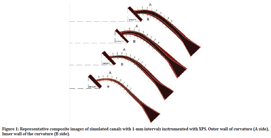
The initial and final images were superimposed using Adobe software program. Measurement scale was also prepared on superimposed images, and the levels of resin removed were calculated at 18 points from two aspects of the canal [9 measurements from the outer wall of curvature (A side) and 9 measurements from the inner wall of curvature (B side)] by using AutoCAD Software (Autodesk, San Rafael, CA). The points of measurement were determined in 1-mm intervals. Apical (point 1, 2, 3), middle (point 4, 5, 6) and coronal (point 7, 8, 9) sections were evaluated (Figure 1). Canal transportation and centering ratio were calculated using the following criteria [8]:

Figure 1. Representative composite images of simulated canals with 1-mm intervals instrumented with XPS. Outer wall of curvature (A side), Inner wall of the curvature (B side).
The amount of resin removal (RR): The value was obtained by combining the widths of resin removal from the 2 aspects of the canal (A+B).
Transportation value (TV): The absolute value of the difference between the widths of resin removal from the two aspects of the canal |A-B|
Centering ability: Calculated by dividing the narrower width of resin removal by the wider one from the 2 aspects of the canal (A/B) or (B/A).
The data were analyzed with Kruskal-Wallis and then Dunn’s multiple comparison tests. The significance level was set at p ≤ 0.05.
Results
The amount of resin removal
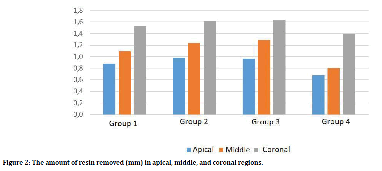
The total amount of resin removed by XPS was presented in Table 1 and Figure 2. Among the groups, there was no statistically significant difference between group 2 and 3 in all tested regions (p>0.05). Also, resin removal was higher in group 2 and 3 than group 1 and 4 in all regions. XPS removed a statistically significantly higher amount of resin from all canal regions in group 1 compared to group 4 (p<0.05). Resin removal efficiency decreased but continued at apical size #50. The total amount of resin removed increased from the apical part to the coronal part within all 4 groups.

Figure 2. The amount of resin removed (mm) in apical, middle, and coronal regions.
| 1-3 mm* Mean ± SD | 4-6 mm* Mean ± SD | 7-9 mm* Mean ± SD | p value | |
|---|---|---|---|---|
| Group 1 | 0.878 ± 0.074 | 1.092 ± 0.073 | 1.526 ± 0.095 | 0 |
| Group 2 | 0.982 ± 0.078 | 1.236 ± 0.098 | 1.616 ± 0.101 | 0 |
| Group 3 | 0.964 ± 0.68 | 1.291 ± 0.226 | 1.633 ± 0.120 | 0 |
| Group 4 | 0.683 ± 0.102 | 0.798 ± 0.094 | 1.389 ± 0.101 | 0 |
| p value | 0 | 0 | 0 |
*Different capital letters in the same column represent statistically significant difference between groups.
Table 1: The mean ± standard deviation values of the total amount of resin removed in apical (1-3 mm), middle (4-6 mm) and coronal (7-9 mm) regions.
Transportation
The amount of transportation was shown in Table 2. There was no statistically significant difference between groups 1, 2, and 3 in terms of TV (middle region, points 4-6). However, transportation decreased in group 4 in the same region (points 4-6). TV increased in the curved section within the groups. XPS created less transportation in group 4 than the other groups in all regions.
| 1-3 mm Mean ± SD | 4-6 mm* Mean ± SD | 7-9 mm* Mean ± SD | |
|---|---|---|---|
| Group 1 | 0.125 ± 0.011 | 0.194 ± 0.027 | 0.074 ± 0.016 |
| Group 2 | 0.172 ± 0.029 | 0.177 ± 0.030 | 0.049 ± 0.017 |
| Group 3 | 0.145 ± 0.013 | 0.186 ± 0.039 | 0,071 ± 0.029 |
| Group 4 | 0.081 ± 0.017 | 0.091 ± 0.033 | 0.045 ± 0.025 |
| p value | 0 | 0 | 0 |
*Different capital letters in the same column represent statistically significant difference between groups.
Table 2: The mean and standard deviation (SD) values of transportation in the apical, middle, and coronal region.
Centering ability
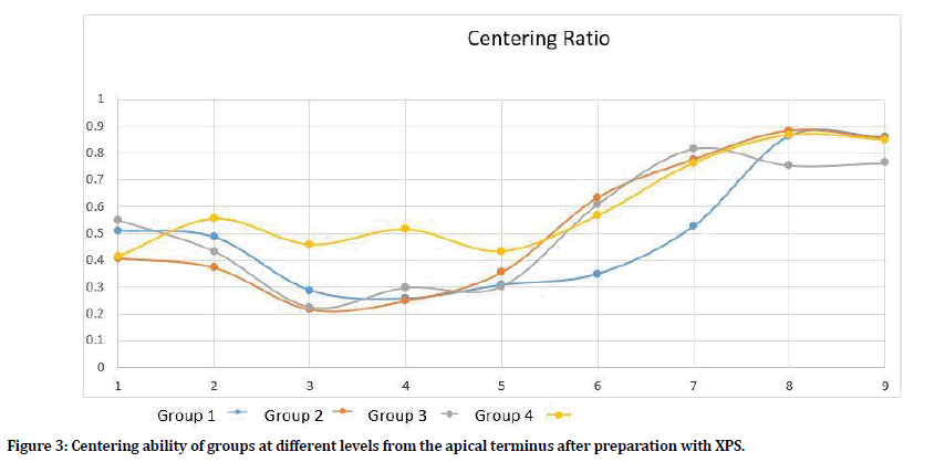
CA was evaluated between groups and reported in Figure 3. The difference between the groups was found significant in all the reference points (p<0.05). There was no statistically significant difference between groups 2, 3, and 4 at points 6, 7 (p>0.05). Centering ratio was statistically similar across group 2 and group 3 at 2, 3, 5, 6, and 7 mm from the apex (p>0.05). The centering ratio had the highest values at points 8 and 9 for all groups.

Figure 3. Centering ability of groups at different levels from the apical terminus after preparation with XPS.
Discussion
The present study compared the shaping ability of XPS in simulated J-shaped canals with various apical diameters. The XPS, a single file system, is the only instrument on the market which is produced with MaxWire alloy. According to the manufacturer, the instrument has a unique mechanism that can adapt to the root canal morphology three-dimensionally. The adaptation is facilitated by the expansion of MaxWire alloy at body temperature [5]. However, no study gives information about the maximum expansion of the file. Azim et al. [8] mentioned that the final preparation taper of the XPS will vary from one case to the other, depending on the original canal anatomy. Also, they found out that it was impossible to predict the final enlargement of the canal operated with XPS because of its adaptive core technology. These findings are also in accordance with Arıcan et al. [9]. In the present study, we evaluated the centering ability and transportation values of XPS in simulated canals with various apical sizes ranging from 0.15 mm to 0.50 mm. By this way, it was aimed to examine the relationship between the adaptation ability of the instrument while increasing apical diameter. According to our results, XPS continued removing resin from root canal walls even in apical diameter #0.50. However, RR decreased gradually from group 1 to group 4. De-Deus et al. [10] observed that activation time of XPS affects the amount of removed dentin in their study. Thus, further studies are required to evaluate the efficiency of XPS in 0.50 mm and larger apical diameter canals with extending the preparation time.
To assess the shaping ability of the instruments, natural extracted teeth and resin blocks have been widely used [11]. Resin blocks have several advantages, namely, reproducibility of results, direct visual comparison, standardization of root canal diameter, length, and curvature in terms of angle and radius between groups [12,13]. Weine et al. [14] recognized that it was not possible to have standardization with extracted teeth while comparing the shaping abilities of files because of their variable anatomy and dentin thickness. Extracted human teeth can simulate clinical situations better than resin blocks. Although blocks do not mimic dentin microhardness and do not provide three-dimensional information, it is possible to determine the whole canal rather than the specified measurement levels, like in CBCT studies [15,16]. Thus, researchers should be careful while assessing the results in block studies [17,18]. Besides its disadvantages, several studies have shown the usability of blocks in endodontics [19,20]. According to our knowledge, blocks with large canal diameter have not been produced and not studied before in endodontic field. In this study, blocks were used to maintain balance and have a standardization between groups. The three-dimensional drawings of these blocks were made by an industrial engineer.
It is well known that one of the most important factors which affect the expansion and flexibility of the instrument is temperature. MaxWire alloy enables the XPS instrument to transform from the martensitic phase to a predetermined austenitic shape at body temperature [8,21]. Therefore, all shaping procedures were done at body temperature in the present study. Arican et al. [9] used XPS in extracted human teeth with large apical diameter at body temperature and showed that it could expand more than #30/.04. Also, it was reiterated by Tabbara et al. [22], who notified that the file had maximum expansion up to 0.08 taper at 34° C. We obtained the data by measuring the size of the root canal per millimeter. When taking into consideration that XPS could continue its shaping ability even in 50/0.02 root canals, we may presume that XPS could expand more than the minimum expansion limit, which was determined by the manufacturer. However, the expansion amount was different between the groups. The mean increase in width of instrumented root canals was higher at group 1 than group 4. However, it was similar in group 2 and group 3. These results demonstrated that the shaping ability of XPS still continued but highly reduced in group 4.
Wu et al. [23] reported that if the canal transportation were less than 0.3 mm, the prognosis of endodontically treated teeth would not be affected negatively. Reham et al. [24] evaluated the shaping ability of XPS in extracted teeth and concluded that the transportation values were less than 0.3 mm in all experimental groups. Their results were also in accordance with Poly et al. [15]. In the present study, transportation was seen in all groups but changed between regions. Although it was shown that low microhardness of resin blocks caused more transportation values than extracted teeth [25], we obtained less canal transportation when compared with Poly et al. [15] and Reham et al. [24]. The possible explanation for these results could be the increased canal enlargement that helped the file to be positioned centrally and caused less transportation. Within groups, the highest transportation value was observed in the curved section and minimal transportation was seen in the coronal region. The obtained results were in accordance with the Silva et al. which compared the different instrument systems in J shaped canals [26].
One of the most important steps in resin block studies is the irrigation protocol. It is overly critical to use continuous irrigation to prevent resin deposition during the shaping procedures of the simulated canals. NaOCl and EDTA irrigants, which are routinely used in endodontic clinics were not preferred for use in the present study. Distilled water was preferred to be used during irrigation because these solutions do not create the same effects in blocks like dentin [27].
Conclusion
Under the limitations of this study, XPS had acceptable transportation and centering values in large and J shaped canals. Although the manufacturer claims that XPS could expand at least # 30/.04 in root canals, in the present study, it was shown that the instrument could remove resin even in apical diameter # 50/.02. From this point of view, it can be suggested that XPS can be used efficiently and safely in root canals, which have more than the apical diameter of # 30. To obtain more specific results, further studies carried on extracted teeth and comparison with new generation instruments will be needed.
Author contributions
Conceptualization: Atav Ateş A, Data curation: Atav Ateş A, Arıcan B, Formal analysis: Atav Ateş A, Arıcan B, Funding acquisition: Atav Ateş A, Arıcan B, Investigation: Atav Ateş A, Arıcan B, Methodology: Atav Ateş A, Arıcan B, Işık V, Project administration: Atav Ateş A, Arıcan B, Resources: Atav Ateş A, Arıcan B, Software: Atav Ateş A, Supervision: Atav Ateş A, Validation: Atav Ateş A, Visualization: Atav Ateş A, Arıcan B Writing - original draft: Atav Ateş A, Arıcan B, Writing - review & editing: Atav Ateş A, Arıcan B, Işık V.
Conflict of Interest
No potential conflict of interest relevant to this article was reported.
Acknowledgments
The authors thank to Assist. Prof Semih Yalçındağ for statistical analysis.
The authors thank to Selen Bayraktar for teaching AutoCAD Software program.
References
- Schilder H. Cleaning and shaping the root canal. Dent Clin North Am 1974; 18:269-296.
- Kishen A. Mechanisms and risk factors for fracture predilection in endodontically treated teeth. Endod Topics 2006; 13:57-83.
- Siqueira JF, Lopes H. Treatment of endodontic infections: Quintessence London 2011.
- ElAyouti A, Dima E, Judenhofer MS, et al. Increased apical enlargement contributes to excessive dentin removal in curved root canals: A stepwise microcomputed tomography study. J Endod 2011; 37:1580-1584.
- https://www.fkg.ch/index.php
- Elnaghy A, Elsaka S. Torsional resistance of XP-endo Shaper at body temperature compared with several nickel titanium rotary instruments. Int Endod J 2018; 51:572-576.
- Lacerda MF, Marceliano-Alves MF, Pérez AR, et al. Cleaning and shaping oval canals with 3 Instrumentation systems: A correlative micro–computed tomographic and histologic study. J Endod 2017; 43:1878-1884.
- Azim AA, Piasecki L, da Silva Neto UX, et al. A novel adaptive core rotary instrument: Micro–computed tomographic analysis of its shaping abilities. J Endod 2017; 43:1532-1538.
- Arican Öztürk B, Atav Ateş A, Fişekçioğlu E. cone-beam computed tomographic analysis of shaping ability of XP-endo shaper and ProTaper next in large root canals. J Endod in press 2020; 46:437-443.
- De-Deus G, Belladonna FG, Simões-Carvalho M, et al. Shaping efficiency as a function of time of a new heat-treated instrument. Int Endod J 2019; 52:337-342.
- Hasheminia SM, Farhad A, Sheikhi M, et al. Cone-beam computed tomographic analysis of canal transportation and centering ability of single-file systems. J Endod 2018; 44:1788-1791.
- Hiran‐us S, Pimkhaokham S, Sawasdichai J, et al. Shaping ability of ProTaper NEXT, ProTaper Universal and iRace files in simulated S‐shaped canals. Aust Endod J 2016; 42:32-36.
- Lim YG, Park SJ, Kim HC, et al. Comparison of the centering ability of WaveOne and Reciproc nickel-titanium instruments in simulated curved canals. Restor Dent Endod 2013; 38:2-5.
- Weine FS, Kelly RF, Lio PJ. The effect of preparation procedures on original canal shape and on apical foramen shape. J Endod 1975; 1:255-262.
- Poly A, AlMalki F, Marques F, et al. Canal transportation and centering ration after preparation in severly curved canals: Analysis by micro-computed tomography and double-digital radiography. Clin Oral Invest 2019; 23:4255-4262.
- Moe MMK, Ha JH, Jin MU, et al. Root canal shaping effect of ınstruments with offset mass of rotation in the mandibular first molar: A micro–computed tomographic study. J Endod 2018; 44:822-827
- Bonaccorso A, Cantatore G, Condorelli GG, et al. Shaping ability of four nickeltitanium rotary instruments in S-shaped canals. J Endod 2009; 35:883-886.
- Burroughs JR, Bergeron BE, Roberts MD, et al. Shaping ability of three nickeltitanium file system in simulated S-shaped root canals. J Endod 2012; 38:1618-1621.
- Ceyhanli K, Kamaci A, Taner M, et al. Shaping ability of two M‑wire and two traditional nickel‑titanium instrumentation systems in S‑shaped resin canals. Niger J Clin Pract 2015; 18:713-717.
- Wu H, Peng C, Bai Y, et al. Shaping ability of ProTaper Universal, WaveOne and ProTaper Next in simulated L-shaped and S-shaped root canals. BMC Oral Health 2015; 15:27.
- de Vasconcelos RA, Murphy S, Carvalho CAT, et al. Evidence for reduced fatigue resistance of contemporary rotary instruments exposed to body temperature. J Endod 2016; 42:782-787.
- Tabbara A, Grigorescu D, Yassin MA, et al. Evaluation of apical dimension, canla taper an maintenance of root canal morphology using XP-endo shaper. J Comtemp Dent Pract 2019; 20:136-144.
- Wu MK, Fan B, Wesselink PR. Leakage along apical root fillings in curved root canals. Part I: effects of apical transportation. J Endod 2000; 26:210-216.
- Reham H, Roshdy N, Issa N. Comparison of canal transportation and centering ability of XP Shaper, WaveOne and one shape: A cone beam computed tomograhpy study of curved root canals. Acta Odontol Latinoam 2018; 31:67-74.
- Khalilak Z, Fallahdoost A, Dadresanfar B, et al. Comparison of extracted teeth and simulated resin blocks on apical canal transportation. Iran Endod J 2008; 3:109-112.
- Silva EJNL, Tameirão MDN, Belladonna FG, et al. Quantitative transportation assessment in simulated curved canals prepared with an adaptive movement system. J Endod 2015; 41:1125-1129.
- Christofzik D, Bartols A, Faheem MK, et al. Shaping ability of four root canal instrumentation systems in simulated 3D-printed root canal models. PloS One 2018; 13:1-14.
Author Info
Ayfer Atav Ates*, Burçin Arıcan and Vasfiye Işık
Department of Endodontics, Istanbul Okan University, TurkeyCitation: Ayfer Atav Ates, Burçin Ar?can, Vasfiye I??k, Comparison of SHAPING Ability of XP-Endo Shaper in Simulated J-Shaped Canals with Various Sizes, J Res Med Dent Sci, 2020, 8 (4):176-181.
Received: 26-Jun-2020 Accepted: 24-Jul-2020
