Review - (2021) Volume 9, Issue 9
Cone-beam Computed TomographyâA New Vision in Endodontics-A Review
Radhika Gupta*, Aditya Patel, Pradnya Nikhade and Manoj Chandak
*Correspondence: Radhika Gupta, Department of Conservative Dentistry and Endodontics, Sharad Pawar Dental College and Hospital, Datta Meghe Institute of Medical Sciences, Deemed to be University (DU) Sawangi, India, Email:
Abstract
The discovery of X-Rays was done by Roentgen in 1895, since then the dental radiography has extensively played an appealing and analytical diagnostic role in dentistry. The management of endodontic complications is truly dependent on dental radiographs to evaluate the anatomy of the respected tooth and its surrounding structures. The key to success in the management of endodontic complications intensely depends upon the diagnostic imaging procedures to deliver the analytical information related to the teeth under consideration and their surrounding anatomy. Though, many of these images may have certain limitations and drawbacks. Recent advancements in the field of radiography have been introduced such as computerized tomography scans, magnetic resonance imaging, ultrasonography, and several imaging techniques that have been transformed diagnosis in the medical and dental field. Primary computerized tomography scans which are used in medical diagnosis are called Medical Computerized Tomography whereas the newer technique used considerably in the field of dentistry is called Cone Beam Computed Tomography (CBCT). The usage of Cone-beam computed tomographic imaging for analysis and management of endodontic complications is rapidly being employed in the field of dentistry. It is useful to detect several endodontic conditions like vertical root fracture, root resorption, to obtain detailed knowledge about the anatomy of the root canal. The usage of cone-beam computed tomographic imaging technique is highly recommended in the situation of missed canals and calcified canals. It can be used in all stages of treatment from preoperative to postoperative assessment and follow-up. The CBCT imaging is considered in those conditions where no adequate data is obtained from conventional radiographic techniques.
Keywords
Cone-beam computed tomography, root resorption, root fracture, dental trauma.
Introduction
Radiographic investigation aids in endodontic diagnosis and treatment planning which is of utmost importance. Intraoral and panoramic radiographic evaluation has a certain drawback in that the three-dimensional anatomy is trampled in a two-dimensional image. This limitation can be overcome by using Cone-beam computed tomography imaging (CBCT) by producing three-dimensional images of teeth and surrounding areas [1,2]. Cone-beam computed tomographic imaging is a newer method of computerized tomography scan that creates three-dimensional data at a lower cost and less absorbed doses than the conventional computerized tomography scan. Cone-beam computed tomographic imaging systems do not require a huge area and can effortlessly fit into most dental clinics [3]. For diagnosis and treatment planning of multifaceted endodontic problems, cone-beam computed tomographic imaging has been proved to be effective equipment. This article aims to evaluate all the topographies of CBCT. It also shows the importance of newer technology which has been undertaken to advance the diagnosis and treatment planning to evaluate the results of endodontic treatment.
CBCT imaging
In the late 1990s, Cone-beam computed tomography, an extra-oral imaging apparatus was discovered to harvest three-dimensional imaging scans of the facial bones at a substantially lesser exposure of radiation as compared to computerized tomography. This uses an effortless, straight association between the radiation source and the sensor which revolves concurrently around the patient’s head. Based upon the cone-beam computed tomographic imaging scanner used, the sensor and the radiation origin revolve between 180 degrees and 360 degrees around the patient's head. The prime benefit of cone-beam computed tomographic imaging over computerized tomographic scanners is that there is a considerable reduction in the radiation dose. This is because of quick scanning and pulsed radiation source with advanced image receptor devices. The pulsed radiation source concludes up to 570 projections. Cone-beam computed tomographic imaging scanners are effortless to operate and it takes a lesser amount of space as a panoramic radiographic unit, which makes cone-beam computed tomographic imaging scans more suitable for the dental practice. The radiation exposure can be minimized by reducing the size of the field of view, increasing the voxel size, and reducing the number of projection pictures taken. The picture quality of the cone-beam computed tomographic imaging technique is considered to be superior to that of computed tomographic scans when used to assess the dental hard tissues. The scanning time for this machine is about 10-40 seconds long, this truly depends upon the type of scanners used and the selection of exposure parameters.
Cone-beam computed tomographic imaging comprises a single revolution of an X-ray through the dental object. The data which is obtained can be viewed in 3 conventional planes namely; the Axial plane, Sagittal plane, and Coronal plane. Image accomplishment is quick and due to its advanced technology, it is comparatively inexpensive and has lower radiation exposures as compare to computerized tomographic scans. The obtained data is adequate for the treatment planning and management of the involved teeth and their surrounding structures, which cannot be viewed in conventional, 2D, plain dental film imaging [4].
Cone-beam computed tomographic imaging comprises an X-ray source with the sensor which is fixed to the revolving gantry. Through the central area of concern, the radiation source with the sensor rotates along the secured fulcrum in the region of interest (ROI). In between the preparation of the exposure, there are hundreds of planar projection pictures which are attained in the field of view (FOV) within the range of a minimum of 180â?¦. CBCT in single revolution gives precise, immediate, and correct 3D radiographic images. Since the exposure of CBCT consist of the entire FOV, a single revolution gantry sequence is essential for acquiring adequate data for the reconstruction of the image [5-9].
Equipment for the CBCT
They are classified based on the positioning of the patient in the process of image procurement, scan volume, and clinical applications.
Patient positioning
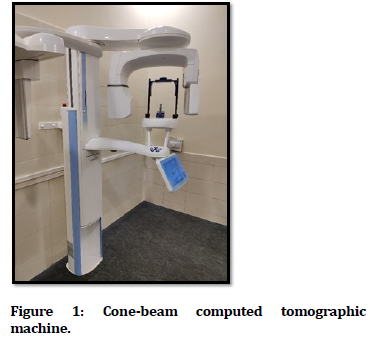
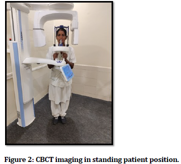
The standing units can be adjusted up to a certain height. This type of unit has been shown difficulty in accommodation for the patients on wheelchairs. The seated units are readily more comfortable compared to standing units, though this unit has certain limitations, its fixed seats don't allow complete scanning of the patient who is physically disabled or the patient who is bound to the wheelchair. Figure 1 shows the cone-beam computed tomographic machine. Figure 2 shows the standing position of the patient while imaging.
Figure 1. Cone-beam computed tomographic machine.
Figure 2. CBCT imaging in standing patient position.
Scan volume
The dimensions of the sensor, geometry of the projection beam, and capability of the beam collimation are considered as an important factor for the accurate dimensions of FOV or scan volume. FOV is available in spherical or cylinder shapes (e.g., New-Tom 3G).
Limitation of field size
Depending upon the appearance and the extension of the disease with the region selected to be imaged, ensures an optimal FOV selection for each patient. Depending on the existing height of scan volume, the use of units can be planned as mentioned-
The confined region is too known as focused, or the limited field is roughly 5 centimetres or less, one arch is 5 centimeters-7 centimetres, inter-arch is 7 centimetres-10 centimetres, whereas the maxilla-facial is 10 centimeters-15 centimetre’s, the craniofacial is greater than 15 centimetre’s. Generally, the lesser the scan volume, the more will be the spatial resolution for the picture. Since the initial sign for the peri-radicular pathology is the presence of discontinuity of the lamina dura and widening in the PDL space, the ideal resolution for any Cone-beam computed tomographic system used in cases of endodontics must not be more than 200 micrometres which is the average width of the PDL [10].
Multimodality
The hybrid multimodal systems associated with a digital panoramic view having a comparatively small range to medium FOV cone-beam computed tomographic system [10]. For an appreciation of the details of the root canal and the periodontal tissues, in endodontics, CBCT imaging needs remarkably high resolution and details. Due to high image resolution, patient radiation exposure is more often increased. The small FOV Cone-beam computed tomographic imaging scans have been suggested for the diagnosis and treatment of endodontic problems. To decrease the volume of exposed tissue the small FOV scans are used, and henceforth, the radiation dose is effective which also improves the image quality. Due to the subtle patient movement, the generated images can get easily degraded; thereby the most preferable machine to maintain the patient constancy is the area where the patients are used to sit, or even in a supine position than the standing position. The most important advantage of cone-beam computed tomographic imaging is that the dedicated cone-beam computed tomographic imaging units are usually considered for a seating or the supine position of the patient, whereas the hybrid panoramic or the CBCT units frequently have the patient in a standing position [4].
Radiation dosages and their reduction
The significant advantage of the CBCT imaging unit is that this equipment is well-adjustable during scanning, with a drawback of greater levels of radiation exposure risks when compared to routine radiographic imaging. The mean effective doses for a large field of view are 212 Sv, for the medium is 177 Sv, and for small is 84 Sv, measured by cone-beam computed tomography. The normal range of 5 – 146 Sv is considered for a small field of view. The normal range of exposure for panoramic radiographic imaging is between 16 and 20 Sv taken into account for comparison [4]. The effective method to decrease the radiation exposure dose can be obtained by the use of a smaller field of view, lesser projections (180 degrees), and using a larger voxel size. Radiation risks for the female population are significantly higher than that of the male population.
Endodontic applications of CBCT
Endodontic usage in CBCT consists of the diagnosis for the periapical pathologies because of inflammation of the pulp, localization of root canals of teeth, identification of internal resorption & external resorption, and root fractures. Current two-dimensional technology is film and digital-based. The study performed by Bender and Seltzer [11,12] concluded that intraoral radiography has certain drawbacks for the detection of periapical pathologies. This study reported that the cortical plate of the bone should be involved for a lesion to be evident radiographically.
Certain suggestions had been given which promises to overcome these drawbacks through cone-beam computed tomographic imaging. It is reviewed as follows:
Recently the review published by Nair et al. [13] on digital and three-dimensional applications of CBCT for endodontic uses summarized as this technology is beneficial for identification of canals of the teeth, management in periapical surgery, and identification of fractured roots in extracted teeth. In vitro study performed by Mora et al [14], used a high-resolution cone-beam tomographic imaging system which revealed the superiority of this newer technique over regular two-dimensional view. To differentiate solid from fluid-filled pathologies i.e., periapical cysts from periapical granulomas, cone-beam computed tomographic imaging was considered to be useful using grayscale values in the lesions, this clinical study was conducted by Simon and co-workers [15].
Rigolone et al. demonstrated the clinical implementation of Cone-beam computed tomographic imaging [16] in the case of apicoectomy surgical procedure including the palatal root of a maxillary first molar. The scientific study was carried on thirty-one patients, which demonstrated that Cone-beam computed tomographic imaging was found to be superior in recognizing a substitute and more conservative surgical approach using a vestibular approach in combination with an operating endodontic microscope.
Patel et al. [17], systematically reviewed the implementation of Cone-beam computed tomographic imaging in endodontics and determined that CBCT is clinically more superior as compared to a conventional periapical radiographic technique for the recognition of periapical lesions.
The important information from radiographic images in clinical endodontics are needed in two phases of the treatment:
a) Preoperative assessment and b) Postoperative assessment.
Preoperative Assessment
Assessment of tooth morphology and anatomy of the root canal
The accuracy of endodontic treatment is based upon the detection of entire root canals of teeth leading to proper assessment, proper biomechanical preparation, and obturation of the root canals [18]. The frequency for the 2nd mesiobuccal canal (MB2) in cases of the upper1st molar had been stated to differ from 69% to 93%. This inconsistency is seen in the buccolingual plane where the structural density changes can occur because of the superimposition of anatomic structures [19,20]. Ramamurthy et al. [21] stated that the estimation of various two-dimensional film modalities was hardly able in the detection of more than the presence of 50% MB2 canals. Since periapical radiographs are two-dimensional in nature, leading to the underestimation of the exact anatomical intricacy for the root canal system of teeth. The optical surface scans and CBCT data allow 3D print of guided sleeves with the stents. This resulted in guided access cavity preparation which is more conservative. The advantage of this guided access cavity preparation consists of conservative and less invasive access cavity preparation, reduced chairside period for patients, and reduced iatrogenic risks of injuries.
Periapical pathology of teeth
Teeth with pulpal and periapical inflammatory pathology of tissues are most commonly seen pathologic conditions. Estrela et al. [22] studied the sample size of eight hundred eighty-eight imaging examinations of patients having an endodontic infection (1,508 teeth) in the detection of apical periodontitis (AP) with the CBCT, panoramic and periapical radiographs. They concluded that the incidence of apical periodontitis is suggestively higher with CBCT imaging. In the study conducted by Low et al. [23] the preoperative evaluation of the apical condition of thirty-seven premolars and molars in the maxilla (156 total roots) which were suggestive for probable apical surgery were compared using periapical radiographs and CBCT imaging. They concluded that CBCT imaging demonstrates more lesions (34%) in comparison to conventional radiographic techniques. Patel et al. [24] in an ex vivo model that comprises of 2 mm diameter defects located in the cancellous bone at the apices of 10 first molar teeth on six partially dentate intact human dry mandibles, reported a detection rate for the intraoral radiograph and CBCT imaging as 24.8% and 100% respectively.
Root fracture
Root fractures are less common than crown fractures. They occur in only 7% or rarer of dental injuries [25,26] moreover root fractures are difficult to diagnose precisely using the conventional radiographic technique. Many authors suggested the worth and importance of CBCT in the diagnosis and management of dentoalveolar trauma, especially in cases of root fractures [27], luxation, displacement, and alveolar fractures [28]. Bernardes et al. [29] performed a retrospective study on conventional periapical radiographic and CBCT imaging for 20 patients with root fractures. They concluded that CBCT imaging showed significant fractures in 18 (90%) of patients as compared to conventional periapical radiographs. This suggests that the CBCT can be considered superior to a conventional radiographic technique for the analysis of root fractures.
Root resorption
Differentiation of Diagnosis of Internal Root resorption (IRR) and External root resorption (ERR) is commonly confused and often remains misdiagnosed especially with conventional radiographic techniques. External resorption usually presents with irregular radiolucency and intact root canals whereas the case of internal resorption presents with defined borders with no canal radiographically visible in the defect [30]. Cone-beam computed tomographic imaging has been used efficaciously to confirm the presence of IRR. It also helps to differentiate IRR from ERR. Periapical radiograph has certain drawbacks, as it results in misdiagnosis, inadequate assessment, and poor management of root resorption [4].
Assessment of dental trauma
Cone-beam computed tomography has also been shown to be useful in the diagnosis and management of dentoalveolar trauma. The exact nature and severity of alveolar and luxation injuries can be assessed from just one scan from which multiplanar views can be selected and assessed with no geometric distortion or anatomical noise. It has been reported that CBCT can be used to detect horizontal root fractures [4]. The same fracture may need multiple periapical radiographs taken at several different angles to detect it and even then, maybe difficult to visualize. As CBCT is an extra-oral technique it is also far more comfortable for the patient who has recently sustained dental trauma when compared to several intra-oral radiographs taken using a beam aiming device. Cohenca et al. used CBCT technology to aid their management of three patients who had sustained dental trauma. In addition to detecting the true nature of the injuries sustained by the tooth, the CBCT scans were able to detect cortical bone fractures, which were not diagnosed from the clinical or conventional radiographic examination [4].
Postoperative Assessment
Quality assessment of root canal treatment
Detection of voids in root canal fillings was carried out employing different radiographic imaging techniques which include intraoral analogic radiographic technique, intraoral digital radiographic technique, and cone-beam computed tomographic imaging. It resulted that the large voids (>300 mm) were noticed in all the imaging techniques whereas, for the smaller void detection, the digital intraoral radiographic technique showed better performance than intraoral analogic and cone-beam computed tomographic imaging [31]. An alternative study was conducted in which the cone-beam computed tomographic imaging was compared with micro-computed tomographic imaging for recognition of voids, it resulted that in various cases the number of voids in root canal fillings was seen in CBCT imaging. The exact working length estimation by using CBCT imaging varied between 0.41 and 0.51 millimeters if compared with the electronic apex locators which are considered as the ‘‘gold standard’’ [32-34]. All these studies emphasized that cone-beam computed tomographic imaging is more advantageous when used as a diagnostic tool for endodontic treatment planning.
In endodontic surgery
Often in cases of endodontic surgery, there may be complications in the posterior region of the jaw because of the proximity of posterior teeth to their relative anatomical structures. The mandibular teeth may lie close to the mandibular canal whereas maxillary molars are frequently close to the maxillary sinus.
The significance of Cone-beam computed tomographic imaging in endodontic surgery was described by Rigolone et al. [35]. They considered forty-three maxillary first molars on thirty-one patients; these patients were referred for retreatment. The mean distance of the palatine root from the external vestibular cortex was measured at 9.73 millimetres. They concluded that Cone-beam computed tomographic imaging may play a significant role in enhancing palatine root apicoectomy by direct vestibular access.
Limitations of CBCT
The factor which affects the image quality and diagnostic precision of cone-beam computed tomographic images is scattered and the presence of beam hardening due to high-density neighboring materials and structures [36-39]. Quality of image is predisposed by numerous technical factors like FOV, voxel size, device, tube voltage, number of projections, and current [40]. Broken endodontic instruments and filling materials of a root canal may lead to the formation of possible artifacts [41]. Patient age also contributes to the image quality of Cone-beam computed tomographic imaging. A possible interlink may be seen between the age factor and the number of subsequent artifacts. With increasing age, the identification of anatomic morphology, like nasal floor, mental foramen, and mandibular canal, appears to diminish. Metallic restorations like silver amalgam restorations, metal posts, stainless-steel crowns, implants, and even gutta-percha can cause possible radiographic artifacts, which is adequate to negotiate the details of root canal anatomy and pertinent pathologies such as root resorption and root fractures. To overcome these limitations Metal artifact reduction algorithms (MAR) are becoming more common in working and observing software to avoid these artifacts [41].
Conclusion
For diagnosis and management of endodontic treatment radiographic examination is considered to be the most appropriate method. Besides conventional radiographic techniques, CBCT imaging provides greater reliability and validity and in the recognition of periapical conditions. CBCT imaging provides accurate location and extent of periapical lesions. CBCT imaging has become the first choice for diagnosis and management of endodontic conditions, especially when a new digital scanner with fewer amounts of radiation doses and improved resolutions becomes available. The value of the cone-beam computed tomographic imaging system can no longer be doubtful—Cone-beam computed tomographic imaging is a valuable task-specific imaging modality and a significant technology in comprehensive endodontic assessment.
References
- Patel S, Dawood A, Ford TP, et al. The potential applications of cone-beam computed tomography in the management of endodontic problems. Int Endod J 2007; 10:818–30.
- Patel S. New dimensions in endodontic imaging: Part 2. Cone-beam computed tomography. Int Endod J 2009; 6:463–475.
- Ito K, Gomi Y, Sato S, et al. Clinical application of a new compact CT-system to assess 3-D images for the preoperative treatment planning of implants in the posterior mandible. A case report. Clin Oral Implants Res 2001; 12:539–542.
- Patel S, Durack C, Abella F, et al. Cone beam computed tomography in Endodontics–a review. Int Endodont J 2015; 48:3-15.
- Farman AG. Image guidance: The present future of dental care. Pract Procedures Aesthet Dent 2007; 18:342–344, 2006.
- Farman AG, Levato CM, Scarfe WC. A primer on cone-beam computed tomography. Inside Dent 2007; 3:90–92.
- Scarfe WC, Farman AG, Sukovic P. Clinical applications of cone-beam computed tomography in dental practice. J Canadian Dent Assoc 2006; 72:75–80.
- Scarfe WC, Farman AG. Cone-beam computed tomography: A paradigm shift for clinical dentistry. Australasian Dent Practice 2007; 102–110.
- Hayakawa Y, Sano T, Sukovic P, et al. Cone-beam computed tomography: a paradigm shift for clinical dentistry. Nippon Dent Rev 2005; 65:125– 132.
- Scarfe WC, Levin MD, Gane D, et al. Use of cone-beam computed tomography in endodontics. Int J Dent 2009; 2009.
- Bender B, Seltzer S. Roentgenographic and direct observation of experimental lesions in bone I. J Am Dent Assoc 1961; 62:152–160.
- Bender B, Seltzer S. Roentgenographic and direct observation of experimental lesions in bone II. J Am Dent Ass 1961; 62:708–16.
- Nair M, Nair U. Digital and advanced imaging in endodontics: A review. J Endod 2007; 33:1–6.
- Mora MA, Mol A, Tyndall DA, et al. In vitro assessment of local computed tomography for the detection of longitudinal tooth fractures. Oral Surg Oral Med Oral Pathol Oral Radiol Endod 2007; 103:825–9.
- Simon J, Reyes E, Malfaz J-M, et al. Differential diagnosis of large periapical lesions using cone-beam computed tomography measurements and biopsy. J Endod 2006; 32:833–7.
- Rigolone M, Pasqualini D, Bianchi L, et al. Vestibular surgical access to the palatine root of the superior first molar: ‘‘low-dose cone-beam’’ CT analysis of the pathway and its anatomic variations. J Endod 2003; 29:773–5.
- Patel S, Dawood A, Pitt Ford T, et al. The potential applications of cone-beam computed tomography in the management of endodontic problems. Int Endod J 2007; 40:818–30.
- Vertucci FJ. Root canal anatomy of the human permanent teeth. Oral Surg Oral Med Oral Pathol 1984; 58:589–599, 1984.
- Pineda F. Roentgenographic investigation of the mesiobuccal root of the maxillary first molar. Oral Surg Oral Med Oral Pathol 1973; 36:253–260.
- Nance R, Tyndall D, Levin LG, et al. Identification of root canals in molars by tuned-aperture computed tomography. Int Endodont J 2000; 33:392–396.
- Ramamurthy R, Scheetz JP, Clark SJ, et al. Effects of the imaging system and exposure on accurate detection of the second mesiobuccal canal in maxillary molar teeth. Oral Surg Oral Med Oral Pathol Oral Radiol Endodont 2006; 102:796–802.
- Estrela C, Bueno MR, Leles CR, et al. Accuracy of cone-beam computed tomography and panoramic and periapical radiography for detection of apical periodontitis. J Endodont 2008; 34:273–279.
- Low KMT, Dula K, Burgin W, et al. Comparison of periapical radiography and limited cone-beam tomography in posterior maxillary teeth referred for apical surgery. J Endodont 2008; 34:557–562.
- Patel S, Dawood A, Mannocci F, et al. Detection of periapical bone defects in human jaws using cone-beam computed tomography and intraoral radiography. Int Endodont J 2009; 42:507–515.
- Andreasen JO, Andreasen FM. Classification, etiology and epidemiology in textbook and color atlas of traumatic injuries to the teeth. 3rd Edn, Munksgaard, Copenhagen, Denmark 1994; 151–216.
- Cvek M, Tsilingaridis G, Andreasen JO. Survival of 534 incisors after intra-alveolar root fracture in patients aged 7–17 years. Dent Traumatol 2008; 24:379–387.
- Tyndall DA, Rathore S. Cone-beam CT diagnostic applications: caries, periodontal bone assessment, and endodontic applications. Dent Clin North Am 2008; 52:825–841.
- Cohenca N, Simon JH, Roges R, et al. Clinical indications for digital imaging in dento-alveolar trauma—part 1: traumatic injuries. Dent Traumatol 2007; 23:95–104.
- Bernardes RA, de Moraes IG, Hungaro Duarte MA, et al. Use of cone-beam volumetric tomography in the diagnosis of root fractures. Oral Surg Oral Med Oral Pathol Oral Radiol Endodont 2009; 108:270–277.
- Gulabivala K, Searson LJ. Clinical diagnosis of internal resorption: An exception to the rule. Int Endodont J 1995; 28:255–260.
- Huybrechts B, Bud M, Bergmans L, et al. Void detection in root fillings using intraoral analogue, intraoral digital, and cone-beam CT images. Int Endod J 2009; 8:675–85.
- Jeger FB, Janner SF, Bornstein MM, et al. Endodontic working length measurement with preexisting cone-beam computed tomography scanning: A prospective, controlled clinical study. J Endod 2012; 7:884–8.
- Connect T, H€ulber-J M, Godt A, et al. Accuracy of endodontic working length determination using cone-beam computed tomography. Int Endod J 2014; 47:698–703.
- Liang YH, Jiang L, Chen C, et al. The validity of cone-beam computed tomography in measuring root canal length using a gold standard. J Endod 2013; 12:1607–10.
- Rigolone M, Pasqualini D, Bianchi L, et al. Vestibular surgical access to the palatine root of the superior first molar: Low-dose cone-beam. CT analysis of the pathway and its anatomic variations. J Endodont 2003; 29:773–775.
- Lofthag-Hansen S, Thilander-Klang A, Gr€ondahl K. Evaluation of subjective image quality about the diagnostic task for cone-beam computed tomography with different fields of view. Eur J Radiol 2011; 2:483–8.
- Costa FF, Gaia BF, Umetsubo OS, et al. Detection of horizontal root fracture with small-volume cone-beam computed tomography in the presence and absence of intracanal metallic post. J Endod 2011; 10:1456–9.
- Bueno MR, Estrela C, De Figueiredo JA, et al. Map-reading strategy to diagnose root perforations near metallic intracanal posts by using cone-beam computed tomography. J Endod 2011; 1:85–90.
- Ritter L, Mischkowski RA, Neugebauer J, et al. The influence of body mass index, age, implants, and dental restorations on image quality of cone-beam computed tomography. Oral Surg Oral Med Oral Pathol Oral Radiol Endod 2009; 3:e108–16.
- Kamburoglu K, Murat S, Kolsuz E, et al. Comparative assessment of subjective image quality of cross-sectional cone-beam computed tomography scans. J Oral Sci 2011; 4:501–8.
- Eskandarloo A, Mirshekari A, Poorolajal J, et al. Comparison of cone-beam computed tomography with intraoral photostimulable phosphor imaging plate for diagnosis of endodontic complications: a simulation study. Oral Surg Oral Med Oral Pathol Oral Radiol
Author Info
Radhika Gupta*, Aditya Patel, Pradnya Nikhade and Manoj Chandak
Department of Conservative Dentistry and Endodontics, Sharad Pawar Dental College and Hospital, Datta Meghe Institute of Medical Sciences, Deemed to be University (DU) Sawangi, IndiaCitation: Radhika Gupta, Aditya Patel, Pradnya Nikhade, Manoj ChandakCone-beam Computed Tomography–A New Vision in Endodontics-A Review, J Res Med Dent Sci, 2021, 9(9): 54-59
Received: 08-Aug-2021 Accepted: 06-Sep-2021


