Research - (2021) Volume 9, Issue 9
Cosmetic Enhancement of Maxillary Central Incisors Using Rigid Matrix Technique
Joyeeta Mahapatra* and Pradnya Nikhade
*Correspondence: Joyeeta Mahapatra, Department of Conservative Dentistry and Endodontics, Sharad Pawar Dental College and Hospital, India, Email:
Abstract
Sustaining injury in the anterior tooth is a frequent finding in clinical dental practice. Such injuries are frequently seen to be associated with maxillary incisors due to their susceptible position in the upper jaw that makes them prone to fractures. Due to the changing trends, esthetic procedures are frequently sought after treatments in clinical practice. There is an increasing demand among patients regarding the replication of the actual tooth form both anatomically as well as esthetically. This reproduction of the finer details can be undertaken with the help of the "Matrix Technique" that aid in the accurate replication of the contacts and contours. The putty index technique is one such technique that employs a rigid index for the fabrication of esthetic composite restoration. The present case deals with a fracture in the anterior teeth due to traumatic occlusion. This was corrected using the "Rigid Index Technique".
Keywords
Matrix Technique, Rigid Index Technique, Putty Index Technique
Introduction
Sustaining injury in the anterior tooth is a frequent finding in clinical dental practice. Such injuries are frequently seen to be associated with maxillary incisors due to their susceptible position in the upper jaw that makes them prone to fractures. Various etiology of such fractures include road traffic accidents, sports mishaps, trauma from malocclusion due to anterior tooth crowding, etc. [1]. Due to the changing trends, esthetic procedures are frequently sought after treatments in clinical practice. There is an increasing demand among patients regarding the replication of the actual tooth form both anatomically as well as esthetically. This reproduction of the finer details can be undertaken with the help of the "Matrix Technique" that aid in the accurate replication of the contacts and contours [2]. Different types of matrices are available for restoration in the esthetic zone. It is classified into two types- flexible and rigid. The putty index technique is one such technique that employs a rigid index for the fabrication of esthetic composite restoration. The present case deals with a fracture in the anterior teeth due to traumatic occlusion. This was corrected using the "Rigid Index Technique".
Case Report
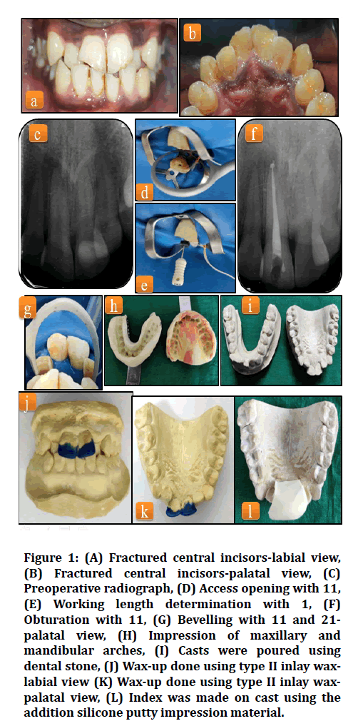
A 23years old male patient came to us with the chief complaint of broken teeth with mild pain in the upper front tooth region for 1 year. The patient was apparently alright 1 year back. Then he noticed broken teeth in the upper front tooth region. It was associated with mild pain which aggravated on intake of cold beverages and was relieved on the removal of stimuli. There is no history of night pain, swelling, or pus discharge. The patient then reported to us for the needful. The past medical and dental history was non-contributory. On clinical examination, Ellis Class II fracture was seen with 21 11 (Figures 1A and 1B). Tenderness on percussion was negative for both teeth. The provisional diagnosis was given as reversible pulpitis with 21 11. Radiograph showing incisal fracture with 21 which involved enamel and dentin and did not involve the pulp (Figure 1C).

Figure 1: (A) Fractured central incisors-labial view, (B) Fractured central incisors-palatal view, (C) Preoperative radiograph, (D) Access opening with 11, (E) Working length determination with 1, (F) Obturation with 11, (G) Bevelling with 11 and 21- palatal view, (H) Impression of maxillary and mandibular arches, (I) Casts were poured using dental stone, (J) Wax-up done using type II inlay waxlabial view (K) Wax-up done using type II inlay waxpalatal view, (L) Index was made on cast using the addition silicone putty impression material.
On radiographic examination, intraoral periapical radiograph showed incisal fracture with 11 which involved enamel, dentin, and pulp in the coronal portion. In the apical portion, PDL widening was seen associated with 11 with ill-defined periapical radiolucency (Figure 1C). On neural sensibility testing, a positive response was seen with heat test using gutta-percha with 21. No response was seen with 11. The final diagnosis was given as chronic irreversible pulpitis with 11 with apical periodontitis and reversible pulpitis with 21.
In the first visit, Rubber dam isolation was done. Difficulties were faced during rubber dam placement due to the presence of anterior crowding in both maxillary and mandibular arches. Access opening was done using round bur (BR- 45, Mani, Japan) and safe end bur (EX 24, Mani, Japan), and pulp extirpation were carried out (Figure 1D). Working length was determined using a radiograph and electronic apex locator(J Morita Root Zx Mini, Tokyo, Japan)which came out to be 23.5mm with 11 (Figure 1E).
Canals were irrigated with 3% sodium hypochlorite (Parcan, Septodont, India) and 0.9% saline for the removal of remaining pulp tissue and debris. Then calcium hydroxide dressing((Prime RC Cal, India)) was placed within the canal and a temporary pack(T-fill, Belgium) was placed. The patient was recalled for the second visit.
In the second visit, a temporary dressing was removed and the canal was irrigated using saline and the endoactivator to remove calcium hydroxide from the canal. Biomechanical preparation was carried out up to No. 60 K-file (Mani, Japan) of 0.02 taper.
Step back was done up to No. 80 K-file (Mani, Japan) of 0.02 taper. This was followed by obturation with 11 (Figure 1F). The cavity was sealed with glass ionomer cement (GC Fuji, Japan).
Bevelling of 45 degrees was given on the surface of the tooth to be restored i.e., 11 21 (Figure 1G). Shade selection was done from the available composite kit (Spectrum Micro hybrid composite, Dentsply, USA). The chosen composite shade was A1 at the incisal aspect and A2 at the cervical aspect. Impression of maxillary and mandibular arches was made (Figure 1H).
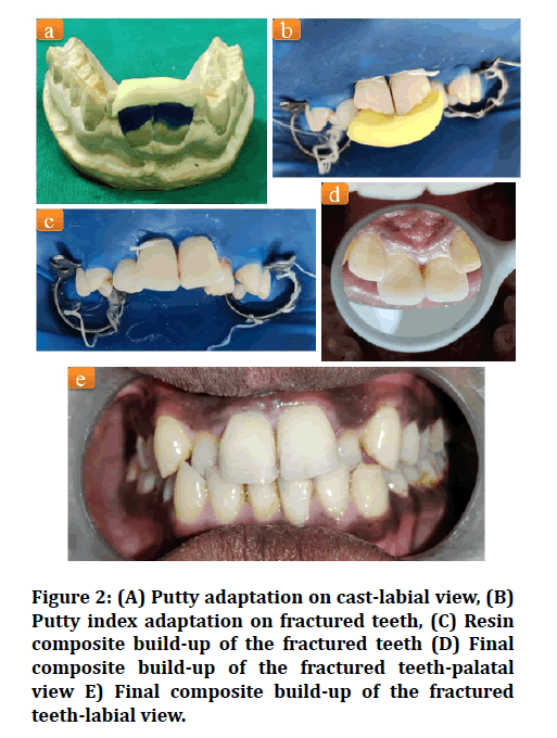
Casts were poured using dental stone (Figure 1I). Waxup was done using Type II inlay wax (Figure 1J and 1K). The putty index was made on the cast using the addition silicone putty impression material (Photosil, DPI) (Figure 1L). Their labial surface was cut off using BP blade No. 11, keeping the palatal surface intact (Figure 2A). Matrix was placed on the palatal surface of the teeth (Figure 2B). Etching was done with 37% phosphoric acid (Prime Dental) for 15 seconds.

Figure 2: (A) Putty adaptation on cast-labial view, (B) Putty index adaptation on fractured teeth, (C) Resin composite build-up of the fractured teeth (D) Final composite build-up of the fractured teeth-palatal view E) Final composite build-up of the fractured teeth-labial view.
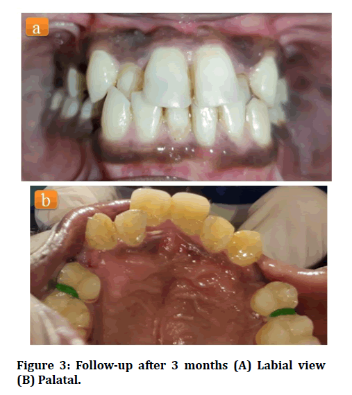
The teeth are then washed for the next 10-15 seconds. A bonding agent (3M ESPE–Adper Single Bond 2, USA) was applied on 11 21 and cured for 20 seconds. A palatal shelf was created with resin composite (Spectrum Micro hybrid composite, Dentsply, USA). The final build-up was done using the composite resin of the selected shade (Figure 2C). Finishing of the composite restoration was done using composite finishing burs(Mani, Japan). Polishing was undertaken the following day using the Shofu Super-Snap polishing system (Figure 2D and 2E). The patient was later sent for orthodontic treatment related to the anterior crowding in maxillary and mandibular arches. A follow-up was taken after 3months for evaluation of the restoration (Figure 3A and 3B).

Figure 3: Follow-up after 3 months (A) Labial view (B) Palatal.
Discussion
Esthetic replication of the maxillary central incisors requires a skilled protocol-oriented approach. This is a chair-side challenge for most clinicians [3]. So, a requirement of restoring the tooth by indirect technique arises. The result is very promising and satisfactory for both the patient and the clinician [4]. Resin composite is the preferred material of choice for such restorations because of its superior esthetics and handling properties [5,6].
Resin composite has an appearance similar to that of the tooth enamel. In addition to that, the success rate of composite restoration has shown to be up to 88% in 10- years follow up [7].
It is an easily repairable material showing the property of good polishability [8]. It can be used for various purposes such as esthetic restorations involving diastema closures, building up of fractured anterior tooth, reattachment of fractured tooth segments, and other restorations such as building up of proximal walls, for post endodontic restorations, for luting fibre posts, as preventive resin restorations, etc. [8–12].
Among the various techniques used for restoring a fractured incisor, the most conventional one is the rigid matrix technique, which utilizes a silicone putty matrix for restoring the fractured anterior [13]. Replication of the original anatomy of the tooth becomes much easier using this technique. The process involves an initial wax build-up using Type II inlay was. The is followed by fabrication of putty index using silicon putty [3].
Conclusion
Restoration of a fractured central incisor is a complicated yet satisfactory task that should be carried out meticulously using a systematic approach. This enables the clinician to deliver optimal results to the patient by restoring the aesthetics, form, and function accurately that was previously present in the patient.
References
- de Oliveira Scudine KG, Moreira KM, Cardoso M, et al Aesthetic and functional rehabilitation of anterior teeth after trauma: A case report. Clin Lab Res Dent 2017.
- Felippe LA, Monteir S, De Caldeira Andrada CA, et al. Clinical Strategies for success in proximoincisal composite restorations. Part II: Composite application technique. J Esthetic Restor Dent 2005; 17:11–21.
- Hasan A, Shahid O. Esthetic restorations, the putty matrix technique. J Dow University Health Sci 2013; 7:122-5.
- Ritter AV. Posterior composites revisited. J Esthet Restor Dent 2008; 20:57–67.
- Mair LH. Ten-year clinical assessment of three posterior resin composites and two amalgams. Quintessence Int 1998; 29.
- McCracken MS, Gordan VV, Litaker MS, et al. A 24-month evaluation of amalgam and resin-based composite restorations. J Am Dent Assoc 2013; 144:583–93.
- Lempel E, Lovász BV, Meszarics R, et al. Direct resin composite restorations for fractured maxillary teeth and diastema closure: A 7 years retrospective evaluation of survival and influencing factors. Dent Material 2017;33:467–76.
- Frese C, Schiller P, Staehle HJ, et al. Recontouring teeth and closing diastemas with direct composite buildups: A 5-year follow-up. J Dent 2013; 41:979–85.
- Shahdad SA, Kennedy JG. Bond strength of repaired anterior composite resins: an in vitro study. J Dent 1998; 10.
- Rathi S, Nikhade P, Bajaj P, et al. Reattaching the fractured fragment in ellis class 3, without extraction/removal of that fragment. Medl Sci 2020; 24:2445-2451.
- Dey R, Priti D D, Mazumdar P, Ghorai L. Aesthetic management of fractured anteriors-A conservative approach. JEMDS 2018; 7:3923–3925.
- Kabbach W, Sampaio CS, Hirata R. Diastema closures: A novel technique to ensure dental proportion. J Esthet Restor Dent 2018; 30:275–80.
- De Araujo EM, Fortkamp S, Baratieri LN. Closure of diastema and gingival recontouring using direct adhesive restorations: A case report. J Esthetic Restorative Dent 2009; 21:229-240.
Author Info
Joyeeta Mahapatra* and Pradnya Nikhade
Department of Conservative Dentistry and Endodontics, Sharad Pawar Dental College and Hospital, DMIMS, Sawangi(M), Wardha-442001, Maharashtra, IndiaCitation: Joyeeta Mahapatra, Pradnya Nikhade, Cosmetic Enhancement of Maxillary Central Incisors Using Rigid Matrix Technique, J Res Med Dent Sci, 2021, 9(9): 231-234
Received: 01-Sep-2021 Accepted: 14-Sep-2021
