Research - (2022) Volume 10, Issue 1
Dental Age Estimation of Upper Left Third Molar (28) Using Willems Method
Harita Ravikumar and Abirami Arthanari*
*Correspondence: Abirami Arthanari, Department of Forensic Odontology, Saveetha Dental College, Saveetha Institute of Medical and Technical Sciences (SIMATS), Saveetha University, India, Email:
Abstract
Introduction: Dental age estimation is a very important procedure done to obtain a person’s age in various situations such as during an unknown death, highly decomposed body etc. age estimation is an important identity of a person and it shouldn't be exploited. Various methods are known, such as Cameriere, Demirjian, Nolla, Willems etc. Aim: To estimate age of the upper left third molar using Willems method Materials and method: Orthopantomogram was obtained and the age was estimated using the chart and the results were tabulated. Data was analysed using spss software and the graphs were plotted. Results and discussion: It was found that the mean of males was 1.82 and for the females it was found to be 1.80. The standard deviation of the males and Female staging was done and it was found to be 1.674 and 1.990 respectively. The association graph was also plotted and the conclusion was made. Conclusion: It was concluded that Willems method showed less discrepancy and it was found to be more accurate.
Keywords
Age estimation, Willems, Forensics, Innovative technology
Introduction
Age is the most essential factor to estimate a person’s maturity. Personal identification is an essential factor in forensic medicine and dentistry. Dental maturity often not only depends on the eruption of the tooth but also the space between the teeth and many more [1]. Age estimation is the important factor in identification of a person. Age estimation can be done using various parameters. It is used in the wider range in forensic medicine as it is used to identify not only the unknown person but also in association with crimes and crime scenes [2,3]. Dental aging seemed to be in two forms: tooth mineralisation and tooth eruption pattern. The definition of eruption is the emergence of teeth in the gums rather than the bone. So this factor makes it impossible to be considered. Different methods of dental age estimation have been used widely such as Nollas’s, Demirjian’s, modified Demirjian by Willems [2]. The third molar eruption can be seen at the age between 12-25 years. Under the consideration of radiographs it can be extended from 9-25 years since it involves the crown and root development.
Kathleen in her study analysed the third molar of the Hispanic population and estimated the dental age using Dimirjian’s method and observed a statistically significant increase. It was also observed that hispanian males reach a faster developmental stage than hispanian females and the maxillary molar reach faster development than mandibular molar [4]. The study conducted by Prabhakaran, used the previously done surveys to check the age estimation in the molars. Third molars are the unique teeth which have a unique pattern of development [5]. According to Indra Priyadarshini, orthopantomogram were collected from the patients of age 14-30 years and the assessment was done [6].
The present study was adopted to overcome the limitations such as the previous studies didn’t mainly concentrate on the upper left third molar in dental age estimation. Our team has extensive knowledge and research experience that has translate into high quality publications [7-26]. The aim of this study is to estimate the age of the upper left third molar using Willem’s method.
Materials and Methods
100 digital orthopantomogram of the randomised controlled subjects were collected and categorized into 50 males and 50 females. The age group was classified as 10-10.9, 11-11.9, 12-12.9, 13-13.9, 14-14.9, 15-15.9, 16-16.9, 17.17.9, 18-18.9, 19-19.9. The study was beforehand approved by the institutional review board of Saveetha dental college and ethical approval was obtained. The data samples collected were then uploaded in excel sheets along with age and tooth development staging. Willem’s staging was done to 1 to 8 which is the modification of A to H for our convenience purpose. The data collected and entered and are transported to SPSS software where the results were studied, analysed along with statistical significance, p-value and regression analysis.
Results
Results are explained in the Tables (Tables 1 to 2) and Figures 4 to 6.
| Criteria | Staging of male | Staging of female |
|---|---|---|
| Standard deviation | 1.674 | 1.99 |
| mean | 1.82 | 1.8 |
| Standard deviation error | 0.237 | 0.281 |
Table 1: Mean standard deviation and standard deviation error of the tooth staging of 28 for both males and females. The standard deviation value of tooth staging of 28 was found to be for males, 1.674 and for females, 1.990. The mean value for male was found to be 1.82 and for females it was 1.80. The standard deviation error was found to be 0.237 and for the females it was 0.281.
| The staging of males and females | The chronological age of males and females | |
| P- value | 0.275 | 0.029 |
Table 2: Showing the p-value of the staging and the chronological age of the males and females. It was found that the p-value of staging of teeth was found to be 0.275 and for the chronological age was found to be 0.029.
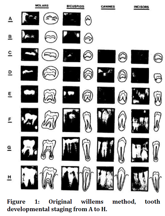
Figure 1. Original willems method, tooth developmental staging from A to H.
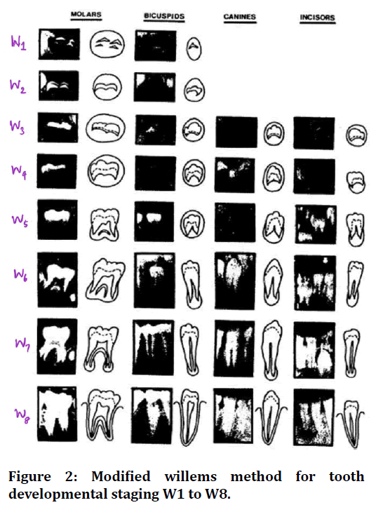
Figure 2. Modified willems method for tooth developmental staging W1 to W8.
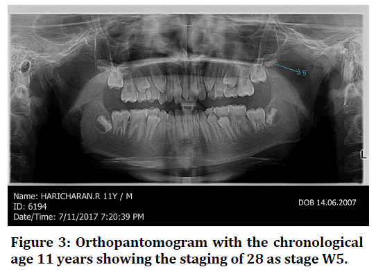
Figure 3. Orthopantomogram with the chronological age 11 years showing the staging of 28 as stage W5.
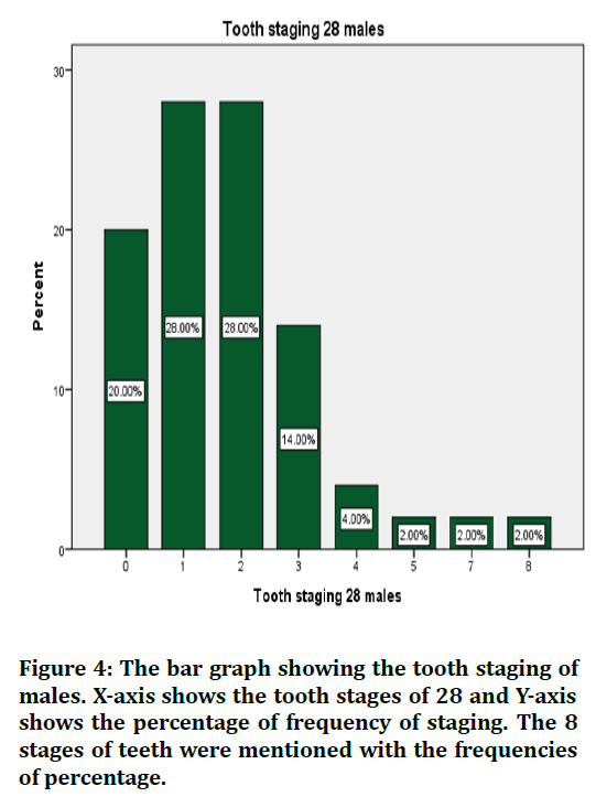
Figure 4. The bar graph showing the tooth staging of males. X-axis shows the tooth stages of 28 and Y-axis shows the percentage of frequency of staging. The 8 stages of teeth were mentioned with the frequencies of percentage.
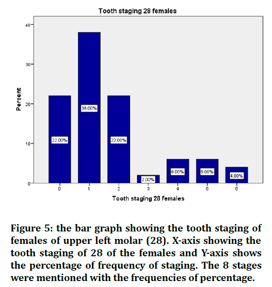
Figure 5. the bar graph showing the tooth staging of females of upper left molar (28). X-axis showing the tooth staging of 28 of the females and Y-axis shows the percentage of frequency of staging. The 8 stages were mentioned with the frequencies of percentage.
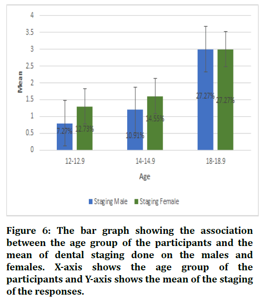
Figure 6. The bar graph showing the association between the age group of the participants and the mean of dental staging done on the males and females. X-axis shows the age group of the participants and Y-axis shows the mean of the staging of the responses.
Discussion
After obtaining the results, it can be seen that in Table 1, the standard deviation of the tooth staging of 28 of the males was found to be 1.674 and the tooth staging of 28 of the females was found to be 1.990. The mean was also calculated for the tooth staging of 28 of males and females, it was found to be 1.82 and 1.80 respectively. The standard deviation error for tooth staging of 28 of the males was 0.237 and 0.281. Table 2 shows the pvalue of the staging of teeth and the p-value of chronological age of both males and females. It was found that the p-value of staging teeth (28) of both males and females was found to be 0.275 which can be seen to be statistically insignificant. The p-value of the chronological age of both males and females was found to be 0.029 which is statistically significant. According to the tooth staging, there were 8 stages in total. From the observations made, graphs were plotted and the value was obtained. In figure 1, it was seen that stage 0 was found to be 20% and stage 1 was 28% stage 2 was 28% stage 3 was 14% stage 4 was 4% stage 5 was 2%. The stage 6 was 0%; stage 7 was 2% and stage 8 was found to be 2%. The tooth staging of the females was seen in figure 2. The results found to be stage 0 was 22%, stage 1 was 38%, stage 2 was 22%, stage 3 was 2%, stage 4 was 6%, stage 5 was 0%, stage 6 was 6% and stage 7 was 0% and stage 8 was 4%. The association graph was plotted between the tooth staging of 28 males and females and the chronological age. Figure 3 shows the tooth staging of 28 females and it was found to be 12.73% and the tooth staging of the 28 males was found to be 7.27% from the age 12-12.9 years, tooth staging of females was 14.55% and tooth staging of males was 10.91% for age 14-14.9 years. Tooth staging 28 female and male was 27.27% was the age 18-18.9 years.
The study conducted by Mani et al. was carried by taking 214 male and female participants and the cross sectional study was carried out by taking random samples of the orthopantomogram and then comparing the two methods of dental age estimation taking one as Willems and the other was Demirjian. The author found that the results of Willems method were less overestimated when compared to Demirjian method of dental age estimation [27].
Galić et al in his study evaluated the most accurate method of estimating the dental age by taking 591 girls and 498 boys by using 3 different dental age estimation methods i.e. Cameriere method, haavikko method and Willems method. It was found that the absolute accuracy was least for the cameriere method then for haavikko and Willems was highest. Hence it was said that the cameriere method was most preferred and least was Willems method of dental age estimation [28].
Franco et al. in their study took panoramic radiographs of people and tried to estimate the age group by comparing 2 different dental age groups which were Willems and Demirjian. The demirjian method showed greater discrepancies when compared to Willems method. It was also mentioned that both the methods had sometimes seemed to be not giving the expected results but in comparison, Willems method showed greater accuracy and hence preferred [29,30].
Zhai in her study mentioned that 1004 samples were used and the age was estimated using 2 methods, Demirjian and Willems and the more accurate results were obtained. It was found that the Demirjian method's mean absolute error was 1.08 years and 1.22 years for the Willems method. Hence he concluded that the Demirjian method was more accurate in providing the age [31].
The study conducted by Franco was taken up by Brazilian children where the samples collected showed the reliability of Willems method and the newly developed Brazilian method of dental age estimation. The kappa and the weighted kappa tests were performed to analyze the inter and interdependence of the method. It was found that the newly developed dental age seemed to be giving similar results as Willems method [29]
The limitation of this study was that the sample size was less and the criterion that was involved in this was limited. To obtain a greater result, more sample size should be taken and the wider criteria to be involved to generalise the study.
Conclusion
Willems method of age estimation is widely used around the world and its accuracy of age estimation on upper left third molar 28 was done. From the results obtained, it can be concluded that Willems method is preferred in estimating age as there were less discrepancies and the accuracy of the estimation of age was found to be closer.
Acknowledgement
We thank Saveetha Dental College and Hospitals for providing us the support to conduct the study.
Source of Funding
Study is funded and supported by
• Saveetha Dental College, Saveetha Institute of Medical and Technical Sciences
• Sarkav Health Services.
References
- De Donno A, Angrisani C, Mele F, et al. Dental age estimation: Demirjian’s versus the other methods in different populations. A literature review. Med Sci Law 2021; 61:125–9.
- Mohammed R, Krishnamraju PV, Prasanth PS, et al. Dental age estimation using Willems method: A digital orthopantomographic study. Contemp Clin Dent 2014; 371.
- Bedek I, Dumančić J, Lauc T, et al. New model for dental age estimation: Willems method applied on fewer than seven mandibular teeth. Int J Legal Med 2020; 134:735–43.
- Mesotten K, Gunst K, Carbonez A, et al. Dental age estimation and third molars: A preliminary study. Forensic Sci Int 2002; 129:110–5.
- Phrabhakaran N. Age estimation using third molar development. Malays J Pathol 1995; 17:31–4.
- Priyadharshini KI, Idiculla JJ, Sivapathasundaram B, et al. Age estimation using development of third molars in South Indian population: A radiological study. J Int Soc Prev Community Dent 2015; 5:S32–8.
- Princeton B, Santhakumar P, Prathap L. Awareness on preventive measures taken by health care professionals attending COVID-19 patients among dental students. Eur J Dent 2020; 14:S105–9.
- Mathew MG, Samuel SR, Soni AJ, et al. Evaluation of adhesion of Streptococcus mutans, plaque accumulation on zirconia and stainless steel crowns, and surrounding gingival inflammation in primary molars: Randomized controlled trial. Clin Oral Investig 2020; 24:3275–80.
- Sridharan G, Ramani P, Patankar S, et al. Evaluation of salivary metabolomics in oral leukoplakia and oral squamous cell carcinoma. J Oral Pathol Med 2019; 48:299–306.
- Hannah R, Ramani P, Ramanathan A, et al. CYP2 C9 polymorphism among patients with oral squamous cell carcinoma and its role in altering the metabolism of benzo [a] pyrene. Oral Surg Oral Med Oral Pathol Oral Radiol 2020; 130:306-12.
- Antony JVM, Ramani P, Ramasubramanian A, et al. Particle size penetration rate and effects of smoke and smokeless tobacco products: An invitro analysis. Heliyon 2021; 7:e06455.
- Sarode SC, Gondivkar S, Sarode GS, et al. Hybrid oral potentially malignant disorder: A neglected fact in oral submucous fibrosis. Oral Oncol 2021; 105390.
- Hannah R, Ramani P, WM Tilakaratne, et al. Author response for “critical appraisal of different triggering pathways for the pathobiology of pemphigus vulgaris—A review”. Wiley 2021.
- Chandrasekar R, Chandrasekhar S, Sundari KKS, et al. Development and validation of a formula for objective assessment of cervical vertebral bone age. Prog Orthod 2020; 21:38.
- Subramanyam D, Gurunathan D, Gaayathri R, et al. Comparative evaluation of salivary malondialdehyde levels as a marker of lipid peroxidation in early childhood caries. Eur J Dent 2018; 12:67–70.
- Jeevanandan G, Thomas E. Volumetric analysis of hand, reciprocating and rotary instrumentation techniques in primary molars using spiral computed tomography: An in vitro comparative study. Eur J Dent 2018; 12:21–6.
- Ponnulakshmi R, Shyamaladevi B, Vijayalakshmi P, et al. In silico and in vivo analysis to identify the antidiabetic activity of beta sitosterol in adipose tissue of high fat diet and sucrose induced type-2 diabetic experimental rats. Toxicol Mech Methods 2019; 29:276–90.
- Sundaram R, Nandhakumar E, Haseena Banu H. Hesperidin, a citrus flavonoid ameliorates hyperglycemia by regulating key enzymes of carbohydrate metabolism in streptozotocin-induced diabetic rats. Toxicol Mech Methods. 2019 ;29:644–53.
- Alsawalha M, Rao CV, Al-Subaie AM, et al. Novel mathematical modelling of Saudi Arabian natural diatomite clay. Mater Res Express 2019; 6:105531.
- Yu J, Li M, Zhan D, et al. Inhibitory effects of triterpenoid betulin on inflammatory mediators inducible nitric oxide synthase, cyclooxygenase-2, tumor necrosis factor-alpha, interleukin-6, and proliferating cell nuclear antigen in 1, 2-dimethylhydrazine-induced rat colon carcinogenesis. Pharmacogn Mag 2020; 16:836.
- Shree KH, Hema Shree K, Ramani P, et al. Saliva as a diagnostic tool in oral squamous cell carcinoma–A systematic review with meta analysis. Pathol Oncol Res 2019; 125:447–53.
- Zafar A, Sherlin HJ, Jayaraj G, et al. Diagnostic utility of touch imprint cytology for intraoperative assessment of surgical margins and sentinel lymph nodes in oral squamous cell carcinoma patients using four different cytological stains. Diagn Cytopathol 2020; 48:101–10. Indexed at. Google Scholar, Cross Ref
- Karunagaran M, Murali P, Palaniappan V, et al. Expression and distribution pattern of podoplanin in oral submucous fibrosis with varying degrees of dysplasia: An immunohistochemical study. J Histotechnol 2019; 42:80–86.
- Sarode SC, Gondivkar S, Gadbail A, et al. Oral submucous fibrosis and heterogeneity in outcome measures: A critical viewpoint. Future Oncol 2021; 17:2123–6.
- Raj Preeth D, Saravanan S, Shairam M, et al. Bioactive Zinc(II) complex incorporated PCL/gelatin electrospun nanofiber enhanced bone tissue regeneration. Eur J Pharm Sci 2021; 160:105768.
- Prithiviraj N, Yang GE, Thangavelu L, et al. Anticancer compounds from starfish regenerating tissues and their antioxidant properties on human oral epidermoid carcinoma kb cells. In: Pancreas. Lippincott williams wilkins two commerce 2001; 155.
- Mani SA, Naing L, John J, Samsudin AR. Comparison of two methods of dental age estimation in 7-15-year-old Malays. Int J Paediatr Dent 2008; 18:380–8.
- Galić I, Vodanović M, Cameriere R, et al. Accuracy of cameriere, haavikko, and willems radiographic methods on age estimation on bosnian–herzegovian children age groups 6–13. Int J Legal Med 2011; 125:315–21.
- Franco A, Thevissen P, Fieuws S, et al. Applicability of Willems model for dental age estimations in Brazilian children. Forensic Sci Int 2013; 231:401.e1–4.
- Djukic K, Zelic K, Milenkovic P, et al. Dental age assessment validity of radiographic methods on Serbian children population. Forensic Sci Int 2013; 231:398.e1–5.
- Zhai Y, Park H, Han J, et al. Dental age assessment in a northern Chinese population. J Forensic Legal Med 2016; 38:43–9.
Indexed at, Google Scholar, Cross Ref
Indexed at, Google Scholar, Cross Ref
Indexed at, Google Scholar, Cross Ref
Indexed at, Google Scholar, Cross Ref
Indexed at, Google Scholar, Cross Ref
Indexed at, Google Scholar, Cross Ref
Indexed at, Google Scolar, Cross Ref
Indexed at, Google Scholar, Cross Ref
Indexed at, Google scholar, Cross Ref
Inexed at, Google Scholar, Cross Ref
Indexed at, Google Scholar, Cross Ref
Indexed at, Google Scholar, Cross Ref
Indexed at, Google Scholar, Cross Ref
Indexed at, Google Scholar, Cross Ref
Indexed at, Google Scholar, Cross Ref
Author Info
Harita Ravikumar and Abirami Arthanari*
Department of Forensic Odontology, Saveetha Dental College, Saveetha Institute of Medical and Technical Sciences (SIMATS), Saveetha University, Chennai, IndiaReceived: 13-Dec-2021, Manuscript No. JRMDS-21-41706; , Pre QC No. JRMDS-21-41706 (PQ); Editor assigned: 15-Dec-2021, Pre QC No. JRMDS-21-41706 (PQ); Reviewed: 29-Dec-2021, QC No. JRMDS-21-41706; Revised: 03-Jan-2022, Manuscript No. JRMDS-21-41706 (R); Published: 10-Jan-2022
