Case Report - (2025) Volume 13, Issue 6
Dental Implications of Morphea: A Case Report
*Correspondence: Reema Abdulfattah Sharaf, Department of Restorative Dentistry, Prince Sultan Medical Military City, Riyadh, South Africa, Email:
Abstract
Morphea or localized scleroderma, primarily involves skin induration but can have significant oral and maxillofacial implications. This article reviews the pathophysiology, clinical manifestations, diagnostic considerations and management strategies for morphea in the context of dental health.
Keywords
Localized scleroderma, Skin, Morphea, Dental implications
Introduction
Morphea, (localized scleroderma), is a chronic autoimmune connective tissue disorder characterized by inflammation and fibrosis primarily affecting the skin and sometimes the underlying structures such as subcutaneous tissue, bone and mucosa [1,2]. Since its initial clinical descriptions, understanding of morphea has evolved to recognize various subtypes, including linear Morphea and en coup de Sabre, which may involve the orofacial region [3,4]. The disease’s clinical manifestations in the oral cavity, although uncommon, can lead to gingival recession, alveolar bone loss and mucosal sclerosis, posing challenges for dental practitioners [5,6].
Epidemiological data indicate an incidence ranging from 0.4 to 2.7 cases per 100,000 individuals, with higher prevalence in females and children, underscoring the importance of early recognition and management [1,7].
Morphea is a rare connective tissue disorder characterized by localized skin hardening. Although its systemic form, scleroderma, is well-documented, the localized variant's impact on oral and dental health is less understood. This article aims to elucidate the effects of morphea on oral structures, emphasizing diagnostic and therapeutic considerations for dental professionals.
Case Presentation
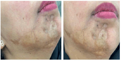
A 36-year-old female with a history of morphea for four years. No known allergies. She was referred by dermatology department to dental clinics to rule out bone involvement. She is complaining of gingival recession and mobility related to lower right teeth. She is also complaining from tissue hardening on the right check with deep scar tissue on the chin.
She is following with dermatology clinics. Received methotrexate for approximately three years without any response. She also received tocilizumab injections (immunosuppressants medication) for around one year with no improvement. And has been treated with topical tacrolimus and topical calcipotriene betamethasone and emollients (Figures 1 and 2).

Figure 1: Hypopigmented atrophic patches on the lower check. A): Lip and chin; B): With erythema surrounding.

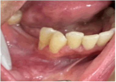
Figure 2: Missing molars and gingival recession around premolars.
She had dental extraction of #45 one year ago followed by bone augmentation. Biopsy has been done confirming the diagnosis.
Extra-oral examination (Figure 1): Multiple well defined white-brown plaques with atrophy and erythema of the right check, distal mandible and chin with sever deformity in the chin.
Intra-oral examination (Figure 2) revealed missing posterior teeth and gingival recession.
Investigations done including Orthopantomogram (OPG) and ultrasound. No significant abnormality has been detected. Liver function test and lipid profile were normal.
Patient is not taking any active medications.
Soft tissue biopsy has been done. Diagnosis: Morphea plaque type. Dental diagnosis: Reduced periodontium.
Treatment involved patient education of soft tissue biopsy, explaining possible effects of facial morphea on the gingival tissue which appears clinically as hyperkeratosis and fibrosis of tissue.
Results and Discussion
Morphea (localized scleroderma), is a rare autoimmune connective tissue disorder characterized by excessive collagen deposition in the skin, resulting in thickened, hard and discolored patches. Morphea mainly affects the skin and, in some cases, it may extend to underlying tissues, bones or muscles. Involvement of oral mucosa and teeth has been rarely reported.
Morphea can affect individuals of any age, but it is most commonly diagnosed in children and middle aged adults.
The precise cause of morphea is unknown, but current researches suggest that it involves an abnormal immune response leading to collagen overproduction and fibrosis. Several factors may contribute to morphea, autoimmune mechanisms: The immune system mistakenly targets the skin’s connective tissue. Genetic predisposition: Family history may increase susceptibility. Environmental triggers: Infections, radiation, trauma or certain medications have been implicated as potential triggers.
Vascular abnormalities: Blood vessel dysfunction might contribute to the localized inflammation and fibrosis.
The interplay of these factors results in inflammation followed by excess collagen deposition, leading to the characteristic sclerotic plaques.
In our case, the patient was living in a rural area and relocated to the city to access hospital follow-up after experiencing symptoms. Unsatisfied with the initial diagnosis, she continued to visit multiple dental clinics, believing her condition was related to her dental health, leading her to extract teeth unnecessarily. It wasn’t until she consulted the dermatology department that her condition was correctly identified. Up on clinical examination, there was no involvement of her oral tissues; however, she requires ongoing monitoring and regular dental visits to ensure comprehensive care.
Diagnosis of morphea involves medical history and physical exam: Evaluation of lesion appearance, distribution and progression. Skin biopsy: Crucial for confirming diagnosis, revealing thickened collagen bundles, flattened epidermis and an inflammatory infiltrate. Laboratory tests: Often normal, but autoimmune markers such as Antinuclear Antibodies (ANA) may be present. There is no specific laboratory test definitively confirms morphea.
Differential diagnosis may include: Scleroderma (systemic), linear dermatoses, ligament or joint-related conditions mimicking skin changes and other fibrosing skin diseases.
Currently, there is no consensus on the treatment approach for localized scleroderma. Various therapeutic strategies have been explored in clinical studies, including the use of topical or oral vitamin D,D-penicillamine, phenytoin, cyclosporine, corticosteroids, methotrexate and interferon therapies, along with psoralen-UVA photochemotherapy [8].
There is also no standard treatment for intraoral scleroderma. Topical calcineurin inhibitors, such as 0.1% tacrolimus ointment, have been effective in reducing skin thickening, hyperpigmentation, hardness, erythema and contraction. According to German guidelines, using topical and intralesional corticosteroids for 1 to 3 months has yielded positive outcomes. Additionally, good results have been reported with the application of a vitamin D analogue combined with phototherapy using low-dose ultraviolet A1, alongside 0.005% calcipotriol ointment applied twice daily [9].
The diagnosis of morphea or localized scleroderma, is based on the rapid and advanced progression of skin lesions. Systemic treatment may be necessary, with methotrexate and steroids being the first-choice drugs. Therefore, evaluating disease activity and subtype is crucial before initiating treatment.
Conclusion
While morphea is a chronic condition without a definitive cure, individualized treatment strategies can help manage symptoms effectively. Consultation with healthcare professionals specializing in dermatology is crucial for optimal care.
References
- Papara C, de Luca DA, Bieber K, et al. Morphea: The 2023 update. Front Med 2023; 13:1108623.
[Crossref] [Google Scholar] [PubMed]
- Mertens JS, Seyger MM, Thurlings RM, et al. Morphea and eosinophilic fasciitis: An update. Am J Clin Dermatol 2017; 18:491-512.
[Crossref] [Google Scholar] [PubMed]
- Niklander S, MarÃn C, MartÃnez R, et al. Morphea âen coup de sabreâ: An unusual oral presentation. J Clin Exp Dent 2017; 9:315.
[Crossref] [Google Scholar] [PubMed]
- Sartori-Valinotti JC, Tollefson MM, Reed AM. Updates on morphea: Role of vascular injury and advances in treatment. Autoimmune Dis 2013; 2013:467808.
[Crossref] [Google Scholar] [PubMed]
- AlSahman L, AlBagieh H, AlSahman R. Oral health-related quality of life in temporomandibular disorder patients and healthy subjects-a systematic review and meta-analysis. Diagnostics 2024; 14:2183.
[Crossref] [Google Scholar] [PubMed]
- Tang MM, Bornstein MM, Irla N, et al. Oral mucosal morphea: A new variant. Dermatology 2012; 224:215-220.
[Crossref] [Google Scholar] [PubMed]
- Peterson LS, Nelson AM, Su WP, et al. The epidemiology of morphea (localized scleroderma) in Olmsted County 1960-1993. J Rheumatol 1997; 24:73-80.
[Google Scholar] [PubMed]
- Shalaby SM, Bosseila M, Fawzy MM, et al. Fractional carbon dioxide laser versus low-dose UVA-1 phototherapy for treatment of localized scleroderma: A clinical and immunohistochemical randomized controlled study. Lasers Med Sci 2016; 31:1707-1715.
[Crossref] [Google Scholar] [PubMed]
- Kreuter A, Krieg T, Worm M, et al. German guidelines for the diagnosis and therapy of localized scleroderma. J Dtsch Dermatol Ges 2016; 14:199-216.
[Crossref] [Google Scholar] [PubMed]
Author Info
Department of Restorative Dentistry, Prince Sultan Medical Military City, Riyadh, South AfricaCitation: Reema Abdulfattah Sharaf, Dental Implications of Morphea: A Case Report, J Res Med Dent Sci, 2025, 13 (06): 001-003.
Received: 02-Jul-2025, Manuscript No. JRMDS-25-170058; , Pre QC No. JRMDS-25-170058 (PQ); Editor assigned: 07-Jul-2025, Pre QC No. JRMDS-25-170058 (PQ); Reviewed: 21-Jul-2025, QC No. JRMDS-25-170058; Revised: 21-Aug-2025, Manuscript No. JRMDS-25-170058 (R); Published: 28-Aug-2025
