Research - (2020) Advances in Dental Surgery
Developmental Defects of Enamel in Children withand Without Cleft Lip:A Case Control Study
Kuzhalvaimozhi P1, Vignesh Ravindran2* and Subhashini VC2
*Correspondence: Vignesh Ravindran, Department of Pediatric and Preventive dentistry, Saveetha Dental college and hospitals, Saveetha Institute of Medical and Technical sciences, Saveetha University, Chennai, India, Email:
Abstract
Orofacial clefts is the most common craniofacial birth defect and the fourth common congenital malformation in humans which require multidisciplinary care. Enamel defects are commonly seen in deciduous and permanent teeth especially maxillary incisors in an individual with cleft lip, palate,and alveolus. Hence a study was conducted to analyse the developmental defects of enamel in children with cleft lip and compare with children without cleft lip. Retrospective data collected from 89,000 case records from June 2019 to March 2020 were taken for the study. Based on the inclusion and exclusion criteria, the present study consisted of 6 children divided into two groups: children with cleft lip and children without cleft lip. In both groups, presence of developmental defects of enamel in the tooth were verified and data was tabulated. The data was subjected to Mann-Whitney test using SPSS software. Children in both the groups (with and without cleft lip) did not have any developmental defect of enamel. Within the limitations of the present study, there was no difference in the occurrence of developmental defects of enamel in children with and without cleft lip.
Keywords
Cleft lip, Central incisor, Developmental Defects, Enamel, Opacity
Introduction
Orofacial cleft (OFC) is the most common craniofacial birth defect in humans. Orofacial clefts exhibit both ethnic and geographic variation. The estimated prevalence is 1.7 in 1000 live births in India. Incidence of cleft lip and palate (CLP) varies from 0.25 to 2.29 per 1000 births in India [1-3]. The craniofacial structure development is a coordinated process which involves the growth of multiple independently derived embryologic prominences called primordia. Incomplete fusion of primordia during 4th to 8th week of embryological life which leads to cleft lip, cleft of primary or secondary palate or both [3,4].
Clefts can be caused by various factors which include infection,toxicity, poor diet, hormonal imbalances, and genetic interference. Among these factors, genetics play an important role in cleft lip and palate. Previous study conducted by Shaw et al. [5] presented evidence that women above 35 years of age had a doubled risk of having a child with cleft lip/palate, and women above 39 years of age had a tripled risk of having a child with cleft lip/palate. Consanguineous marriages also have an increased risk of developing cleft lip or cleft palate in the children. Dental complications include congenital missing teeth, neonatal teeth, ectopic eruption, supernumerary teeth, anomalies of shape and size of tooth, macrodontia, microdontia,fused teeth, enamel hypoplasia, deep bite, cross bite which can be anterior or posterior, crowding and spacing of the teeth [6].
Enamel defects are commonly seen in deciduous and permanent maxillary incisors in patients with cleft lip, and palate and has been associated with the cleft especially when the alveolus is involved. Depending upon the macroscopic appearance, defects of enamel may be classified into enamel hypoplasia and Hypomineralised enamel. Among these two defects, hypoplasia is a quantitative defect whereas Hypomineralised enamel is a qualitative defect which is seen as an abnormality in enamel translucency [7-10]. Defects in enamel formation may cause aesthetic alterations and compromise tooth enamel structure. Severe defect leads to early enamel loss, consequently, results in tooth wear and impaired functioning. Less mineralized enamel or enamel with an irregular surface may become more susceptible to caries development thereby leading to pulpal involvement [11-13]. Such defects are commonly noticed in children with cleft lip and/or palate [13]. However, studies conducted in the south Indian population are limited. The present study was done to assess the presence of developmental defects of enamel in patients with cleft lip and compare with children without cleft lip and to create awareness among the cleft lip patients.
Materials and Methods
This is a retrospective study. This study was carried out in a hospital-based university setting. This study was evaluated and ethically approved by an institutional ethical review committee. Retrospective data collected from 89,000 case records from June 2019 to March 2020. Informed consent was obtained from the parents or guardian before starting the treatment. Inclusion criteria: Children with cleft lip, children aged from 3 years to 18 years, children with at least one or two erupted teeth, children with at least one or two erupted teeth, complete photographic and written records regarding the complete intra-oral examination of the patient. Age and gender matched controls i.e. children without cleft lip, were taken according to the relevant cases obtained from the inclusion criteria. The exclusion criteria were incomplete and censored dental records, children below the age of 6 months and improper photographs.
Total cases acquired for this study were patients 6 which includes 3 children with cleft lip and 3 children without cleft lip (age, gender matched controls). Selected case and control group were examined by three people: one reviewer, one guide and one researcher. Patient's case sheets were reviewed thoroughly. Cross checking of data including digital entry and intraoral photographs was done by an additional reviewer, and as a measure to minimize sampling bias, samples for the group were picked by the simple random sampling method. Digital entry of clinical examination and intraoral photographs were assessed. For both groups, presence, or absence of developmental defects of enamel were noted by a researcher, entered Microsoft excel (MS Excel) and then transferred into Statistical Package for the Social Sciences (SPSS) Software for statistical analysis. A correlation test (Mann- Whitney test) was done between the children with cleft lip and children without cleft lip. The difference was statistically significant when the p-value was less than 0.05.
Results and Discussion
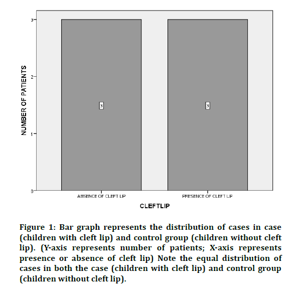
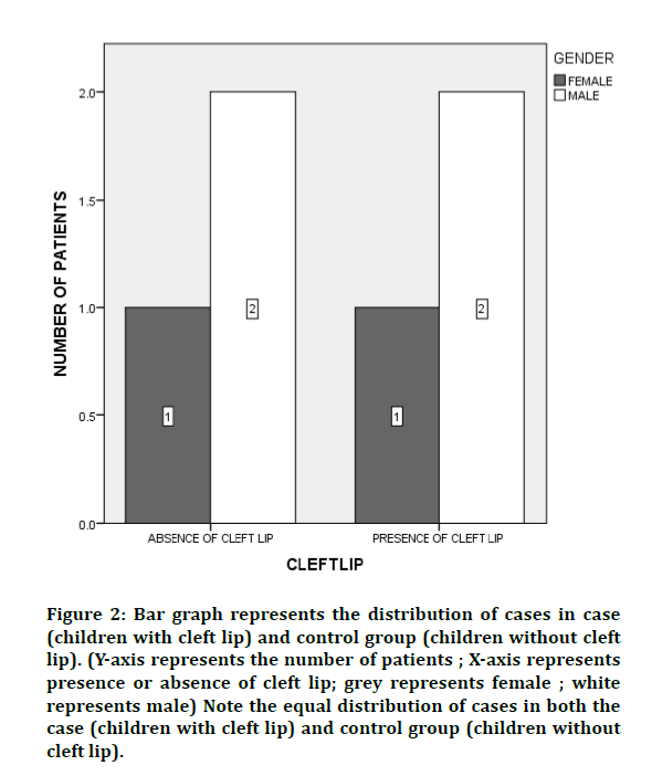
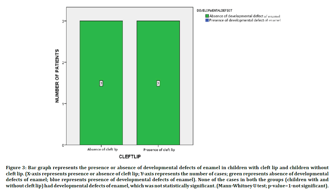
The final study sample size included a total of 6 children with 3 children with cleft lip (case group) and 3 children without cleft lip (control group). In this study, the control group was matched based on age and gender as like the case group (Figures 1 and 2). Absence of developmental defect of enamel was noticed in all children in both the groups i.e. children with and without cleft lip. On comparison of the results using Mann- Whitney test, the results were not statistically significant (p-value=1) (Figure 3).

Figure 1: Bar graph represents the distribution of cases in case (children with cleft lip) and control group (children without cleft lip). (Y-axis represents number of patients; X-axis represents presence or absence of cleft lip) Note the equal distribution of cases in both the case (children with cleft lip) and control group (children without cleft lip).

Figure 2: Bar graph represents the distribution of cases in case (children with cleft lip) and control group (children without cleft lip). (Y-axis represents the number of patients ; X-axis represents presence or absence of cleft lip; grey represents female ; white represents male) Note the equal distribution of cases in both the case (children with cleft lip) and control group (children without cleft lip).

Figure 3: Bar graph represents the presence or absence of developmental defects of enamel in children with cleft lip and children without cleft lip. (X-axis represents presence or absence of cleft lip; Y-axis represents the number of cases; green represents absence of developmental defects of enamel; blue represents presence of developmental defects of enamel). None of the cases in both the groups (children with and without cleft lip) had developmental defects of enamel, which was not statistically significant. (Mann-Whitney U test; p-value=1-not significant).
Dysfunction of ameloblasts results in changes in the appearance of the enamel in permanent dentition. These developmental defects of enamel (DDE) may range from slight change in the tooth colour to a complete absence of enamel. Developmental defects of enamel cause tooth sensitivity and an increased risk of dental caries [14]. Assessment of such enamel defects would be necessary for early detection and warning of caries risk in such individuals which would help the practitioner to provide preventive measures [7,8].
The results of the present study show that children with and without cleft lip did not show any developmental defects in the enamel of the teeth. This was contradictory to majority of the studies which suggests a prevalence in children with cleft lip [15-17]. However, there were a few studies that supports the results of the current study with minimal presence of defects in the enamel of children with cleft lip [18,19]. This difference could be due to the smaller sample size, that would have affected the results of the current study in a unidirectional manner.
Fluoride use has been recommended to prevent dental caries [20]. Decreased concentration of fluoride also results in increased incidence of dental caries [21]. Prevalence of enamel defects increases with increasing fluoride level in drinking water. Good attitude of parents reflects as a good oral health in children and vice versa [22]. Preservation of primary teeth in the dental arch is important to guide the eruption of the permanent teeth in the optimal position. Grossly decayed primary teeth which are extracted before exfoliation causes space in the dental arch which causes malocclusion if space maintainer was not given [23,24]. Bacteria play a vital role in the initiation and progression of dental caries which eventually causes pulpal and periapical disease [25]. Defect in enamel lead to early caries which would lead to pulpal involvement and even extraction [26-31]. Fluoride and regular toothbrushing can help in maintaining proper oral hygiene [32-34].
Maciel et al. [35] reported that there is a high incidence ofenamel defects on the cleft side for both deciduous and permanent dentition. Chappleet al. [36] reported that 24% of cleft lip patients had enamel hypoplasia. Chia- An- Shen et al. [37] in 2019 reported that 87.9% of cleft lip patients had enamel defects. Amandeep Chopra et al [38] reported that the prevalence of developmental defects of enamel was higher in children with cleft. This is contradictory to our study.
Advantages of this study were that this was a case control study with age and gender matched controls to provide best results with high internal validity, reasonable data, Disadvantage of the study was that this was a unicentric study with geographic limitations, limited sample size and has lower external validity. Future scope for this study includes larger sample size which is not confined to a particular geographic area and to assess the presence of developmental defects if enamel by clinically examining the cleft lip patients.
Conclusion
Within the limitations of the present study, there is no evidence of developmental defect of enamel in children with and without cleft lip.
Acknowledgement
The authors of this study acknowledge the institute, for their help towards collecting all the patient case records and other datas in relevance to the current study.
Conflict of Interests
The authors declare that there were no conflicts of interest.
References
- Mossey PA, Little J, Munger RG, et al. Cleft lip and palate. Lancet 2009; 374:1773–1785.
- Jugessur A, Farlie PG, Kilpatrick N. The genetics of isolated orofacial clefts: From genotypes to subphenotypes. Oral Dis 2009; 15:437–453.
- Szabo GT, Tihanyi R, Csulak F, et al. Comparative salivary proteomics of cleft palate patients. Cleft Palate Craniofac J 2012; 49:519–523.
- Zainab J, Salih BA. Oral health status and treatment needs among 3-12 years old children with cleft lip and/or palate in Iraq. J Baghdad College Dent 2012; 24:145–151.
- Shaw WC, Semb G. Current approaches to the orthodontic management of cleft lip and palate. J R Soc Med 1990; 83:30–33.
- Kaul R, Jain P, Saha S, et al. Cleft lip and cleft palate: Role of a pediatric dentist in its management. Int J Pedodont Rehab 2017; 2:1.
- Jälevik B. Prevalence and diagnosis of molar-incisor- hypomineralisation (MIH): A systematic review. Eur Arch Paediatr Dent 2010; 11:59–64.
- Suckling GW. Developmental defects of enamel--historical and present-day perspectives of their pathogenesis. Adv Dent Res 1989; 3:87–94.
- Robinson C, Brookes SJ, Shore RC, et al. The developing enamel matrix: Nature and function. Eur J Oral Sci 1998; 106:282–291.
- Smith CE. Cellular and chemical events during enamel maturation. Crit Rev Oral Biol Med 1998; 9:128–161.
- Gasse B, Grabar S, Lafont AG, et al. Common SNPs of amelogeninx (AMELX) and dental caries susceptibility. J Dent Res 2013; 92:418–424.
- Olszowski T, Adler G, Janiszewska-Olszowska J, et al. MBL2, MASP2, AMELX, and ENAM gene polymorphisms and dental caries in Polish children. Oral Dis 2012; 18:389–395.
- Bailleul-Forestier I, Molla M, Verloes A, et al. The genetic basis of inherited anomalies of the teeth. Part 1: Clinical and molecular aspects of non-syndromic dental disorders. Eur J Med Genet 2008; 51:273–291.
- Wong HM, McGrath C, King NM. Dental practitioners’ views on the need to treat developmental defects of enamel. Community Dent Oral Epidemiol 2007; 35:130–139.
- Suckling G. Defects of enamel in sheep resulting from trauma during tooth development. J Dent Res 1980; 59:1541–1548.
- Suckling GW, Purdell-Lewis DJ. The pattern of mineralization of traumatically induced developmental defects of sheep enamel assessed by microhardness and microradiography. J Dent Res 1982; 61:1211–1216.
- Suckling G, Elliott DC, Thurley DC. The production of developmental defects of enamel in the incisor teeth of penned sheep resulting from induced parasitism. Arch Oral Biol 1983; 28:393–399.
- Andreasen JO, Ravin JJ. Enamel changes in permanent teeth after trauma to their primary predecessors. Scand J Dent Res 1973; 81:203–209.
- Hall SR, Iranpour B. The effect of trauma on normal tooth development. Report of two cases. J Dent Child 1968; 35:291–295.
- Ramakrishnan M, Bhurki M. Fluoride, fluoridated toothpaste efficacy and its safety in children-Review. Int J Pharma Res 2018; 10:109–114.
- Somasundaram S, Ravi K, Rajapandian K, et al. Fluoride content of bottled drinking water in Chennai, Tamilnadu. J Clin Diagn Res 2015; 9:ZC32–ZC4.
- Gurunathan D, Shanmugaavel AK. Dental neglect among children in Chennai. J Indian Soc Pedod Prev Dent 2016; 34:364–369.
- Ravikumar D, Jeevanandan G, Subramanian EMG. Evaluation of knowledge among general dentists in treatment of traumatic injuries in primary teeth: A cross-sectional questionnaire study. Eur J Dent 2017; 11:232–237.
- Panchal V, Jeevanandan G, Subramanian E. Comparison of instrumentation time and obturation quality between hand K-file, H-files, and rotary Kedo-S in root canal treatment of primary teeth: A randomized controlled trial. J Indian Soc Pedod Prev Dent 2019; 37:75–79.
- Jeevanandan G, Kedo S. Paediatric rotary files for root canal preparation in primary teeth-Case report. J Clin Diagn Res 2017; 11:ZR03–ZR05.
- Subramanyam D, Gurunathan D, Gaayathri R, et al. Comparative evaluation of salivary malondialdehyde levels as a marker of lipid peroxidation in early childhood caries. Eur J Dent 2018; 12:67–70.
- Govindaraju L, Jeevanandan G, Subramanian EMG. Comparison of quality of obturation and instrumentation time using hand files and two rotary file systems in primary molars: A single-blinded randomized controlled trial. Eur J Dent 2017; 11:376–379.
- Govindaraju L, Jeevanandan G, Subramanian EMG. Knowledge and practice of rotary instrumentation in primary teeth among indian dentists: A questionnaire survey. J Int Oral Health 2017; 9:45.
- Jeevanandan G, Govindaraju L. Clinical comparison of Kedo-S paediatric rotary files vs manual instrumentation for root canal preparation in primary molars: a double blinded randomised clinical trial. Eur Archives Paediatr Dent 2018; 19:273–278.
- Govindaraju L, Jeevanandan G, Subramanian E. Clinical Evaluation of quality of obturation and instrumentation time using two modified rotary file systems with manual instrumentation in primary teeth. J Clin Diagn Res 2017; 11:ZC55–ZC58.
- Lakshmanan L, Mani G, Jeevanandan G, et al. Assessing the quality of obturation and instrumentation time using Kedo-S files, Reciprocating files and Hand K-files. Br Dent Sci 2020; 23.
- Christabel SL, Gurunathan D. Prevalence of type of frenal attachment and morphology of frenum in children, Chennai, Tamil Nadu. World J Dent 2015; 6:203–207.
- Packiri S, Gurunathan D, Selvarasu K. Management of paediatric oral ranula: A systematic review. J Clin Diagn Res 2017; 11:ZE06–ZE09.
- Govindaraju L. Effectiveness of chewable toothbrush in children-A prospective clinical study. J Clin Diagnostic Res 2017; 11:ZC31.
- Maciel SP, Costa B, Gomide MR. Difference in the prevalence of enamel alterations affecting central incisors of children with complete unilateral cleft lip and palate. Cleft Palate Craniofac J 2005; 42:392–395.
- Chapple JR, Nunn JH. The oral health of children with clefts of the lip, palate, or both. Cleft Palate Craniofac J 2001; 38:525–528.
- Shen C-A, Guo R, Li W. Enamel defects in permanent teeth of patients with cleft lip and palate: A cross-sectional study. J Int Med Res 2019; 47:2084–2096.
- Chopra A, Lakhanpal M, Rao NC, et al. Oral health in 4-6 years children with cleft lip/palate: a case control study. N Am J Med Sci 2014; 6:266–269.
Author Info
Kuzhalvaimozhi P1, Vignesh Ravindran2* and Subhashini VC2
1Department of Pediatric and Preventive dentistry, Saveetha Dental college and hospitals, Saveetha Institute of Medical and Technical sciences, Saveetha University, Chennai, India2Department of Pediatric and Preventive dentistry, Saveetha Dental college and hospitals, Saveetha Institute of Medical and Technical sciences, Saveetha University, Chennai, India
Citation: Kuzhalvaimozhi P, Vignesh Ravindran, Subhashini VC, Developmental Defects of Enamel in Children with and Without Cleft Lip: A Case Control Study, J Res Med Dent Sci, 2020, 8 (7): 298-302.
Received: 29-Sep-2020 Accepted: 02-Nov-2020 Published: 09-Nov-2020
