Review Article - (2022) Volume 10, Issue 7
Digital Dental Photography: A Review
Akash Sibal1*, Shriya Singi1, Akshay Jaiswal1 and Anuja Ikhar2
*Correspondence: Akash Sibal, Department of Dentistry, Meghe Institute of Medical Sciences, Sawangi, Maharashtra, India, Email:
Abstract
Dental photography has always been an important aspect of the practice. The journey begins with film photography, which was only used for record keeping and referral purposes before being replaced by digital photography. Its use in dental practice is straightforward, quick, and incredibly effective for documenting work procedures, patient education, and clinical research, resulting in several benefits for patients and dentists. The article discusses the advantages of dental photography above film photography, the basic equipment needed to take decent photos, and how digital dental photography can help dentists.Keywords
Dental photography, Digital camera, Digital photographyIntroduction
It has been said a picture is worth a thousand words, and this phrase certainly rings true in the practice of dentistry. Photography is important in today’s practice as other traditional methods of diagnosis such as radiographs and diagnostic casts. It is estimated that approximately 65% of the population are visual learners. Confucius was quoted as having said “tell me and I will forget. Show me and I may remember. Involve me and I will understand”. Photography is one of our best tools to involve patients in their own diagnosis and care and help to motivate them to accept the treatment they want, and often times need, to preserve the integrity of their oral health and appearance. It's difficult to imagine our lives without digital photography, which has invaded every aspect of fashion design, science, medicine, communication, and industry. Film photography, which was introduced over a century ago, was a huge stride forward. The transition from film to digital imaging is the most significant advancement in dental photography. Digital photography is significant on multiple levels and has become synonymous with modern dentistry. Its use in dental practice is straight forward, quick, and incredibly effective in documenting work procedures, educating patients, and conducting clinical research, resulting in several benefits for dentists and patients.
Literature Review
History
In 1840, the world's first dentistry school and the world's first photographic gallery opened, both of which were administered by a dentist who also happened to be a photographer. The operator can capture patient information, events, and scientific findings in a unique way using photography. Alexander Wolcott (1804–1844) was a pioneer in the field of photography. In 1840, he received a patent for his camera inventions and devised a system for photographic studio lighting. He created history by founding the first commercial photographic studio in less than a month later [1]. W Elde of Columbus and R Thompson used before and after images of dental procedures for the first time in 1848, and an article was published, ushering in a new era.
The following are some of the reasons why you should utilize dental photography: Dental photography has now become simpler and more convenient, with the emergence of newer technologies. Dental practice is not just about the dental practice, as experienced dentists understand; it's also about persuading patients and avoiding legal complications. Whether you've been practicing for a while or are just getting started, one thing remains constant: the significance of documentation.
It's critical to keep track of everything:
- Keep a detailed record of your and the patient's trip from beginning to end.
- Assist in the presenting of cases.
- To furnish proof in the event of a dispute.
Improved patient communication and education
There have been auditory and visual aids for patient education in the past, such as movies, models, and pamphlets, however none of these approaches has completely addressed the material. A demonstration on dental operations could be prepared utilizing filmed cases, electronic display and PowerPoint presentation. These specific images of surgical steps, anatomy, finished cases and materials can aid in patient education about the diagnosis and suggested therapy, boosting their understanding and acceptance of the procedure [2,3].
Verification of insurance: Insurance companies require periodontal charting, radiographs, or a narrative before disbursing benefits to consumers. As a result, a digital photograph can be used to support a narrative.
Consultation with an expert: The only way to present our patients to other clinicians was through charted radiographs and written reports. With the use of images, a completely new dimension has been established. An over the phone consultation with an expert may be sufficient with a comprehensive case history and high resolution pictures [4]. Similarly, images of mutual patients and their recent achievements from the referring dentist might be transferred or received so that the operator could analyses the situation without having to be physically present in the office.
Communication inside the laboratory: A shade guide is often used to convey information regarding the character, shade, or color of teeth or gingiva [5]. This technique has a number of drawbacks, including a failure to adequately describe the shadowing and depth that a tooth reveals. As a result, a color corrected photograph can be used to construct a final prosthesis with more precise dimensions of color (Hue, Value and Chroma).
Advertising and marketing by professionals: Pre and post-operative images are effective motivators for patients to accept treatment plans or to demonstrate a certain talent.
Professional guidance: When it comes to discussing dental ideas or specific surgical procedures, words and bullets are frequently insufficient. A picture is worth a thousand words and generates more attention and debate than handwritten content.
Work ethic and treatment philosophy: Cleaning surgical sites for pictures takes time and energy, and it requires endurance and meticulous attention. This mentality motivates us to complete our work with extreme precision. As a result, preparing the patient for pictures helps us increase our own abilities.
Basic armamentarium: The following is a list of the essential dental photography equipment.
- Digital camera
- Light and electronic flash systems
- Contrastors
- Intra-oral Mirrors
- Three Angles
- Monochrome
- Macro lens
Digital camera: Cameras are broadly classified into three main categories:
- Those based on the Single Lens Reflex (SLR camera) design with interchangeable lenses.
- Those based on a compact design where the lenses are not interchangeable-Digital camera and intra-oral camera.
- Digital SLR camera (combination of Digital and SLR camera).
Single Lens Reflex (SLR) camera system: The body and lens of a SLR camera used in dental photography are the two primary components. Manually controlled cameras will be sufficient; however, auto exposure cameras allow you to concentrate more on the patient than the camera. The most essential feature is that it can utilize interchangeable lenses [6]. It's tiny and easy to use.
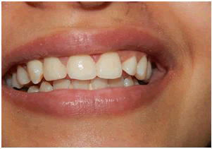
Digital camera system: Digital photography has gained a lot of traction, and its use in dentistry has a lot of benefits. This gives photographers more creative freedom, allows for instant image assessment, and is the most cost-effective option (Figure 1). They contain a LCD screen on which you can check your photos and eliminate any that are of low quality.
Figure 1: Shows picture taken by digital camera.
Compared to traditional cameras, digital cameras are more technologically advanced. The CCD is the brains of any digital camera. Photodiodes, often known as pixels, are discrete regions on the CCD that register the light shining on them. As a result, megapixels are made up of millions of pixels. The smaller the pixels, the greater the MP (Megapixels) value, and the better the solution [7]. A digital zoom lens is not a genuine zoom lens; instead, it crops the image, discarding information at the corners and increasing the lens apparent magnification. The term "optical zoom" refers to a shift in focal length. It works by refracting light and magnifying the picture on the CCD using a set of lenses. Unlike digital zoom, optical zoom enlarges image quality as well as the details and clarity that result.
Digital SLR camera: These cameras combine the advantages of digital camera and SLR systems. These features are as follows:
- An interchangeable lens that enables extraordinary telephoto shots that would be hard or even impossible to obtain with a small digital camera.
- Noise will be substantially lower in digital SLRs with large sensors than in tiny cameras. This improves the ability to recover from exposure errors, shadow detail and fine detail.
- Digital SLRs features a phase detection autofocus technology, which is faster and more efficient, and have shorter shutter lag times in general, making it simpler to capture movement.
Photographic equipment
Cheek retractors: These are used to retract the buckle mucosa, labial mucosa, and lips from the field to be imaged, allowing lighter to enter the oral cavity and enhancing visibility. Clear plastic or metal cheek retractors are available. Metal retractors have a less appealing appearance, although they can be sterilized by autoclave. Translucent plastic-retractors are more visually appealing, and the original tissue color that appears through them lessens distraction. There are two types of retractors: single-ended and double-ended. Both a small and a large curvature can be accommodated by retractors with two ends. This enables a wide variety of mouth sizes to be accommodated. Retractors' ends serve as handles to aid in retraction. Intraoral photography, like any other dental treatment in which pathogenic germs could be transferred to dental professionals or patients, necessitates stringent aseptic procedures [8]. Chemical sterilization is required since plastic retractors cannot be autoclaved.
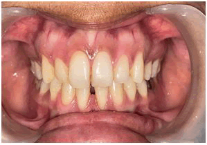
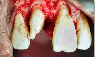
Light and electronic flash systems: When a mobile camera's flash is used for intraoral photos, it bounces off the surface of the teeth, creating an eerie glow that can't be edited off, obliterating the image's purpose (Figure 2).
Figure 2: Shows picture taken by light and electronic flash system.
- To highlight the intraoral cavity, always use a selfie light.
- To retract your cheeks and lips, always use cheek retractors.
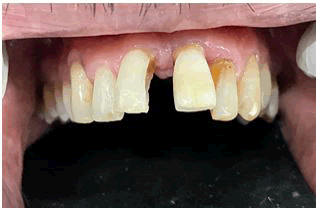
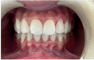
Contractors:Intraoral, these are employed to provide the illusion of a black background. As the name suggests, they provide the backdrop for a great contrast between the pink and the white in the mouth (Figure 3). Contractors are a "must-have" when it comes to taking aesthetic shots especially of the upper arch.
Figure 3: Shows picture taken in contrastor.
- While you click, have the patient/assistant hold it under the top ach.
- Photos must be cropped neatly.
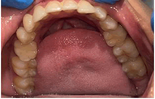
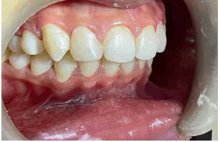
Intra-oralmirrors:The cheap plastic intra-oral mirrors are readily fogged, lack appropriate clarity, and are easily scratched. It is preferable to invest in high-quality glass intra-oral reflectors, as they will become a necessity in daily practice [9]. They normally come in two sizes: small and medium (Figure 4). When not in use, they should be carefully preserved to avoid unintentional damage and undesired scratches, which would degrade the image quality. Because we are utilizing them in a patient's mouth, they must be cleansed after use. A small amount of cotton soaked in isopropyl alcohol should be used.
Figure 4: Shows Picture taken in Intra oral mirror.
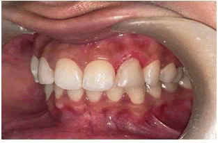
Threeangles:Allthree angles are required for complete protocol and case documentation/presentation, especially in situations involving both anterior and posterior teeth, as well as any type of occlusal rehabilitation. These are the three angles: frontal, left lateral and right lateral (Figures 5a, 5b, 5c). This is to better appreciate the pre-op occlusion, over jet, overbite, and upper anterior inclination [10]. All needed paperwork must show frontal, left, and right lateral views. Pre-op and Post-op photograph are a must.
Figure 5a: Shows Picture taken in Frontal view.
Figure 5b: Shows Picture taken in left lateral view.
Figure 5c: Shows Picture taken in Right lateral view.
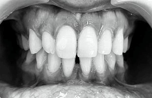
Monochrome:This is a crucial topic in dental photography since it depicts an aesthetic fit between direct and indirect restorations and natural teeth (Figure 6). This is done to show the Value.
Figure 6: Shows picture taken in monochrome.
Macrolens:These are required when capturing macro texture, like as gingival stippling, texture on crowns, composites, and dentures, among other things (Figure 7). Most mobile cameras have a "macro mode," however you may wish to utilize an extra macro lens for more high-end photos.
Figure 7: Capturing macro texture.
Discussion
Dental photography is a valuable and necessary tool for presenting treatment plans and educating patients. Great visual introductions and successful correspondence can immediately improve on the clarification of obscure dental terms, show, or legitimize the requirement for treatment, and empower clinicians and their assistants to introduce finished cases to patients that are visual declarations of their expert capacities. In terms of creating and storing photographic images, digital cameras are simple to operate. Digital photography has made it much easier to document therapy, which is important not only in a therapeutic but also in a legal sense. In clinical photography, familiarity with equipment and commitment to fundamental rules can mean the difference between success and failure. A methodical approach is required. This should include the handling and storage of images in addition to the photography itself. Before engaging in any clinical photography, it's important to think about the legal and ethical implications.
Conclusion
The Clinician can communicate with the patient and other practitioners for referral or treatment purposes using digital pictures. Technical features such as smile breadth, facial profile, smile line, occlusal plane, and emerging profile, compensating curves, gingival architecture and shade matching can be better visualized with precise profile and intraoral photographs. It also makes the image of laboratory cases look more like the image of the actual patient. With additional information available, operators and technicians can provide superior skills with better accuracy and achieve the patient's preferred restoration outcome with realistic results.
References
- Galante DL. History and current use of clinical photography in orthodontics. J Calif Dent Assoc 2009; 37:173-174.
- Bengel W. Mastering Digital Dental Photography. 1st ed. New Maiden, Surry (UK): Quintessence Publishers. 2006.
- Yoo A. 10 reasons why dental photography should be an essential part of your practice. Dent Econ 2014; 104:1-7.
- Geissberger M. Esthetic Dentistry in clinical practice. Wiley- Blackwell Publishers. 2010.
- Desai V, Bumb D. Digital dental photography: A contemporary revolution. Int J Clin Pediatr Dent 2013; 6:193.
- Sreevatsan R, Philip K. Digital photography in general and clinical dentistryâ??technical aspects and accessories. Int Dent J Stud Res 2015; 3:17-24.
[Googlescholar][Indexed]
- Manjunath SG, Raju Ragavendra T, Setty SK, et al. Photography in clinical dentistryâ??A review. Int J Dent Clin 2011; 3:40-43.
- Chandni P, Anupam S, Nitin S, et al. An overview on dental photography. Int J Dent Health Sci 2016; 3:581-589.
- Alberto C. Digital photography and documentation techniques in dentistry and dental technology. Zerodonto 2013; 1:1-103.
- Daniel R. Technical analysis of clinical digital photographs. J Calif Dent Assoc 2009; 37:199-206.
Author Info
Akash Sibal1*, Shriya Singi1, Akshay Jaiswal1 and Anuja Ikhar2
1Department of Dentistry, Meghe Institute of Medical Sciences, Sawangi, Maharashtra, India2Department of Conservative Dentistry and Endodontics, Datta Meghe Institute of Medical Sciences, Sawangi, Maharashtra, India
Citation: Akash Sibal, Anuja Ikhar, Shriya Singi, Akshay Jaiswal, Digital dental photography: A Review, J Res Med Dent Sci, 2022, 10 (7): 117-125.
Received: 09-May-2022, Manuscript No. JRMDS-22-49947; , Pre QC No. JRMDS-22-49947; Editor assigned: 12-May-2022, Pre QC No. JRMDS-22-49947; Reviewed: 26-May-2022, QC No. JRMDS-22-49947; Revised: 02-Jul-2022, Manuscript No. JRMDS-22-49947; Published: 09-Jul-2022

