Research - (2019) Volume 7, Issue 3
Effect of Cementum Extended Composite Filling with Two Different Posts on Fracture Resistance and Fracture Strength of Severely Damaged Primary Anterior Teeth
Razieh Meshki1, Parisa Sarikhani2* and Fateme Zarouni2
*Correspondence: Parisa Sarikhani, Pediatric Dentistry Resident, Department of Pediatric Dentistry, School of Dental Medicine, Ahvaz Jundishapur University of Medical Sciences, Iran, Email:
Abstract
Introduction and Objective: Restoration of the severely damaged anterior teeth is a challenge for many dentists. Therefore, the purpose of this study was to investigate the effect of cementum extended composite filling with two different inter-canal posts on Fracture resistance and Fracture Strength of severely damaged primary anterior teeth.
Method: In this experimental study, 50 severely damaged primary anterior teeth with at least 2/3 of root remaining were used. After decoronization, they were endodontically treated and randomly divided into 5 groups including group 1: composite filling extended 0.5 mm to cementum (ceCF), group 2: composite filling with composite filling with composite post (CP), group 3: composite filling extended 0.5 mm to cementum with composite filling (ceCF+CP), group 4: composite filling with composite filling with omega-shaped post (OP) and group 5: composite filling extended 0.5 mm to cementum with omega-shaped post (ceCF+OP). The bonded surface of each sample was measured by CBCT 1 mm above cement-enamel junction. After thermal cycles, the fracture resistance and fracture strength were measured by using the UTM machine. Data were analyzed using one-way ANOVA and Tukey test.
Results: ceCF+OP groups showed the greater fracture resistance and fracture strength. The mean fracture resistance values of 5 groups were 533.54, 240.37, 546.64, 447.77, 628.85 N. The mean fracture strength values were 21.3, 9.36, 22.44, 17.0, 24.36 N/mm2.
Conclusion: According to the results, all type of composite fillings with different posts that had been evaluated in this study can be used in severely damaged primary anterior teeth.
Keywords
Cementum extended composite filling, Fracture resistance, Fracture strength, Anterior teeth, Composite filling
Introduction
Anterior primary teeth are affected by trauma and early childhood caries [1]. Early childhood caries is a common disease characterized by a great destruction of the tooth structure [2]. According to the definition of the American Academy of Pediatric Dentistry, the disease of early childhood caries (ECC) is presence of 1 or more decayed (non-cavitated or cavitated carious lesions), missing[due to caries), or filling tooth surfaces in any primary tooth in child 71 months of age or younger [3]. The most common involved teeth are central and lateral incisors of maxilla and first mandibular and maxillary molars [4]. Primary lesions occur in the form of white decalcification spots at the gingival margin of incisors, which are rapidly affected by discoloration [5]. These caries progress and destroy the coronal portion [6]. The early loos of this teeth leads to abnormal tongue's position, reduction of bite force, mastication problems, speech impairment, psychological problems due to esthetic concerns, reduction of vertical height and mouth breathing habit [7,8].
Restorative treatments for these teeth have always been a controversial problem for pediatric dentists [7]. Restoration of these teeth is very difficult due to the small size of the coronal portion of teeth; relatively large pulp space and the patient's age [8]. In the restoration of the anterior teeth, in addition to preserving tooth structure and reconstructing of the original form, considering to aesthetic through the use of composite resins is very much considered [9]. Most of these restorative procedures are suitable if sufficient amount of tooth structure remains while these teeth have lost most or their entire coronal portion of teeth structure [5]. In these items, the coronal portion of teeth structure leads to a reduction in support for the composite coronal portion of teeth restoration, and the possibility of breaking through the impact due to the high volume of the mass [5]. In items where incisors do not have a sufficient coronal structure, placing a post in the root canal of the tooth after performing pulpectomy, increases the tooth strength and helps to maintain the coronal portion of teeth's restoration [10].
Various types of posts are available for primary teeth, including prefabricated posts [11-13], omega or alpha [2,14-16] orthodontic wires, cast posts with retention fittings [16], short composite filling with composite posts [4], fiber posts [15], and biological posts [17]. It seems that the clinical extension of the coronal portion of teeth to the cementum may increase the durability of restoration [4]. In patients treated under general anesthetic condition, cautery or any other device can be used to perform a simple gingivectony and remove small part of gingival tissue to increase the clinical coronal portion of teeth length therefore the possibility of bonding with the cementum can be provided and it may increase the survival rate of the tooth restoration [4].
Considering the importance of the items mentioned and the increasing demand for aesthetic restorations, this study was conducted with the purpose of comparing the fracture resistance and fracture strength of five different methods to restored severely damaged primary anterior teeth including cementum extended, composite post, cementum extended with composite post, cementum extended with omega-shaped post and omega-shaped post.
Materials and Methods
This in-vitro, experimental study was conducted on 50 primary anterior and non-restored teeth with at least 2/3 of root length were sound in these teeth, were selected.
Sample preparation
The teeth were cleaned using 2% sodium chlorite (Poland, cerkamed, chloraxid) and kept in distilled water (Shahid Ghazi, Tabriz-Iran) in a refrigerator at 4°C. Distilled water was periodically changed until the completion of the sample [1]. After completing the samples, the crown was cut 1 mm above the CEJ by high speed diamond fissure bur (Tizkavan, Iran). The samples were examined after the removal of coronal portion of teeth so that they had enough structure to conduct research. Access cavity preparation was done. Root canal was instrumented and standard canal preparation was done for each samples. Canals were filled with metapex (Calcium Hydroxide with Iodoform) (METABIOMED, Korea) up to 4 mm beneath the CEJ. 1 mm layer of glass ionomer was placed over the root canal filling as the base. 3 mm empty space remained in the root canal. The teeth were divided into 5 groups by random block method to locate post and restorations types.
Method of calculating sample size and sampling
According to the study by Seraj et al. [4], the mean and standard deviation of fracture resistance were as much as 76.37 ± 601.08 and 76.37 ± 507.5 in the first and second group with a confidence level of 90% and a test power of 80%. The size of the samples was calculated from Equation 1:
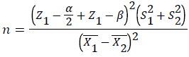 (1)
(1)
Ten teeth were calculated for each group and the total number of samples in this study was 50 teeth.
Implementation method
Stages of samples preparation:
The canals were filled 1 mm shorter than the working length up to file number #40 (Mani, Japan) and were washed with normal saline. The canals were dried with paper point (GAPADENT, China) and filled with metapex (METABIOMED, Korea) 1 mm shorter than the length of the operation.
The teeth were randomly divided into 5 groups; each of them randomly received one type of posts.
Each of the samples then received a code containing the group and sample number.
The samples were mounted in jaw-shaped stone plaster to facilitate the next steps.
Considering the bonded surface effect in fracture resistance of samples, CBCT scans were carried out using Planmeca Promax 3D (Planmeca, Helsinki, Finland). The images were taken at 84 kVp, 6 mA and 12 Seconds exposure. Then, the bonded surface area of the samples was calculated using On Demand3D software.
In the first group, 1 mm lightcure glass ionomer (Glass ionomer Fuji II LC, Japan) were placed on the metapex (1 mm thick) as the base, which were cured for 40 seconds with LED (Woodpecker, LED, China) and its residues were cleaned using a round bur and low speed hand piece. Enamel, dentin and intracanal walls were etched for 15 Seconds. Etched surface were rinsed and dried. Two layer of dentin bond (3M ESPE, St. Paul, MN, USA) were applied and cured for 40 Seconds. Next Composite (Z250, 3M ESPE, USA) was incrementally applied. Restoration with 0.5 mm extension was performed on cementum after the canal entrance seal.
Regarding other groups, 4 mm coronal canal was empty in order to provide post space. 1 mm the glass ionomer (Glass ionomer Fuji II LC, Japan) was placed and cured for 40 seconds with LED (Woodpecker, LED, China). Then, its residues were cleaned as 3 mm space remained for the post. The space remaining was etched in the root canal of each tooth with 35% phosphoric acid (vericam, denfill, Korea) for 15 seconds. After rinsing and drying, the post space walls were doped with microblasted dentin bonding (Single Bond, 3M ESPE, USA) and they are cured for 40 seconds (according to the factory's order) after drying it for 2 seconds-5 seconds [1,2,4,5] (Figures 1 and 2).

Figure 1. Teeth to calculate cross-sectional surface using CBCT radiography mounted

Figure 2. Calculate the cross-sectional area by CBCT
Groups include:
Group 1 (Composite filling extended 0.5 mm to cementum)
The crown build up was performed with composite (Z250, 3M ESPE, USA), so that 1 mm of enamel and 0.5 mm of cementum was covered by the composite (Every 2 mm composite was cured for 40 seconds).
Group 2 (Composite filling with composite post)
The prepared space for the post was filled and compressed using a composite (Z250 3M ESPE, USA) with a high burnished condenser inside the canal, then cured. The complete build-up of the tooth was done in 4 mm length using composite (Z250, 3M ESPE, USA) (Every 2 mm composite was cured for 40 seconds) (Figure 3).

Figure 3. Composite filling with composite post schematic
Group 3 (Composite filling extended 0.5 mm to cementum with composite post)
The prepared post ’ s space was filled with composite (Z250 3M ESPE, USA) and compressed with a high burnished condenser, then cured. The complete reconstruction was done in 4 mm length using composite (Z250, 3M ESPE, USA). The reconstruction was such that 1 mm of enamel and 0.5 mm of cementum was covered by the composite (Every 2 mm composite was cured for 40 seconds) [4].
Group 4 (Composite filling with omega-shaped post)
Orthodontic wire was shaped in 0.7 mm thick stainless steel in the form of Omega with the help of universal plier. Before inserting the composite filling with omegashaped post in the root, the canal of the tooth was formed mushroom-shape preparation by using No. 1 round bur . The omega-shaped post was placed 3 mm inside the canal and 2 mm above CEJ. The post was placed inside the composite flow and then, cured with LED light cure. The complete reconstruction was done in 4 mm length using composite (Z250, 3M ESPE, USA) (Every 2 mm composite was cured for 40 seconds) (Figure 4).

Figure 4. Restored tooth with omega-shaped post
Group 5 (Composite filling extended 0.5 mm to cementum with omega-shaped post)
Orthodontic wire was shaped in 0.7 mm thick stainless steel in the form of Omega with the help of universal plier. Before inserting the composite filling with omegashaped post in the root, the canal of the tooth was formed mushroom-shape preparation by using No. 1 round bur. The omega-shaped post was placed 3 mm inside the canal and 2 mm above CEJ. The post was placed inside the composite flow and then, cured using a LED The complete reconstruction was done in 4 mm length using composite (Z250, 3M ESPE, USA). The reconstruction was such that 1mm of enamel and 0.5 mm of cementum was covered by the composite (Every 2 mm composite was cured for 40 seconds).
After restoration, all teeth were polished with polishing composite bur (Tizkavan, Iran) with high-speed turbine and water cooling. Then, the samples stored in distilled water for 24 hours at 37°C. The samples were mounted in acrylic resin using a cylindrical generator to be placed in acrylic 1mm below CEJ. Subsequently, the samples were subjected to 1500 cycles of thermal expansion of 5 degrees and 55 degrees with 30 seconds expander time and 15 seconds time of displacement. Samples were applied under shear force at 148 degrees and at a speed of 0.5 mm/min in the Universal Testing Machine (DARTEC series HC 10) to evaluate the fracture strength. Applying force in the one-third mid area of tooth palatal close to the antiseptic edge until the break of restoration continued [4] (Figure 5). The numbers obtained were considered as fracture resistance of the samples. The fracture strength was calculated by dividing these numbers into the bonded surfaces.

Figure 5. Applying 148 degrees of force to samples in the UTM device
Results
A total of 50 teeth were randomly divided into 5 groups. Considering the effect of cross-sectional surface on the fracture resistance of teeth, the difference in this factor was assessed in 5 groups by ANOVA analysis which was not statistically significant (p-value>0.05) (pvalue= 0.908). The numbers of fracture resistance and fracture strength of the samples were analyzed by one way ANOVA. The mean of fracture resistance and fracture strength are shown in Table 1.
| Group | Variables | Mean ± Standard Deviation | Standard Error | Study Averages | |
|---|---|---|---|---|---|
| Lower Band | Upper Band | ||||
| Cementum Extended Composite Filling | Fracture Resistance Based on Newton | 533.54 ± 30.17 | 9.54 | 505.3 | 589.9 |
| Fracture Strength Based on Newton per Square Millimeter | 21.30 ± 2.61 | 0.82 | 17.66 | 26.48 | |
| Area of Bonded Surface Based on Newton per Square Millimeter | 25.31 ± 2.79 | 0.88 | 21.01 | 28.67 | |
| Composite Fiiling+Omega-Shaped Post | Fracture Resistance Based on Newton | 447.77 ± 62.20 | 19.67 | 349.81 | 530.83 |
| Fracture Strength Based on Newton per Square Millimeter | 17.02 ± 3.55 | 1.12 | 12.03 | 23.17 | |
| Area of Bonded Surface Based on Newton per Square Millimeter | 26.85 ± 4.36 | 1.37 | 22.11 | 34.9 | |
| Omega-Shaped Post+Cementum Extended Composite Filling | Fracture Resistance Based on Newton | 628.85 ± 104.80 | 33.14 | 447.39 | 772.08 |
| Fracture Strength Based on Newton per Square Millimeter | 24.36 ± 3.77 | 1.19 | 18.49 | 30.63 | |
| Area if Bonded Surface Based in Newton per Square Millimeter | 25.97 ± 3.52 | 1.11 | 21.98 | 31.72 | |
| Composite Post+Cementum Extended Composite Filling | Fracture Resistance Based on Newton | 546.64 ± 78.31 | 24.76 | 442.83 | 658.74 |
| Fracture Strength Based on Newton per Square Millimeter | 22.44 ± 6.287 | 1.988 | 16.82 | 36.66 | |
| Area of Bonded Surface Based on Newton per Square Millimeter | 25.30 ± 4.74 | 1.49 | 21.11 | 31.72 | |
| Composite Filling+Composite Post | Fracture Resistance Based on Newton | 240.37 ± 64.68 | 20.45 | 116.79 | 318.02 |
| Fracture Strength Based on Newton per Square Millimeter | 9.36 ± 2.65 | 0.84 | 5.04 | 13.18 | |
| Area of Bonded Surface Based on Newton per Square Millimeter | 26.79 ± 4.05 | 1.37 | 21.34 | 33.57 | |
Table 1: Central dispersion indicators in the groups for the studied variables
Table 1 represents that the maximum fracture strength and fracture resistance was related to composite filling extended 0.5 mm to cementum with omega-shaped post (ceCF+OP) and the lowest fracture strength and fracture resistance was related to composite post (CF+CP) in groups. Tukey test was used to compare the values of restoration resistance among the groups (Tables 2 and 3).
| Variables | Omega-shaped post+cementum extended composite filling | Composite post+cementum extended composite filling | Cementum extended composite filling | Composite fiiling+Omega-shaped post | Composite filling+composite post |
|---|---|---|---|---|---|
| Omega-shaped post+cementum extended composite filling | - | 1.912 | 3.05 | 7.33 | 14.99 |
| Composite post+ cementum extended composite filling | - | - | 1.138 | 5.422 | 13.08 |
| Cementum extended composite filling | - | - | - | 4.28 | 11.94 |
| Composite fiiling+Omega-shaped post | - | - | - | - | 7.65 |
Table 2: Comparison of fracture strength difference in composite restorations in square in groups.
| Variables | Omega-shaped post+cementum extended composite filling | Composite post+cementum extended composite filling | Cementum extended composite filling | Composite fiiling+Omega-shaped post | Composite filling+composite post |
|---|---|---|---|---|---|
| Omega-shaped post+ cementum extended composite filling | - | 0.82 | 0.44 | 0.002* | 0.0001* |
| Composite post+cementum extended composite filling | - | - | 0.96 | 0.032* | 0.0001* |
| Cementum extended composite filling | - | - | - | 0.13 | 0.0001* |
| Composite fiiling+Omega-shaped post | - | - | - | - | 0.001* |
Table 3: Comparison of fracture strength p-value of composite restorations in square in groups.
The results of this study showed that the fracture strength of the composite filling+composite post (CF+CP) group is statistically significant in compared to other groups (Table 3). There was also a statistically significant difference between the fracture strength of the composite filling+omega-shaped post (CF+OP) group compared to cementum extended composite filling+omega post (ceCF +OP) group (p-value=0.002) and Composite post +cementum extended composite filling (ceCF+CP)) group (p-value=0.032) (Table 3). The fracture strength of the Omega-shaped post+cementum extended composite filling (ceCF+OP) group was greater than the cementum extended composite filling (ceCF) and the Composite post +cementum extended composite filling (ceCF+CP) groups, but this difference was not statistically significant (p-value=0.823, p-value=0.444) (Table 3). Moreover, the fracture strength of Composite post+cementum extended composite filling (ceCF+CP) group was significantly higher than cementum extended composite filling (ceCF), which was not statistically significant (p-value=0.969). In comparison with fracture strength, cementum extended composite filling (ceCF) group showed a greater strength compared to omega-shaped post restored group, but this difference was not statistically significant (pvalue= 0.137).
Discussion
The purpose of this study was to investigate the effect of cementum extended composite filling with two different posts on Fracture resistance and Fracture Strength of severely damaged primary anterior teeth. Fracture resistance is one of the most important properties of restorative materials, especially during mastication [16].
Previous studies indicated that one of the factors affecting fracture resistance is dimensions of tooth cross section [17]. By measuring the mesiodistal and baccolingual width of the tooth in the CEJ, and by matching the samples in different groups, the researchers reduce the effect of this on the results of the study [17,18]. In this study, we have investigated the precision of the cross-section and compared the fracture strength of the samples. For this purpose, the bonded surface area of the samples was measured using CBCT and there was no significant difference between these groups (p-value > 0.05).
Furthermore, the restoration height was equal in all groups so that interventional factors were eliminated as far as possible. In this research, force was applied to the teeth under an angle of 148 degrees. In permanent teeth, this angle is 135 degrees, which imitates the class I to apply the bite forces to maxillary incisors in occlusion [19]. Due to the inclination of the incisors in primary teeth, this angle is considered to be 148 degrees. Vichi et al. suggested this angle in their study on the restoration of primary teeth [20]. One of the advantages of this study, which was not observed in similar studies in primary teeth, is using thermal cycles of 5 and 55 degrees to simulate the study to the oral cavity [21]. Since these cycles in the oral cavity can affect the restoration strength and durability [22], using them while reconstructing the clinical condition can reduce fracture strength and increase the accuracy of the results. The results of the study showed that the cementum extended with omega post’s (ceCF+OP) group with the mean value as much as 628.850 Newtons had the highest fracture resistance and the restored group with composite post (CF+CP) with the mean value as much as 240.372 N had the lowest fracture resistance. The cause of high fracture resistance and strength in the cementum extended with omega post’s (ceCF+OP) group can be due to synergistic effect of composite filling with omega-shaped post located on the canal using a flowable composite, on the other hand the extention of restoration to cementum, involves more tooth structure. Another reason can be the effect of the ferrule. In this study, 0.5 mm of cementum was prepared for restoration to enhance the ferrule effect and involve more surfaces. This preparation may result greater fracture strength and resistance, along with the cementum extended restorations than restoration extended just to the enamel.
The lowest fracture resistance in the study was as much as 240.372 N.mm-2. Rentes et al. conducted a study during 3 years to 5.5 years old; the maximum bite force during mastication was as much as 213.17 N ± 43.97 N on average [21]. It concluded that all methods can be used in children. However, its long-term success requires more studies. Sudula et al. [2] revealed that the fracture resistance of the anterior teeth restored with omegashaped post was as much as 124.643 N ± 40.69 N. The compressive strength of the anterior teeth restored with omega-shaped post was as much as 61.60 N ± 29.59 N in Nilavarsan et al. [1]. The results of this study showed, the anterior teeth that were restored with omega-shaped post were 447.777 N ± 62.20 N, which is higher than the previous two studies. Sudula et al. [2] used 0.6 mm orthodontic stainless steel wire to form omega shaped post and used flowable composite to restore the crown core while in the present study, 0.7 mm orthodontic stainless steel wire was used to form the omega-shaped post. Z-250 composite was used for reconstruction of coronal portion. The reason for the difference between this study and Nilavarasan et al. study can be the difference in the measurement scale [1]. In Nilavarasan et al. study [1], compressive strength has been assessed. Mojarad et al. [5] compared the fracture resistance of three different posts. The lowest fracture resistance was related to the composite post’s group (728.26 ± 248.9) and then the omega-shaped post’s group (31.3 ± 365.6). In the present study, the fracture resistance of the omegashaped post is more than the composite post’s group. Seraj et al. [4] compared the fracture resistance of composite restorations in severely damaged anterior teeth. Among all the groups, the highest fracture resistance was related to the Composite post with cementum extended composite filling (ceCF+CP) (96.04 ± 601.08 N). In the present study, after cementum extended with Omega-shaped post (ceCF+OP), the highest fracture resistance was belongs to Composite post with cementum extended composite filling (ceCF+CP) group, which is consistent with the Seraj et al. study. In Seraj et al. study, the fracture resistance of composite post ’ s group (CF+CP) was greater than the cementum extended composite filling (ceCF) (but not statistically significant), which is inconsistent with our study in this regard [4]. No study was found similar to the current study, so more comprehensive comparison was impossible.
Conclusion
According to the results, all type of composite fillings with different posts that had been evaluated in this study can be used in severely damaged primary anterior teeth according to Rentes et al. study that measure maximum bite force 213.17 ± 43.97 N on average in children with 3 years-5.5 years old.
Acknowledgment
Authors appreciate the workers participating in this study. There was no conflict of interest among authors. This study is a summarized project approved by the Research Council of Gonabad University of Medical Sciences and sponsored by the Research Department of Gonabad University of Medical Sciences.
Conflict of Interest
The authors declare that there is no conflict of interest regarding the publication of this manuscript.
References
- Nilavarasan N, Hemalatha R, Vijayakumar R, et al. Comparsion of compressive strength among three different intracanal post materials in primary anterior teeth: An invitro study. Eur J Dent 2016; 10:464-8.
- Sudula KK, Noorani H, Shivaprakash PK, et al. Comparative evaluation of fracture load resistance and retention of Polyethylene fiber post with enhanced retentive omega-shaped short post in primary anterior teeth: An invivo study. J Dent Sci 2016; 8:215-20.
- Dean J, McDonald R, Avery D. McDonald and Averys dentistry for the child and adolescent, ninth edition. Midwestern US 2016.
- Seraj B, Ehsani S, Taravati S, et al. Fracture resistance of cementum-extended composite filling in severly damaged deciduous incisors: An invitro study. Eur J Dent 2014; 8:445-9.
- Mojarad F, Salah BB. Comparison of failure resistance of three different post types in reconstruction of severely damaged denture insecticides. J Hamadan Univ Med Sci Health Serv 2013; 20:240-6.
- Richardson B, Cleaton-Jones P. Nursing bottle caries. Pediatrics 1977; 60:748-9.
- ChunawallaYK, Zingade S, Ahmed BMN, et al. Glass fiber reinforced composite resin post & core in decayed primary anterior teeth. IJCDS 2011; 2:55-60
- Reisine S, Psoter W. Socioeconomic status and selected behavioral determinants as risk factors for dental caries. J Dent Educ 2001; 65:1009-16.
- Papathanasiou A, Curzon M, Fairpo C. The influence of restorative material on the survival rate of restorations in primary molars. Pediatr Dent 1994; 16:282-8.
- Uekusa S, Yamaguchi K, Miyazaki M. Bonding efficacy of single-step self-etch systems to sound primary and permanent tooth denin. Oper Dent 2006; 31:569-76.
- Pollard M, Curzon J, Fenlon W. Restoration of decayed primary incisors using strip coronal portion of teeths. Dent Update 1991; 18:150-2.
- Seraj B, Ghadimi S, Estaki Z, et al. Fracture resistance of tree different posts in restoration of severely damaged primary anterior teeth: An invitro study. Dent Res J 2015; 12:372-8.
- Viera CL, Ribeiro CC. Polyethylene fiber tape used as a post and core in decayed primary anterior teeth: A treatment option. J Pediatr Dent 2001; 26:1-4
- Wanderley MT, Ferreira SL, Rodrigues CR, et al. Primary anterior tooth restoration using posts with macro retentive elements. Quintessence Int 1999; 30:432-6.
- Ramires-Romito AC, Wanderley MT, Oliveira MD, et al. Biologic restoration of primary anterior teeth. Quintessence Int 2000; 31:405-11.
- Samaha AEW, El-Shabrawy S, El-Meligy O, et al. Clinical and laboratory evaluation of two different types of the post system in restoring destructed primary anterior teeth. Egypt Dent J 2005; 51:1159-77.
- Sirimai S, Riis D, Morgano S. An in vitro study of the fracture resistance and the incidence of vertical root fracture of pulpless teeth restored with six post-and-coresystems. J Prosthet Dent 1999; 81:262-9.
- Kathuria A, Kavitha M, Khetarpal S. Ex vivo fracture resistance of endodontically treated maxillary central incisors restored with fiber-reinforced composite filling with composite post s and experimental dentin posts. J Conserv Dent 2011; 14:401-5.
- Jindal S, Jindal R, Gupta K, et al. Comparative evaluation of the reinforcing effect of different post systems in the restoration of endodontically treated human anterior teeth at two different lengths of post space preparation-An in vitro study. J Dent Child 2013; 10:124-33.
- Vichi A, Ferrari M, Davidson C. Influence of ceramic and cement thickness on the masking of various types of opaque posts. J Prosthet Dent 2000; 83:412-7.
- Rentes AM, Gavião MB, Amaral JR. Bite force determination in children with primary dentition. J Oral Rehabil 2002; 29:1174-80.
- Torabi K, Fattahi F. Fracture resistance of endodontically treated teeth restored by different FRC posts: An in vitro study. Indian J Dent Res 2009; 20:282-7.
Author Info
Razieh Meshki1, Parisa Sarikhani2* and Fateme Zarouni2
1Department of Pediatric Dentistry, School of Dental Medicine, Ahvaz Jundishapur University of Medical Sciences, Ahvaz, Iran2Pediatric Dentistry Resident, Department of Pediatric Dentistry, School of Dental Medicine, Ahvaz Jundishapur University of Medical Sciences, Ahvaz, Iran
Citation: Razieh Meshki, Parisa Sarikhani, Fateme Zarouni, Effect of Cementum Extended Composite Filling with Two Different Posts on Fracture Resistance and Fracture Strength of Severely Damaged Primary Anterior Teeth, J Res Med Dent Sci, 2019, 7(3): 38-45.
Received: 23-Apr-2019 Accepted: 15-May-2019