Case Report - (2021) Volume 9, Issue 7
Esthetic Rehabilitation of Anterior Teeth with Attrited and Abraded DentitionâA Case Report
Neha Alone1*, Snehal Kharwade2, Grishmi Niswade3, Deepa Pazare4 and Praktan Gire5
*Correspondence: Neha Alone, Department of Prosthodontics, Swargiya Dadasaheb Kalmegh Smruti Dental College and Hospital, India, Email:
Abstract
The smile is the most important factor as it affects the social appearance, increases perceived attractiveness and is considered a signal of trustworthiness and intelligence. Dentists use tooth wear to fix smiles, but they are pathologic when it causes discomfort, changes the appearance and function of the dentition, and affects patients’ esthetics. Tooth wear, especially dental ceramics, is widely used to restore and rehabilitate anterior dentition because it restores the shape, texture, lustre and colour of the dentition. All ceramic restorations are most commonly used for the fabrication of the restoration as it gives esthetically excellent and long-term results. Of this, lithium-disilicate glass-ceramics are one of the newest generations which has high strength, adhesively bonded and also esthetically pleasing. In this case report, esthetic rehabilitation of dentition achieved with lithium- disilicate glass-ceramic (IPS e.max Press II) for the patient with abrasion and erosion.
Keywords
Attrition, Abrasion, Erosion, Ceramics, Rehabilitation.
Introduction
Aesthetics play a crucial role among individuals. They are aware of their personality and required treatment for the same. The size and form of anterior teeth is important for aesthetics [1]. Tooth surface loss (TSL) or tooth wear is the most common complaint among individuals and it may be because of the physiologic and pathologic causes like attrition, abrasion and erosion based on various etiological factors. This tooth wear is irreparable, which increases with age of the wellbeing. Attrition is the gradual loss of the dental hard tissues because of functional or para-functional activity of the teeth. In abrasion, there is a pathologic wearing of the teeth, which is caused by the frictional action of a foreign body on the teeth, such as that caused by tooth brushing, whereas erosion is the loss of tooth substance because of the chemical process [2-4]. As a result, there is a loss of enamel which may cause pain and sensitivity and the tooth appears yellow. I know ceramic as the most natural-looking synthetic replacement for missing teeth. A heat pressed monolithic glass ceramic material which comprises 65% lithium disilicate as crystalline structures. In this case report esthetic rehabilitation of anterior teeth was done with all ceramic crowns using lithium-disilicate reinforced glass ceramic (IPS e.max Press II, Ivoclar, Vivadent) in the patient who complained of sensitivity and improper size and shape of the anterior teeth because of erosion and abrasion related to TSL.
Case Report
A 36-year-old male reported to the Department of Prosthodontics for aesthetic rehabilitation of anterior teeth. He complained of worn dentition, improper shape, size and unaesthetic shading of teeth in upper and lower front region.
The patient was non-smoker with high functional demands and moderate esthetic expectations. Patient had a habit of aggressive brushing and consumption of acidic beverages. Patient had a history of chipping off the ceramic veneering prosthesis with 11.
On intraoral examination, attrition and erosion with upper and lower front teeth were seen. Midline diastema was present and discoloration with upper right central incisor (Figure 1). Radiographic examination revealed incomplete root canal treatment with lower anterior and pulp involvement with upper anterior teeth (Figure 2).
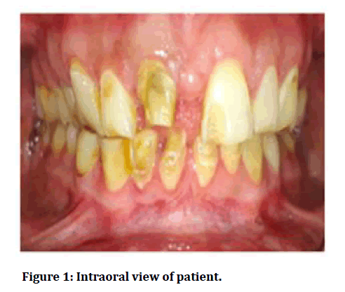
Figure 1: Intraoral view of patient.
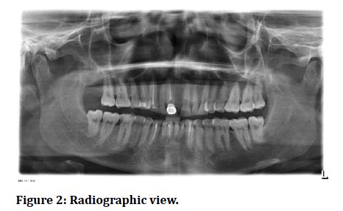
Figure 2: Radiographic view.
The patient was advised root canal treatment with the entire upper and lower anterior teeth followed by gingivectomy from 13 to 23 and from 33 to 43 to make the gingival zenith uniform. Treatment options were discussed with the patient and it was decided to give all ceramic prostheses with upper and lower anterior teeth.
Diagnostic impressions were made and casts were articulated on a semi-adjustable articulator using facebow transfer (Figure 3). A diagnostic wax up was then made (Figure 4). A crown lengthening procedure was then completed (Figures 5A and 5B).
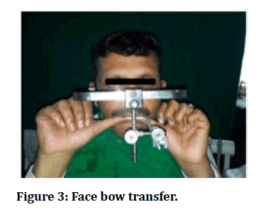
Figure 3: Face bow transfer.
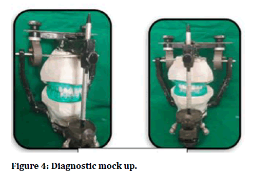
Figure 4: Diagnostic mock up.

Figure 5A and 5B: Crown lengthening with maxillary and mandibular anterior teeth.
Tooth preparation was then done with upper and lower anterior with 11, 12, 13, 21, 22, 23, 31, 32, 33, 41 42, 42 and gingival retraction cord was used for gingival retraction (Figure 6). A full arch impression was made with polyvinyl siloxane impression material (Figures 7A and 7B) for accurate recording of finish line and definitive cast with type IV stone was poured. Provisionalization was carried out using bis-acryl resin (Figures 8A-8C).

Figure 6: Tooth preparation with maxillary and mandibular anterior teeth along with gingival retraction.

Figure 7: Impression with maxillary and mandibular arch.
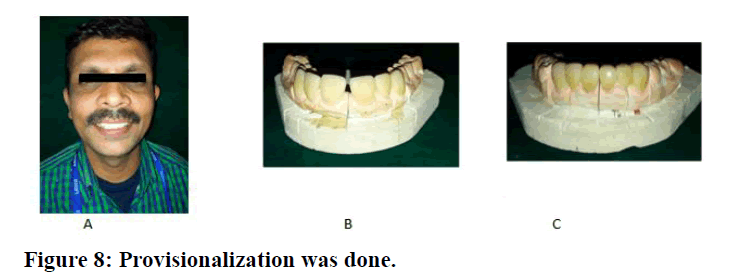
Figure 8: Provisionalization was done.
The IPS e-max monolithic crowns were fabricated (IPS emax Press MT). Trial insertion of the definitive restoration was completed to verify the marginal fit, internal adaptation, overall aesthetics and gauge patient’s satisfaction.
After adjustments, the restorations were sent to the laboratory for sintering, glazing and polishing. Internal surfaces of the restorations were etched with 5% hydrofluoric acid for 20 seconds and washed with an air water spray and air dried. A silane coupling agent (Monobond Plus, Ivoclar Vivadent) was then applied to internal surfaces of restoration for 60 seconds and air dried.
The prepared teeth were etched using 37% Phosphoric Acid (N-etch, IvoclarVivadent) for 15 seconds. After rinsing and air drying, a bonding agent (Tetric N Bond, Ivoclarvivadent) was applied & light cured for 10 seconds. Dual curing luting composite (IvoclarVivadent) was used for cementation.
Following cementation, the occlusion is checked in a centric and eccentric position for any interference. In Figure 9A and Figure 9B showing the preoperative and postoperative photograph of the patient. Follow-up visits were then scheduled for the patient.
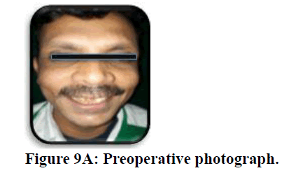
Figure 9A: Preoperative photograph.
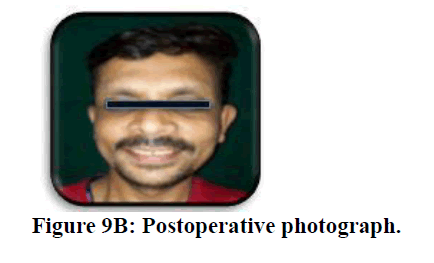
Figure 9B: Postoperative photograph.
Discussion
A cautious clinical examination and an intensive case history (dental, therapeutic, count calories and eating propensities, occupation, bruxism, or any other verbal propensities) are vital for conclusion and treatment planning.
As, in this case diminished tooth structure was displayed, lithium disilicate based ceramic framework (IPS e.max) was used to manufacture crowns of 1.0-1.5 mm thickness. Lithium disilicate has picked up notoriety for front and back single crowns and halfway scope rebuilding efforts since not at all like feldspathic or leucite fortified ceramic framework, it has higher flexural quality (360-400 MPa) and can be used where holding is limited [5,6]. It has great translucency and reacts superior chromatically to little thicknesses than leucite glass-ceramic in cases with discoloured projection teeth [7]. In a think about by Niuet al. [8], 2 mm thick example of A1 LT e max was able to accomplish shade matches underneath the edge of clinical worthiness on all establishment materials tried.
Although the long-term survival of lithium disilicate FDPs is discouraging [9], lithium-disilicate single crowns show an amazing survival rate (2 year CSRl of 100% and a 5 year CSR of 97.8%), particularly within the front locale [10]. Despite glass ceramics showing lower mechanical quality than oxide ceramics, the break resistance has appeared to extend to gum cementation [11]. A cement method using double remedy resin cement was used to bond the rebuilding efforts may be a negligibly obtrusive approach for substitution of misplaced tooth tissue.
References
- Cairo F, Graziani F, Franchi L, et al. Periodontal plastic surgery to improve aesthetics in patients with altered passive eruption/gummy smile: A case series study. Int J Dent 2012; 1-6.
- Sato S, Hotta TH, Pedrazzi V. Removable occlusal overlay splint in the management of tooth wear: A clinical report. J Prosthet Dent 2000; 83:392-395.
- Kelleher M, Bishop K. Tooth surface loss: An overview. Br Dent J 1999; 186:61-66.
- Barlett D, Philips K, Smith B. A difference in perspective-the North American and European interpretations of tooth wear. Int J Prosthodont 1999; 12:401-408.
- Hattab FN, Yassin OM. Etiology and diagnosis of tooth wear: A literature review and presentation of selected cases. Int J Prosthodont 2000; 13:101-107.
- Höland W, Schweiger M, Frank M, et al. A comparison of the microstructure and properties of the IPS Empress 2 and the IPS Empress glass-ceramics. J Biomed Mater Res 2000; 53:297-303.
- Almeida JS, Rolla JN, Edelhoff J, et al. All-ceramic crowns and extended veneers in anterior dentition: A case report with critical discussion. Am J Esthet Dent 2011; 1:60-81.
- Niu E, Agustin M, Douglas RD. Color match of machinable lithium disilicate ceramics: effects of foundation restoration. J Prosthet Dent 2013; 110:501-9.
- Sola-Ruiz MF, Lagos-Flores E, Roman-Rodriguez JL, et al. Survival rates of a lithium disilicate-based core ceramic for three-unit esthetic fixed partial dentures: A 10-year prospective study. Int J Prosthodont 2013; 26:175-80.
- Pieger S, Salman A, Bidra AS. Clinical outcomes of lithium disilicate single crowns and partial fixed dental prostheses: A systematic review. J Prosthet Dent 2014; 112:22-30.
- Pagniano RP, Seghi RR, Rosenstiel SF, et al. The effect of a layer of resin luting agent on the biaxial flexure strength of two all-ceramic systems. J prosthet Dent 2005; 93:459-66.
Author Info
Neha Alone1*, Snehal Kharwade2, Grishmi Niswade3, Deepa Pazare4 and Praktan Gire5
1Department of Prosthodontics, Swargiya Dadasaheb Kalmegh Smruti Dental College and Hospital, Hingna, Nagpur, Maharashtra, India2Department of Prosthodontics, SMBT Institute of Dental Sciences and Research, Dhamangaon, Igatpuri, Nashik, Maharashtra, India
3Department of Periodontics, Swargiya Dadasaheb Kalmegh Smruti Dental College and Hospital, Hingna, Nagpur, Maharashtra, India
4Department of Periodontics, V.Y.W.S Dental College and Hospital, Amrawati, Maharashtra, India
5Mumbai, Maharashtra, India
Citation: Neha Alone, Snehal Kharwade, Grishmi Niswade, Deepa Pazare, Minal Ganvir, Praktan Gire,Esthetic Rehabilitation of Anterior Teeth with Attrited and Abraded Dentition-A Case Report, J Res Med Dent Sci, 2021, 9(7): 274-277
Received: 30-Jun-2021 Accepted: 19-Jul-2021
