Research - (2022) Volume 10, Issue 1
Estimation of Dental Age in the Maxillary Right Third Molar (18) Using Willemâs Method in South Indian Population
Devika B and Abirami Arthanari*
*Correspondence: Abirami Arthanari, Department of Forensic Odontology, Saveetha Institute of Medical and Technical Sciences, Chennai, India, Email:
Abstract
Introduction: Age estimation is an important area of forensic odontology and it has increased its importance in the past few years. There are various methods in the estimation of dental age. Some of them are Atlas approaches, by the use of radiographs, where the morphological distinct stages of mineralization of all the teeth are observed. The scoring method; it was framed by Demirjian. In 2001, Willems et al. assessed the accuracy of Demirjian's method in Belgian Caucasian population; this modified method calculated the overall maturity score by adding the adapted scores for the seven teeth in the mandible which presented the result directly as the estimated age of the individual. This modification has been evaluated among various populations and has been reported to be more accurate than the original Demirjian’s method. Aim and objective: The aim of the study is to estimate the dental age in the maxillary right third molar (18) using Willem's method. Materials and methods: In our study, 100 OPGs were collected, out of which 50 were males and another 50 were females. The OPGs collected were from the age group of 10 to 20 years. The population was divided into 10 groups. The study was approved by an ethical clearance team. The data was collected along with actual age and dental age. Willems' method of stage was used to determine the dental age. The staging in Williams’s method was modified from A to H to (i) to (viii) for easy accessibility. The data was entered in SPSS (version 26.0) for statistical analysis. Descriptive analysis was performed. Results: In this present study the data was derived and estimated with the SD value of 2.22 years in males and 3.89 years in females. Chronological age shows statistically significant value (p=0.054), while tooth staging shows statistically insignificant value (p=0.762). Conclusion: Age estimation plays an important role in forensic, legal and criminal proceedings. In this study, significant relation was found between estimated DA and CA and thus Willems method seems to be applicable in estimating age.
Keywords
Age estimation, Willem's method, Maxillary right third molar, OPGs, South Indian Population, Innovative Method
Introduction
Age estimation is an important area of forensic odontology and it has been gaining increased importance in the past few years. A person’s age can be estimated by the amount of maturation of the various tissue systems [1]. Determining the age of an individual is an important procedure in forensic sciences [2]. Estimating age from teeth is considered to be reliable, as maturational events associated with tooth formation are less variable [3]. Development of a third molar is the key stone as it's the only tooth still in development by age 16. However, previous findings show that the mineralization of the third molar is a population-specific process. The development and eruption of third molars does not seem to occur in every ethnic group at the same age [4]. There are various methods in the estimation of dental age. Some of them are Atlas approaches, by the use of radiographs, where the morphological distinct stages of mineralization of all the teeth are observed [5]. Next is the scoring method; it was framed by Demirjian. He tried to simplify chronological age estimation and restricted the number of stages of tooth development to 8 giving a score of “A” through “H”. He also confined the analysis to the 1st seven teeth in the left quadrant [6]. In 2001, Willems et al. assessed the accuracy of Demirjian's method in Belgian Caucasian population. He modified the scoring system when a significant overestimation was reported [7]. This modified method calculated the overall maturity score by adding the adapted scores for the seven teeth in the mandible which presented the result directly as the estimated age of the individual. This modification has been evaluated among various populations and has been reported to be more accurate than the original Demirjian’s method [8]. The above two methods of age estimation are for children.
In previous studies, age estimation was done using Demirjian’s method between the age group of 13 to 23 years in mandibular molar and concluded that there was a strong relation between dental and chronological age [9]. In another study, 332 subjects participated, and their OPGs were taken, and age was calculated using Willem’s method in the mandibular molar. The study showed significant relation between DA and CA [10]. According to Temitope Ayodeji Esan et al, more precise estimation of CA in different populations was estimated using Willem’s method, while Demirjian’s method has a broad application in terms of determining maturity scores [11]. In a previous research, the sample size was 60 and ranging from 6-16 years was taken and orthopantomography of all the patients was made. The DA was found out to be less than the actual CA of the sample, hence there seems to be an underestimation. A dental growth lag is seen in the Chitwan population [12]. Our team has extensive knowledge and research experience that has translate into high quality publications [13-32]. The aim of the study is to estimate the dental age in the maxillary right third molar using Willem's method.
Materials and Methods
In our study, 100 OPGs were collected, out of which 50 were males and another 50 were females. The OPGs collected were from the age group of 10 to 20 years. The population was divided into 10 groups, 10-10.9, 11-11.9, 12-12.9, 13-13.9, 14-14.9, 15-15.9, 16-16.9, 17-17.9, 18-18.9 and 19-19.9 years of age. The study is approved by the ethical clearance team. The data was collected Descriptive analysis was performed (Figure 3). along with actual age and dental age. Willems' method of stage was used to determine the dental age. The staging in Willems' method was modified from A to H (Figure 1) to (i) to (viii) (Figure 2) for easy accessibility. The data was entered in SPSS (version 26.0) for statistical analysis.
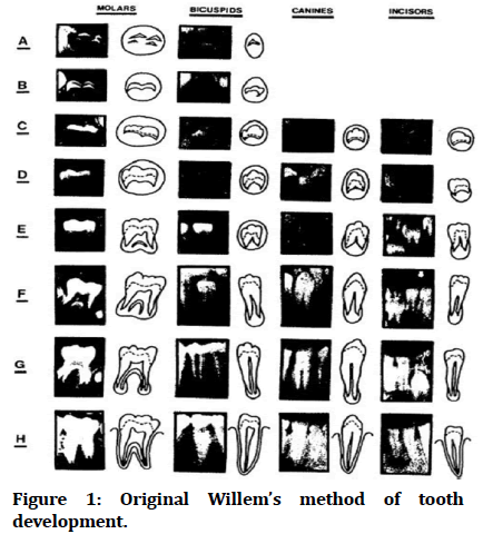
Figure 1: Original Willem’s method of tooth development.
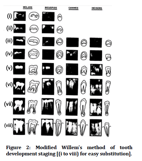
Figure 2: Modified Willem’s method of tooth development staging [(i to viii) for easy substitution].
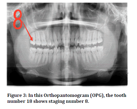
Figure 3: In this Orthopantomogram (OPG), the tooth number 18 shows staging number 8.
Results
This study took the sample size to be 100 OPGs amongst which 50 were females and 50 males. The mean chronological age of 50 male samples are 14.8, while the mean estimated age of 50 male samples are 4.12 (Figure 5). The difference that has been found between the actual chronological age and the estimated tooth staging of male population is statistically significant with a p value <0.05 (0.05) (Table 2). The mean chronological age of 50 female samples are 14.82, while the mean estimated age of 50 female samples are 4.42 (Figure 4). The difference that has been found between the actual chronological age and the estimated tooth staging of the female population is statistically insignificant (0.762). The standard mean error of male population is 0.354 and the female population is 0.335. (Table 1). The association between tooth developmental stages and age group was graphically represented (Figure 6, 7). In the age group of 18-18.9 years, male population had more staging accuracy than females.
| Tooth staging | |||
|---|---|---|---|
| Mean | SD | Std. Mean error | |
| MALE | 4.12 | 2.5042 | 0.354 |
| FEMALE | 4.42 | 2.36548 | 0.335 |
Table 1: Table shows the mean, standard deviation and standard mean error of tooth staging of male and female population.
| Male | Female | |
|---|---|---|
| P-Value | 0.054 | 0.762 |
Table 2: Table shows the P-value of male and female population.
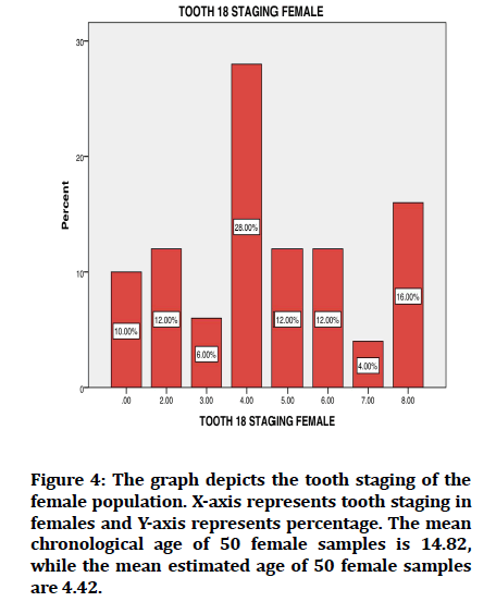
Figure 4:The graph depicts the tooth staging of the female population. X-axis represents tooth staging in females and Y-axis represents percentage. The mean chronological age of 50 female samples is 14.82, while the mean estimated age of 50 female samples are 4.42.
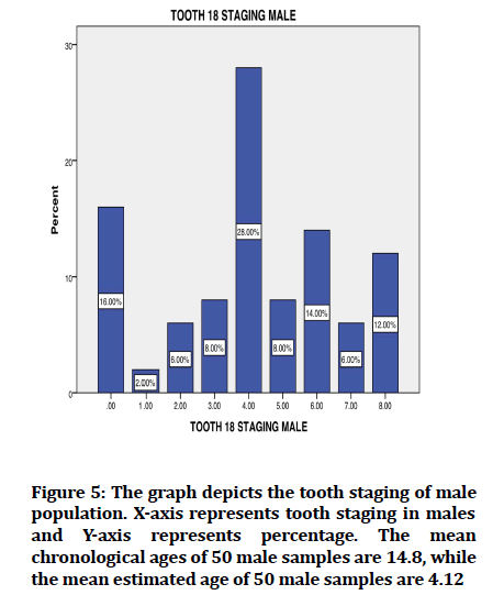
Figure 5:The graph depicts the tooth staging of male population. X-axis represents tooth staging in males and Y-axis represents percentage. The mean chronological ages of 50 male samples are 14.8, while the mean estimated age of 50 male samples are 4.12.
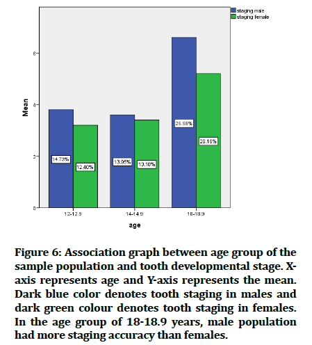
Figure 6:Association graph between age group of the sample population and tooth developmental stage. Xaxis represents age and Y-axis represents the mean. Dark blue color denotes tooth staging in males and dark green colour denotes tooth staging in females. In the age group of 18-18.9 years, male population had more staging accuracy than females.
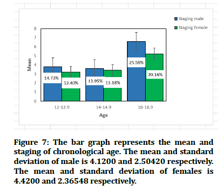
Figure 7:The bar graph represents the mean and staging of chronological age. The mean and standard deviation of male is 4.1200 and 2.50420 respectively. The mean and standard deviation of females is 4.4200 and 2.36548 respectively.
Discussion
Developing teeth in radiographs are frequently used to assess dental maturity and estimate age. Age estimation has become increasingly important to determine the age of living individuals. Identification of age is very important for a variety of reasons, including identifying criminal and legal responsibility, determining the emotional support needed for the victim of a sexual assault and for many other social events such as a birth certificate, marriage, beginning a job, joining the army, and retirement [33]. Dental development is viewed as a more reliable gauge for assessing the age of children and juveniles in forensic and anthropological contexts [34]. The evaluation of mineralization from OPGs is the most suitable method for age estimation using teeth because a single radiograph gives the complete developmental status of dentition. Digital OPGs were used as the images can be magnified to make analysis easier.
In the present study, the age group was divided into 10 groups of 10-10.9 to 18-18.9. This is in accordance with a previous study by Abirami Arthanari et al, where 200 OPGs were taken and a similar age group division was done [35]. In the present study, 4.12 ± 2.50 years was the age of males and 4.42 ± 2.36 years was the age of females. In a previous study, the Willems method reduces the average overestimation, with 0.36 years for boys and 0.02 years for girls. The age group of 15 years was more in males and the age group of 13 and 15 was more in the female population. In our study, the staging number 4 shows a higher proportion in both the population. It signifies that the crown formation is complete till the dentino-enamel junction. Errors in age estimation are smaller in every age group with the Willems method [36]. Moreover, previous studies have failed to consider the tooth as a whole [37]. The results of this study reveal that Willems’s method is more related to the actual age, prone to overestimation but still the best of all methods studied [38].
The “P” value is representative of Pearson's correlation coefficient of dental age and chronological age. The “P” value was 0.054 in males and 0.762 in females, thus suggesting a strong positive correlation between chronological age and dental age of observed samples in males (P ≤ 0.05) and a non-significant correlation in females (P ≰ 0.05). This finding was similar to the findings of Maber et al [39] and Mani et al [40] which reported higher accuracy of Willems method among males than females. The differences in dental maturation between male and female populations that might cause significant mean age differences are more visible in the male population, whereas females experience an earlier growth spurt phase than males. The growth spurt phase in females occurs in early birth periods, 6 until 7 years, and 12 to 14 years old. Some literature has discussed that the mean difference of the onset of a pubertal growth spurt in males and females is about 2 years earlier for girls [41]. However, sexual dimorphism in developmental pace takes place around the completion of the tooth crown and prolongs to increase during the stage of root formation. These findings suggest that tooth formation follows the pattern of general growth and may be affected by hormonal changes [42]. The limitations of the study are small population size and not finding the dental age of the population.
Conclusion
Age estimation plays an important role in forensic, legal and social issues. When Williams method of age estimation has been applied to South Indians, underestimation of age was noted leading to delayed dental maturity. In this study, a significant relation was found between estimated DA and CA among male population than females and thus the Williams method seems to be applicable in estimating age in the South Indian population. As no published data is available regarding the application of Williams method in selected populations, this paper provides an insight in using Williams method in South Indians for estimating mean and standard deviation of the given population. Further studies to formulate an equation where SD can be applied to find the dental age should be carried out.
Acknowledgement
We would like to express our gratitude towards Saveetha Dental college and hospital, Saveetha university, Institute of Medical and Technical Science, Saveetha University for their constant support in completing this research.
Conflict of Interest
Nil
Source of Funding
This study was supported by the following agencies.
• Saveetha Dental College, SIMATS, Saveetha University, India. • Virtusa Consultancy Services.
References
- Hegde RJ, Sood PB. Dental maturity as an indicator of chronological age: Radiographic evaluation of dental age in 6 to 13 years children of Belgaum using Demirjian methods. J Indian Soc Pedod Prev Dent 2002; 20:132-8.
- Sathawane RS, Agrawal N. Applicability of Chaillet-Demirjian’s and Willem’s age assessment methods in Chhattisgarh population: Proposing Chhattisgarh population specific formula. Int J Maxillofac Imaging 2017; 3:8-11.
- Maia MC, Martins MD, Germano FA, et al. Demirjian's system for estimating the dental age of northeastern Brazilian children. Forensic Sci Int 2010; 200:177-1.
- Olze A, Schmeling A, Taniguchi M, et al. Forensic age estimation in living subjects: The ethnic factor in wisdom tooth mineralization. Int J Legal Med 2004; 118:170-3.
- Willems G. A review of the most commonly used dental age estimation techniques. J Forensic Odonto-stomatology 2001; 19:9-17.
- Demirjian A, Goldstein H, Tanner JM. A new system of dental age assessment. Hum Bio 1973; 1:211-27.
- Willems G, Van Olmen A, Spiessens B, et al. Dental age estimation in Belgian children: Demirjian's technique revisited. J Forensic Sci 2001; 46:893-5.
- Grover S, Marya CM, Avinash J, et al. Estimation of dental age and its comparison with chronological age: Accuracy of two radiographic methods. Med Sci Law 2012; 52:32-5.
- Ajmal M, Assiri KI, Al-Ameer KY, et al. Age estimation using third molar teeth: A study on southern Saudi population. J Forensic Dental Sci 2012; 4:63.
- Mohammed RB, Krishnamraju PV, Prasanth PS, et al. Dental age estimation using Willems method: A digital orthopantomographic study. Contemp Clin Dent 2014; 5:371.
- Esan TA, Yengopal V, Schepartz LA. The Demirjian versus the Willems method for dental age estimation in different populations: A meta-analysis of published studies. PLoS One 2017; 12:e0186682.
- Kapoor D. Age estimation using Orthopantomograph and Willems method in Chitwan population: an original study. JCMS-Nepal 2018; 14:98-101.
- Santhakumar P, Prathap L. Awareness on preventive measures taken by health care professionals attending COVID-19 patients among dental students. Eur J Dent 2020; 14:S105-9.
- Mathew MG, Samuel SR, Soni AJ, et al. Evaluation of adhesion of Streptococcus mutans, plaque accumulation on zirconia and stainless steel crowns, and surrounding gingival inflammation in primary molars: Randomized controlled trial. Clin Oral Investig 2020; 24:3275-80.
- Sridharan G, Ramani P, Patankar S, et al. Evaluation of salivary metabolomics in oral leukoplakia and oral squamous cell carcinoma. J Oral Pathol Med 2019; 48:299-306.
- Hannah R, Ramani P, Ramanathan A, et al. CYP2 C9 polymorphism among patients with oral squamous cell carcinoma and its role in altering the metabolism of benzo [a] pyrene. Oral Surg Oral Med Oral Pathol Oral Radiol 2020; 130:306-12.
- Antony JV, Ramani P, Ramasubramanian A, et al. Particle size penetration rate and effects of smoke and smokeless tobacco products–An invitro analysis. Heliyon 2021; 7:e06455.
- Sarode SC, Gondivkar S, Sarode GS, et al. Hybrid oral potentially malignant disorder: A neglected fact in oral submucous fibrosis. Oral Oncol 2021: 105390.
- Ramani P, Tilakaratne WM, Sukumaran G, et al. Critical appraisal of different triggering pathways for the pathobiology of pemphigus vulgaris-A review. Oral Dis 2021.
- Chandrasekar R, Chandrasekhar S, Sundari KS, et al. Development and validation of a formula for objective assessment of cervical vertebral bone age. Prog Orthod 2020; 21:1-8.
- Subramanyam D, Gurunathan D, Gaayathri R, et al. Comparative evaluation of salivary malondialdehyde levels as a marker of lipid peroxidation in early childhood caries. Eur J Dent 2018; 12:067-70.
- Jeevanandan G, Thomas E. Volumetric analysis of hand, reciprocating and rotary instrumentation techniques in primary molars using spiral computed tomography: An in vitro comparative study. Eur J Dent 2018; 12:021-6.
- Ponnulakshmi R, Shyamaladevi B, Vijayalakshmi P, et al. In silico and in vivo analysis to identify the antidiabetic activity of beta sitosterol in adipose tissue of high fat diet and sucrose induced type-2 diabetic experimental rats. Toxicol Mech Methods 2019; 29:276-90.
- Sundaram R, Nandhakumar E, Haseena Banu H. Hesperidin, a citrus flavonoid ameliorates hyperglycemia by regulating key enzymes of carbohydrate metabolism in streptozotocin-induced diabetic rats. Toxicol Mech Methods 2019; 29:644-53.
- Alsawalha M, Rao CV, Al-Subaie AM, et al. Novel mathematical modelling of Saudi Arabian natural diatomite clay. Mater Res Express 2019; 6:105531.
- Yu J, Li M, Zhan D, et al. Inhibitory effects of triterpenoid betulin on inflammatory mediators inducible nitric oxide synthase, cyclooxygenase-2, tumor necrosis factor-alpha, interleukin-6, and proliferating cell nuclear antigen in 1, 2-dimethylhydrazine-induced rat colon carcinogenesis. Pharmacogn Mag 2020; 16:836.
- Shree KH, Ramani P, Sherlin H, et al. Saliva as a diagnostic tool in oral squamous cell carcinoma–a systematic review with meta analysis. Pathol Oncol Res 2019; 25:447-53.
- Zafar A, Sherlin HJ, Jayaraj G, et al. Diagnostic utility of touch imprint cytology for intraoperative assessment of surgical margins and sentinel lymph nodes in oral squamous cell carcinoma patients using four different cytological stains. Diagn Cytopathol 2020; 48:101-10.
- Karunagaran M, Murali P, Palaniappan V, et al. Expression and distribution pattern of podoplanin in oral submucous fibrosis with varying degrees of dysplasia–an immunohistochemical study. J Histotechnol 2019; 42:80-6.
- Sarode SC, Gondivkar S, Gadbail A, et al. Oral submucous fibrosis and heterogeneity in outcome measures: A critical viewpoint. Future Oncol 2021; 17:2123-6.
- Preeth DR, Saravanan S, Shairam M, et al. Bioactive Zinc (II) complex incorporated PCL/gelatin electrospun nanofiber enhanced bone tissue regeneration. Eur J Pharm Sci 2021; 160:105768.
- Prithiviraj N, Yang GE, Thangavelu L, et al. Anticancer compounds from starfish regenerating tissues and their antioxidant properties on human oral epidermoid carcinoma kb cells. Pancreas 2020; 155–6.
- El-Bakary AA, Hammad SM, Ibrahim FM. Comparison between two methods of dentalage estimation among egyptian children. Mansoura J Forensic Med Clin Toxicol 2009; 17:75-87.
- Oziegbe EO, Esan TA, Oyedele TA. Brief communication: Emergence chronology of permanent teeth in Nigerian children. Am J Phys Anthropol 2014; 153:506-11.
- Arthanari A, Doggalli N, Vidhya A, et al. Age estimation from second & third molar by modified gleiser and hunt method: A retrospective study. Indian J Forensic Med Toxicol 2020; 14.
- Urzel V, Bruzek J. Dental age assessment in children: A comparison of four methods in a recent French population. J Forensic Sci 2013; 58:1341-7.
- Galić I, Vodanović M, Cameriere R, et al. Accuracy of cameriere, haavikko, and willems radiographic methods on age estimation on Bosnian–Herzegovian children age groups 6–13. Int J Legal Med 2011; 125:315-21.
- Cortés MM, Rojo R, García EA, et al. Accuracy assessment of dental age estimation with the Willems, Demirjian and Nolla methods in Spanish children: Comparative cross-sectional study. BMC Pediatr 2020; 20:1-9.
- Maber M, Liversidge HM, Hector MP. Accuracy of age estimation of radiographic methods using developing teeth. Forensic Sci Int 2006; 159:S68-73.
- Mani SA, Naing LI, John J, et al. Comparison of two methods of dental age estimation in 7–15‐year‐old Malays. Int J Paediatr Dent 2008; 18:380-8.
- Burstone CJ. Process of maturation and growth prediction. Am J Orthod 1963; 49:907–19.
- Alshihri AM, Kruger E, Tennant M. Dental age assessment of Western Saudi children and adolescents. Saudi Dent J 2015; 27:131-6.
Indexed at, Google Scholar, Cross Ref
Indexed at, Google Scholar, Cross Ref
Indexed at, Google Scholar, Cross Ref
Indexed at, Google Scholar, Cross Ref
Indexed at, Google Scholar, Cross Ref
Indexed at, Google Scholar, Cross Ref
Indexed at, Google Scholar, Cross Ref
Indexed at, Google Scholar, Cross Ref
Indexed at, Google Scholar, Cross Ref
Indexed at, Google Scholar, Cross Ref
Indexed at, Google Scholar, Cross Ref
Indexed at, Google Scholar, Cross Ref
Indexed at, Google Scholar, Cross Ref
Indexed at, Google Scholar, Cross Ref
Indexed at, Google Scholar, Cross Ref
Indexed at, Google Scholar, Cross Ref
Indexed at, Google Scholar, Cross Ref
Indexed at, Google Scholar, Cross Ref
Indexed at, Google Scholar, Cross Ref
Indexed at, Google Scholar, Cross Ref
Indexed at, Google Scholar, Cross Ref
Indexed at, Google Scholar, Cross Ref
Indexed at, Google Scholar, Cross Ref
Indexed at, Google Scholar, Cross Ref
Indexed at, Google Scholar, Cross Ref
Indexed at, Google Scholar, Cross Ref
Indexed at, Google Scholar, Cross Ref
Indexed at, Google Scholar, Cross Ref
Author Info
Devika B and Abirami Arthanari*
Department of Forensic Odontology, Saveetha Institute of Medical and Technical Sciences, Chennai, IndiaCitation: Devika B, Abirami Arthanari, Estimation of Dental Age in the Maxillary Right Third Molar (18) Using Willemâ??s Method in South Indian Population , J Res Med Dent Sci, 2022, 10(1): 9-14
Received: 06-Dec-2021, Manuscript No. JRMDS-21-41540; , Pre QC No. JRMDS-21-41540 (PQ); Editor assigned: 08-Dec-2021, Pre QC No. JRMDS-21-41540 (PQ); Reviewed: 22-Dec-2021, QC No. JRMDS-21-41540; Revised: 27-Dec-2021, Manuscript No. JRMDS-21-41540 (R); Published: 03-Jan-2022
