Research - (2022) Volume 10, Issue 1
Evaluation and Comparison of the Dimensions of Cranial Base in Boys and Girls between 10â14 Years of Age having Skeletal Class-II Malocclusion as Compared to Skeletal Class I Malocclusion
Kamble RH, Anuradha Rajkuwar, Karthika Nambiar*, Toshniwal TG and Sumukh Nerurkar
*Correspondence: Karthika Nambiar, Department of Orthodontics and Dentofacial Orthopaedics, Sharad Pawar Dental College, Datta Meghe Institute of Medical Sciences, Sawangi meghe, Wardha, India, Email:
Abstract
Introduction: As orthodontists we are interested in understanding how the face changes from its embryologic form through childhood, adolescence and adulthood. Of particular interest, an understanding of how and where the growth occurs as the growth and development of cranial base is interrelated with face, directly influencing the growth of the maxilla and mandible and, consequently, the establishment of their anteroposterior relationship. Numerous studies have been conducted in the past using lateral cephalogram- a 2D technique; to determine the dimensions of the cranial base in individuals. With the advent of the 3D methods of evaluation, Orthodontists have started being more aware of the functional spaces and their role in the growth and development. Aim of this study: It was to evaluate and quantify, three dimensional changes in the cranial base using the growth records of children between 10 to 14 years of age using 3D DVT. Methodology: The sample comprised of 20 patients divided into two groups of 10 each having skeletal Class-II and Class-I malocclusion. Each group divided into two subgroups having 5 boys & 5 girls respectively all between the ages of 10-14 years. The parameters depicting the cranial base, anterior cranial base (ACB) length. [N-S]And Posterior cranial base (PCB) length, were quantified using several linear measurements. The data was collected at 6 - month time interval (T0, T1, T2 and T3). Results: The subjects with class-II malocclusion showed an increased anterior cranial base length and a decreased posterior cranial base length as compared to class-I cases. Conclusion: The growth and development of cranial base is interrelated with face, directly influencing the growth of the maxilla and mandible and, consequently, the establishment of their anteroposterior relationship. Any variation at cranial base will have effects on the position of maxilla and maxilla mandibular relationship.
Keywords
Anterior cranial base, Posterior cranial base, 3D- DVT, Class-I malocclusion, Class-II malocclusion
Introduction
The cranial base (CB) is of particular importance in orthodontics, because its growth and development is associated with face, which in turn influences the growth of the maxilla and mandible and, consequently, establishes an anteroposterior relationship. Six separate bones namely- ethmoid, frontal, sphenoid, parietal, occipital and temporal all of which are interconnected by synchondrosis [1-3], together constitute the cranial base. Cranial base synchondroses are important growth centers of the craniofacial skeleton and the last sites in the cranium to terminate growth [4]. The chondrocranium, which acts as a growth plate, gives rise to the cranial base, which is later replaced by bone via endochondral ossification [5].
According to Bjork et al. [6], the cranial base derives primarily from the chondrocranium, and its form varies greatly during development. There is a flattening tendency before birth, which shifts during the early years of life, gradually flexing until it reaches its final form about the age of ten. According to Bishara et al. [7], at two years of age, the cranial base reaches 87 percent of its adult size, 90 percent at five years, and 98 percent at fifteen years. Also, according to Moore et al. [8], the cranial base reaches around 90% of its total size at about five years of age, and it is stable from then on. As a result, it develops quickly throughout the first few years of life before decelerating.
Being orthodontists, a thorough knowledge of the facial changes, starting from the embryonic form to adulthood. Moreover, the site and direction of growth, the amount of growth remaining, the influence of genetic and environmental factors that aid in facial growth and how we, as orthodontists can bring about optimum results through our treatment is of utmost importance.
Any changes at the CB will have its influence on the relative position of the upper denture base and in turn the maxilla mandibular relationship.
Therefore, the current study was conducted in order to evaluate and quantify, three dimensional changes in the cranial base using the growth records of children between 10 to 14 years of age using 3D DVT. This study provides data on the incremental growth changes in the cranial base in 3 dimensions in Skeletal Class I and II malocclusions. Hence the results of this study will help us in understanding the growth changes in Indian population.
Aim
This study aimed at evaluating and comparing the dimensions of cranial base in boys and girls from 10 years to 14 years of age having skeletal Class-II malocclusion as compared to skeletal Class I malocclusion.
Objectives
• To evaluate the dimensions of Cranial base length in skeletal Class-II
• malocclusion.
• To evaluate the dimensions of Cranial base length in skeletal Class-I malocclusion.
• To compare dimensions of cranial base length in skeletal Class-II and Class-I malocclusion at 6 monthly interval period between age 10 to 14 years.
Materials and Methods
The current study was conducted in the “Department of Orthodontics and Dentofacial Orthopedics”, Sharad Pawar Dental College and Hospital, Sawangi (M.), Wardha”, in co-ordination with Department of Radiodiagnosis and Imaging, Acharya Vinoba Bhave Rural Hospital (AVBRH), Datta Meghe Institute of Medical Sciences, Deemed University (DMIMS, DU), Wardha, Maharashtra. The sample comprised of 20 patients divided into two groups of 10 each having skeletal Class- II and Class-I malocclusion. Each group is divided into two subgroups having 5 boys & 5 girls respectively.
The criteria for patient selection were as follows
• Age range between 10 - 14 years for both males and females.
• No H/O previous orthodontic & orthopaedic treatment.
• No history of any congenital deformities, metabolic disorders or syndromes.
Complete case history and clinical examination (Intramural and Extraoral examination) of cases screened was done.
The lateral cephalograms of selected cases were traced, digitized and analysed with computer software to assess the skeletal malocclusion.
Based on these findings the study sample was segregated into two groups:
Group 1- Skeletal Class - I (Angle ANB=0° to 4°)
Boys
Girls
Group 2- Skeletal Class- II (Angle ANB=more than 5°)
Boys
Girls
The parameters depicting the cranial base, Anterior cranial base (ACB) length [N-S] and Posterior cranial base (PCB) length, were quantified using several linear measurements. The data was collected at 6-month time interval (T0, T1, T2 and T3). The results obtained were tabulated and subjected to statistical analysis.
Statistical analysis
The data was analysed using descriptive and inferential statistics. The software which was used in the analysis were SPSS 17.0, EPI-INFO 6.0 version and Graph Pad Prism 6.0 version and p<0.05 is considered as level of significance. The statistical tests used for the analysis of the result were:
Student’s paired t test.
Student’s unpaired t test.
Results and Observations
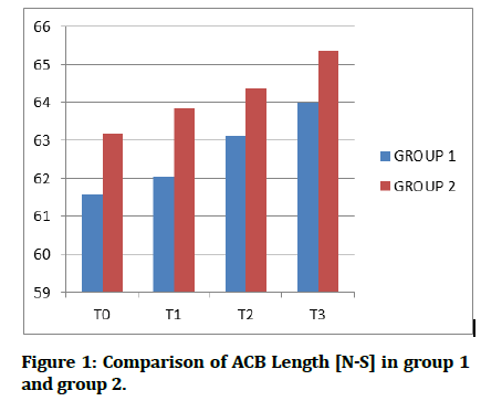
Comparison of ACB Length [N-S] in Group 1 and Group 2 (Table 1 & Figure 1)
Figure 1: Comparison of ACB Length [N-S] in group 1 and group 2.
| Time interval | Group 1 | Group 2 | T-Value | P-Value |
|---|---|---|---|---|
| T0 | 61.58 ± 2.60 | 63.17 ± 2.97 | 1.27 | 0.22, NS |
| T1 | 62.02 ± 2.39 | 63.84 ± 3.07 | 1.47 | 0.15, NS |
| T2 | 63.11 ± 2.85 | 64.36 ± 3.08 | 0.94 | 0.35, NS |
| T3 | 63.99 ± 3.03 | 65.34 ± 3.08 | 0.98 | 0.33, NS |
Table 1: Comparison of ACB Length [N-S] in Group 1 and Group 2.
The mean ACB length in Class I malocclusion was 61.58 mm (SD ± 2.6), 62.02mm (SD ± 2.39), 63.11mm (SD ± 2.85) and 63.99 mm (SD ± 3.03) at time interval T0, T1, T2, and T3 respectively and in Class II malocclusion it was 63.17mm (SD ± 2.97), 63.84 mm (SD ± 3.07), 64.36 mm (SD ± 3.08) and 65.34 mm (SD ± 3.08) at time interval T0, T1, T2, and T3 respectively. No significant variance was observed (P=0.000 i.e. P<0.05) in anterior cranial base length at the entire time interval T0 (P=0.22), T1 (P=0.15), T2 (P=0.35) and T3 (P=0.33).
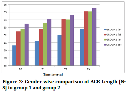
Gender wise comparison of ACB Length [N-S] in Group 1 and Group 2 (Table 2 & Figure 2)
| Time interval | Group 1 | P-Value | Group 2 | P-Value | ||
|---|---|---|---|---|---|---|
| (a) | (b) | (a) | (b) | |||
| T0 | 60.67 ± 2.53 | 62.49 ± 2.60 | 0.29,NS | 62.83 ± 3.11 | 63.50 ± 3.15 | 0.74, NS |
| T1 | 61.27 ± 2.33 | 62.78 ± 2.45 | 0.34,NS | 63.61 ± 3.09 | 64.07 ± 3.40 | 0.82, NS |
| T2 | 62.05 ± 2.42 | 64.17 ± 3.11 | 0.26,NS | 64.05 ± 3.21 | 64.67 ± 3.27 | 0.77, NS |
| T3 | 62.86 ± 1.97 | 65.12 ± 3.70 | 0.26,NS | 65.11 ± 2.96 | 65.57 ± 3.52 | 0.83, NS |
Table 2: Gender wise comparison of ACB Length [N-S] in group 1 and group 2.
Figure 2: Gender wise comparison of ACB Length [NS] in group 1 and group 2.
The mean ACB length in Boys of Class-I malocclusion was 60.67 mm (SD ± 2.53), 61.27mm (SD ± 2.33), 62.05 mm (SD ± 2.42) and 62.86mm (SD ± 1.97) at time interval T0, T1, T2 and T3 respectively and in girls it was 62.49 mm (SD ± 2.6), 62.78mm (SD ± 2.45), 64.17 mm (SD ± 3.11) and 65.12 mm (SD ± 3.7) at time interval T0, T1, T2 and T3 respectively. No significant difference was observed (P=0.000 i.e. P<0.05) in anterior cranial base length at the entire time interval T0 (P=0.29), T1 (P=0.34), T2 (P=0.26) and T3 (P=0.26).
The mean ACB length in boys of Class-II malocclusion was 62.83 mm (SD ± 3.11), 63.61 mm (SD ± 3.09), 64.05 mm (SD ± 3.21) and 65.11 mm (SD ± 2.96) at time interval T0, T1, T2 and T3 respectively and in girls it was 63.5mm (SD ± 3.15), 64.07 mm (SD ± 3.40), 64.67mm (SD ± 3.27) and 65.57 mm (SD ± 3.52) at time interval T0, T1, T2 and T3 respectively. No significant difference was observed (P=0.000 i.e. P<0.05) in Anterior cranial base length at the entire time interval T0 (P=0.74), T1 (P=0.82), T2 (P=0.77) and T3 (P=0.83).
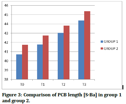
Comparison of PCB length [S-Ba] in Group 1 and Group 2 (Table 3 & Figure 3)
| Time Interval | Group 1 | Group 2 | T-Value | P-Value |
|---|---|---|---|---|
| T0 | 40.70 ± 1.51 | 41.75 ± 4.05 | 0.76 | 0.45, NS |
| T1 | 41.77 ± 2.05 | 42.75 ± 3.93 | 0.69 | 0.49, NS |
| T2 | 43.03 ± 1.76 | 43.81 ± 3.89 | 0.58 | 0.56, NS |
| T3 | 44.37 ± 2.07 | 45.37 ± 3.81 | 0.72 | 0.47, NS |
Table 3: Comparison of PCB length [S-Ba] in Group 1 and Group 2.
Figure 3:Comparison of PCB length [S-Ba] in group 1 and group 2.
The mean Posterior cranial base length in Class I malocclusion was 40.70 mm (SD ± 1.51), 41.77 mm (SD ± 2.05), 43.03mm (SD ± 1.76) and 44.37mm (SD ± 2.07) at time interval T0, T1, T2, and T3 respectively and in Class II malocclusion it was 41.75 mm (SD ± 4.05), 42.75 mm (SD ± 3.93), 43.81 mm (SD ± 3.89) and 45.37 mm (SD ± 3.81) at time interval T0, T1, T2, and T3 respectively. No significant difference was observed (P=0.000 i.e. P<0.05) in Posterior cranial base length at the entire time interval T0 (P=0.45), T1 (P=0.49), T2 (P=0.56) and T3 (t=0.72, P=0.47).
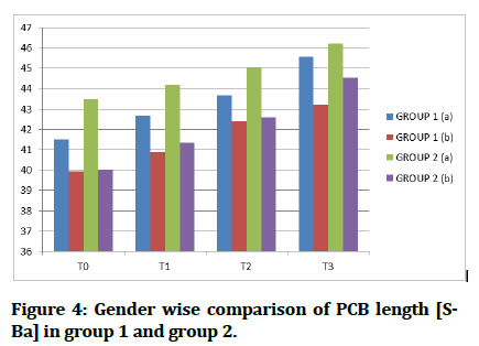
Gender wise comparison of PCB length [S-Ba] in Group 1 and Group 2 (Table 4 & Figure 4
| Time interval | Group 1 | P-Value | Group 2 | P-Value | ||
|---|---|---|---|---|---|---|
| (a) | (b) | (a) | (b) | |||
| T0 | 41.50 ± 0.49 | 39.91 ± 1.83 | 0.09,NS | 43.49 ± 3.96 | 40.02 ± 3.71 | 0.19, NS |
| T1 | 42.64 ± 0.89 | 40.90 ± 2.61 | 0.19,NS | 44.18 ± 4.00 | 41.32 ± 3.70 | 0.27, NS |
| T2 | 43.67 ± 0.96 | 42.38 ± 2.24 | 0.27,NS | 45.05 ± 4.41 | 42.57 ± 3.28 | 0.34, NS |
| T3 | 45.57 ± 1.24 | 43.18 ± 2.15 | 0.06,NS | 46.20 ± 4.00 | 44.54 ± 3.87 | 0.52, NS |
Table 4: Gender wise comparison of PCB length [S-Ba] in group 1 and group 2.
Figure 4:Gender wise comparison of PCB length [SBa] in group 1 and group 2.
The mean Posterior cranial base length in boys of Class-I malocclusion was 41.50 mm (SD ± 0.49), 42.64 mm (SD ± 0.89), 43.67 mm (SD ± 0.96) and 45.57 mm (SD ± 1.24) at time interval T0, T1, T2 and T3 respectively and in girls it was 39.91 mm (SD ± 1.83), 40.90 mm (SD ± 2. 61), 42.38mm (SD ± 2.24) and 43.18 mm (SD ± 2.15) at time interval T0, T1, T2 and T3 respectively.
No significant difference was observed (P=0.000 i.e. P<0.05) in Posterior cranial base length at the entire time interval T0 (P=0.09), T1 (P=0.19), T2 (P=0.27) and T3 (P=0.06).
The mean Posterior cranial base length in boys of Class-II malocclusion was 43.49 mm (SD ± 3.96), 44.18 mm (SD ± 4), 45.05 mm (SD ± 4.41) and 46.20 mm (SD ± 4) at time interval T0, T1, T2 and T3 respectively and in girls it was 40.02 mm (SD ± 3.71), 41.32 mm (SD ± 3.7), 42.57 mm (SD ± 3.28) and 44.54 mm (SD ± 3.87) at time interval T0, T1, T2 and T3 respectively.
No significant difference was observed (P=0.000 i.e. P<0.05) in Posterior cranial base length at all the time interval T0 (P=0.19), T1 (P=0.27), T2 (P=0.34) and T3 (P=0.52).
The mean ACB length in boys of Class-II malocclusion was 62.83 mm (SD ± 3.11), 63.61 mm (SD ± 3.09), 64.05 mm (SD ± 3.21) and 65.11 mm (SD ± 2.96) at time interval T0, T1, T2 and T3 respectively and in girls it was 63.5mm (SD ± 3.15), 64.07 mm (SD ± 3.40), 64.67mm (SD ± 3.27) and 65.57 mm (SD ± 3.52) at time interval T0, T1, T2 and T3 respectively. No significant difference was observed (P=0.000 i.e. P<0.05) in Anterior cranial base length at the entire time interval T0 (P=0.74), T1 (P=0.82), T2 (P=0.77) and T3 (P=0.83).
Comparison of PCB length [S-Ba] in Group 1 and Group 2 (Table 3 & Figure 3)
The mean Posterior cranial base length in Class I malocclusion was 40.70 mm (SD ± 1.51), 41.77 mm (SD ± 2.05), 43.03mm (SD ± 1.76) and 44.37mm (SD ± 2.07) at time interval T0, T1, T2, and T3 respectively and in Class II malocclusion it was 41.75 mm (SD ± 4.05), 42.75 mm (SD ± 3.93), 43.81 mm (SD ± 3.89) and 45.37 mm (SD ± 3.81) at time interval T0, T1, T2, and T3 respectively. No significant difference was observed (P=0.000 i.e. P<0.05) in Posterior cranial base length at the entire time interval T0 (P=0.45), T1 (P=0.49), T2 (P=0.56) and T3 (t=0.72, P=0.47).
Gender wise comparison of PCB length [S-Ba] in Group 1 and Group 2 (Table 4 & Figure 4
The mean Posterior cranial base length in boys of Class-I malocclusion was 41.50 mm (SD ± 0.49), 42.64 mm (SD ± 0.89), 43.67 mm (SD ± 0.96) and 45.57 mm (SD ± 1.24) at time interval T0, T1, T2 and T3 respectively and in girls it was 39.91 mm (SD ± 1.83), 40.90 mm (SD ± 2. 61), 42.38mm (SD ± 2.24) and 43.18 mm (SD ± 2.15) at time interval T0, T1, T2 and T3 respectively.
No significant difference was observed (P=0.000 i.e. P<0.05) in Posterior cranial base length at the entire time interval T0 (P=0.09), T1 (P=0.19), T2 (P=0.27) and T3 (P=0.06).
The mean Posterior cranial base length in boys of Class-II malocclusion was 43.49 mm (SD ± 3.96), 44.18 mm (SD ± 4), 45.05 mm (SD ± 4.41) and 46.20 mm (SD ± 4) at time interval T0, T1, T2 and T3 respectively and in girls it was 40.02 mm (SD ± 3.71), 41.32 mm (SD ± 3.7), 42.57 mm (SD ± 3.28) and 44.54 mm (SD ± 3.87) at time interval T0, T1, T2 and T3 respectively.
No significant difference was observed (P=0.000 i.e. P<0.05) in Posterior cranial base length at all the time interval T0 (P=0.19), T1 (P=0.27), T2 (P=0.34) and T3 (P=0.52).
Discussions
As orthodontists, we are fascinated by how the face evolves from its embryonic state to infancy, adolescence, and adulthood. Understanding how and where growth happens is particularly interesting since the cranial base's growth and development is associated with the face, directly affecting the growth of the upper and lower denture bases and, as a result, the establishment of their anteroposterior relationship.9 The most frequently used cephalometric landmarks include the Nasion (Na), Sella turcica (S) and Basion (Ba). There are two limbs of this measurement. The anterior limb, consisting of the maxillary attachment extends from S to frontal-nasal suture. The posterior limb, which marks the mandibular attachment, extends from the S to the anterior border of the foramen magnum i.e Ba. There have been various controversies in the literature regarding the selection of cranial base landmarks, wherein articulare (Ar) has been suggested instead of Ba because of its apparent ease of identification. In favour of Ba, it is apparently closer to the cranial base and therefore more valid to be considered as landmark. Glenoid fossa, the part of occipital bone that articulates with temporal bone and the post-sphenoid part of the sphenoid bone together formulate the PCB. The growth of the PCB is governed by sphenooccipital synchondrosis which controls the vertical height of the jaws and the mandibular position [5]. This will aid in unlocking the mandible from below the maxilla leading to its growth. It places the glenoid fossa anteriorly or posteriorly according to the growth of the PCB and also provides space for the vertical maxillary growth. Numerous studies have been conducted in the past using lateral cephalogram- a 2D technique; in order to assess the dimensions of the CB in individuals. These studies suffered from several flaws such as two dimensional interpretations of three dimensional structures, distortion, low reproducibility due to difficulties in identifying the skeletal landmarks etc.
With the advent of the 3D methods of evaluation, Orthodontists have started being more aware of the functional spaces and their role in the growth and development. Also a more accurate evaluation of these structures is now possible.
The patients selected for the current study were selected from the OPD of the “Department of Orthodontics and Dentofacial Orthopaedics”,” Sharad Pawar Dental College” within the age group of 10 to 14 years. The parameters depicting the cranial base-ACB Length [N-S], PCB length [S-Ba] were quantified using several linear and angular measurements. The data was collected at 6-month time interval (T0, T1, T2 and T3). Comparisons were also made based on gender of the subjects, and from the results the following deductions can be made.
Assessment of the ACB Length (S-N)
In this study; an incremental increase was observed in the anterior cranial base length in both groups.
Although there was no statistical significance amongst the two, the mean value obtained in Group 2 was greater than that of Group 1 at various time intervals. The anterior cranial base length was found to be greater in Group 2 as compared to Group 1, whereas in both the groups, girls showed greater length as compared to boys. Since the nasomaxillary complex is attached to the anterior cranial base region, growth of SOcc synchondrosis may have an influence on the depth of the upper face. Therefore, an increase in the anterior cranial base length could be the cause of a prognathic maxilla and a convex profile which is observed in a Class II Division 1 malocclusion.
On comparing the ACB length of both groups at different time intervals, it was observed that there was a significant increase in this dimension over the period of time in both groups.
Findings of this study can be explained by the research done by Dibbets et al. [10] who found that the CB angle was reduced and the (S–N) and (S–Ba) limbs were shortened from Class II, via Class I, to Class III malocclusions. Similar findings were obtained by Thiesen et al. [11] wherein they observed a greater CB length in subjects with Class II malocclusion as opposed to Class I malocclusion.
The evaluation of the PCB length (S-Ba)
The length of the PCB showed an incremental increase in both groups. Although a statistical significance was not observed, between both the groups, the mean value obtained for Group 2 was greater than that of Group 1 at various time intervals. Gender wise comparison in boys and girls.
Group 1 and 2 respectively depicted an increase in the posterior cranial base length in Group 2 as compared to Group 1, whereas in both the groups boys showed greater length as compared to girls.
Table 3 shows comparison of PCB length (SBa) at various time interval in Group 1 and 2 respectively.
The posterior cranial base length (S-Ba) was found to be significantly increasing over. The period of time in both the groups.
Findings of this study can be explained by the research done by Wilhelm et al [12] in 2001. They found that the posterior cranial base length is shorter in Class II (45.2 ± 3.3 mm) as compared to Class I (46.2 ± 3.41 mm) malocclusion.
Similar findings were obtained by Sayin et al [13] (2005). They found that PCB length was shorter in Class II malocclusion (33.92 ± 2.83 mm) as compared to Class I (36.88 ± 2.68 mm).
Conclusion
A detailed understanding of the skeletal and dental components that contribute to a specific malocclusion is important in the subject of orthodontics and dentofacial orthopaedics because these elements may influence the treatment approach to achieve the best results for every individual seeking treatment. The growth and development of cranial base is interrelated with face, directly influencing the growth of the maxilla and mandible and, consequently, the establishment of their anteroposterior relationship. Any variation at cranial base will have effects on the position of maxilla and maxilla mandibular relationship.
Conclusions that can be drawn from this study is as follows
• The subjects with Class II malocclusion had a tendency towards having large anterior cranial base length as compared to individuals with Class I malocclusion.
• The subjects with Class II malocclusion had a tendency towards having small posterior cranial base length as compared to individuals with Class I malocclusion.
• In gender wise comparison, females had tendency towards having large anterior cranial base length; whereas males had tendency towards having large posterior cranial base length.
• At various time intervals, there was a potential increase in the anterior and posterior cranial base length of both the groups.
References
- Anderson D, Popovich F. Relation of cranial base flexure to cranial form and mandibular position. Am J Phys Anthropol 1983; 61:181-7.
- Björk A. Cranial base development: A follow-up x-ray study of the individual variation in growth occurring between the ages of 12 and 20 years and its relation to brain case and face development. Am J Orthod Dentofacial Orthop 1955; 41:198-225.
- Ferner AG, Staubesand RT, Sobotta. Atlas de anatomia humana. Rio de Janeiro: Guanabara Koogan 1983.
- Dorenbos J. In vivo cerebral implantation of the anterior and posterior halves of the spheno-occipital synchondrosis in rats. Arch Oral Biol 1972; 17:1067-11.
- Cendekiawan T, Wong RW, Rabie AB. Relationships between cranial base synchondroses and craniofacial development: A review. Ann Anat 2010; 2.
- Bishara SE. Ortodontia. São Paulo Edn. Santos 2004.
- Moore WJ, Lavelle CL. Growth of the facial skeleton in the hominoidea. Academic Press 1974.
- Alves PV, Mazzuchelli J. Cranial base angulation in Brazilian patients seeking orthodontic treatment. J Craniofac Surg 2008; 19:334-8.
- Dibbets JMH. Morphological association between the angle classes. Eur J Orthod 1996; 18:111–118.
- Thiesen G, Pletsch G, Zastrow MD, et al. Comparative analysis of the anterior and posterior length and deflection angle of the cranial base, in individuals with facial Pattern I, II and III. Dental Press J Orthod 2013; 18: 69-75.
- Wilhelm BM, Beck FM, Lidral AC, et al. A comparison of cranial base growth in Class I and Class II skeletal patterns. Am J Orthod Dentofac Orthop 2001; 119:401–405.
- Sayin MO, Turkkahraman H. Cephalometric evaluation of nongrowing females with skeletal and dental Class II, division 1 malocclusion. Angle Orthod 2005; 75:656–660.
Indexed at, Google Scholar, Cross Ref
Indexed at, Google Scholar, Cross Ref
Indexed at, Google Scholar, Cross Ref
Indexed at, Google Scholar, Cross Ref
Indexed at, Google Scholar, Cross Ref
Indexed at, Google Scholar, Cross Ref
Indexed at, Google Scholar, Cross Ref
Author Info
Kamble RH, Anuradha Rajkuwar, Karthika Nambiar*, Toshniwal TG and Sumukh Nerurkar
1Department of Orthodontics and Dentofacial Orthopaedics, Sharad Pawar Dental College, Datta Meghe Institute of Medical Sciences, Sawangi meghe, Wardha, IndiaCitation: Kamble RH, Anuradha Rajkuwar, Karthika Nambiar, Toshniwal NG, Sumukh Nerurkar, Evaluation and Comparison of the Dimensions of Cranial Base in Boys and Girls between 10â??14 Years of Age having Skeletal Class-II Malocclusion as Compared to Skeletal Class I Malocclusion, J Res Med Dent Sci, 2022, 10(1): 358-363
Received: 19-Dec-2021, Manuscript No. Jrmds-21-44722; , Pre QC No. Jrmds-21-44722 (PQ); Editor assigned: 21-Dec-2021, Pre QC No. Jrmds-21-44722 (PQ); Reviewed: 04-Jan-2022, QC No. Jrmds-21-44722 ; Revised: 07-Jan-2022, Manuscript No. Jrmds-21-44722 (R); Published: 14-Jan-2022


