Research - (2022) Volume 10, Issue 3
Evaluation of Commonly Treated Maxillary Teeth with Preventive Resin Sealant among Children with Permanent Dentition-A Retrospective Study
Ashwin kumar R and Vignesh Ravindran*
*Correspondence: Vignesh Ravindran, Department of Pedodontics and Preventive Dentistry, Saveetha Dental College and Hospitals Saveetha Institute of Medical and Technical and Sciences, Saveetha University, India, Email:
Abstract
Dental sealants were introduced in the 1960s to help prevent dental caries, mainly in the pits and fissures of occlusal tooth surfaces. Sealants act to prevent bacteria growth that can lead to dental decay. Evidence suggests that fissure sealants are effective in preventing caries in children and adolescents compared to no sealants. Effectiveness may, however, be related to caries incidence level of the population. Pit and fissure sealants reduce occlusal caries when proper patient selection and application techniques are followed. The aim of this study was to investigate the most commonly treated primary maxillary teeth with pit and fissure sealants for caries prevention. Data was collected from patients’ dental records in the department of pediatric dentistry to meet the inclusion and exclusion criteria. A total of 171 records of children who had undergone pit and fissure sealants in primary maxillary teeth were evaluated. Descriptive analysis and chi-square tests were performed. Most commonly treated tooth with sealant in the maxillary arch was 55 (40.4%). Sealant application was more common on the maxillary left side (51.5%).
Keywords
Maxillary teeth, Innovative technique, Sealants, Prevention, Permanent teeth
Introduction
Dental caries or tooth decay is a multifactorial chronic oral disease that affects most of the populations throughout the world and it has been considered as the most important global oral health burden. Caries is interplay between specific acidogenic bacteria in the dental plaque biofilm, fermentable carbohydrates and tooth structure. The biofilm bacteria produce organic acids that can cause mineral loss from the tooth surface which causes demineralization. In favorable conditions, a reversal, that is, a mineral gain, is possible which leads to remineralization. If the demineralization process prevails, visually detectable caries lesions occur. Development of a caries lesion is a dynamic process that may progress, stop or reverse. Assessment of the grade and activity of the lesion is challenging [1].
Definition of dental caries and a system to measure the caries process is integrated by the International Caries Detection and Assessment System [2,3]. In ICDAS II, the codes for coronal caries range from 0 to 6, depending on the severity of the lesion: codes 0 to 3 involve a sound tooth surface to caries in enamel; codes 4 to 6 involve caries in dentine.
Since the 1970s, caries prevalence has declined in most industrialized countries, and has been attributed to general factors, such as improvements in living conditions and oral hygiene, and public health measures, such as widespread use of fluorides and better disease management. Dental sealants were introduced in the 1960s to help prevent dental caries, mainly in the pits and fissures of occlusal tooth surfaces. Sealants act to prevent bacteria growth that can lead to dental decay. Evidence suggests that fissure sealants are effective in preventing caries in children and adolescents compared to no sealants. Occlusal surfaces of molars are highly susceptible to dental decay. Smooth surfaces and interproximal surfaces have benefited to the greatest extent from the caries' preventive effects of various fluoride agents. Young permanent molars exhibit occlusal characteristics, which increase the risk of caries [4]. Although topical fluoride treatments are most effective in preventing smooth surface caries, they are less effective in preventing pit and fissure caries. Caries in pits and fissures remain a major problem. Occlusal surfaces account for only 12.5% of all tooth surfaces, but they account for about 50% of the caries in school-aged children. Sealant can play an important role in prevention of caries in non-carious pits and fissures. However, deep pits and fissures with incipient caries may require a combination treatment of prevention and restoration.
Dental sealant is applied to a tooth surface to provide a physical barrier that prevents growth of biofilm by blocking nutrition. Although sealants were introduced for preventing caries on occlusal surfaces, they are now considered active agents in controlling and managing initial caries lesions on occlusal surfaces and, recently, on approximal surfaces as well [5].
Options of occlusal sealant materials are numerous but resins/composites and glass ionomers comprise the main material types. A resin, Bisphenol A glycidyl methacrylate (BIS-GMA), forms the basis for numerous resinbased dental sealants and composites that are available [6]. The effectiveness of resin‐based sealants is closely related to the longevity of sealant coverage which is also known as clinical retention. The resin‐based sealants can be divided into generations according to their mechanism for polymerisation or their content. The development of sealants has progressed from first‐generation sealants, which were activated with ultraviolet light, through to second‐ and third‐generation sealants, which are auto polymerized and visible‐light activated, and fourth‐generation sealants which contain fluoride. First‐generation sealants are no longer marketed. Our team has extensive knowledge and research experience that has translate into high quality publications [7–26]. The aim of the study is to evaluate commonly treated maxillary teeth with preventive resin sealant among children with permanent dentition [27].
Materials and Methods
This type of participants to this study includes Saveetha dental college patients who are below 18 years and above 12 years of age. Data is collected from under the category of patients who have done treatment for pit and fissure sealants, over 300 patient details were collected. The collected details were organized in excel sheets by doing tabulation and sent to spss software, the data collection and analysis was done by a single examiner. Type of outcomes: dentine caries in permanent molars which is measured dichotomously as incidence of carious lesions on treated occlusal surfaces of molars or premolars (yes or no). Caries was defined as caries in the dentine but if scored using the ICDAS II scale, in addition to codes 4 to 6, code 3 was also accepted as caries (localised enamel breakdown on occlusal surface reflecting established decay). It is measured continuously as changes in decayed, missing and filled (DMF) rates at occlusal surface. The extracted data was tabulated in a spreadsheet (excel 2017) and analysis using SPSS 19.0 version software. Descriptive statistics and chi square tests were performed with the level of significance at 5% (p<0.05).
Results
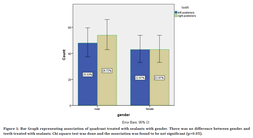
The total study population was 300 patients of the age group of 12-17 years. In this study 54.25% belonged to male gender and 45.74% belonged to the female gender.There was no difference between gender and teeth treated with sealants. Chi square test was done and the association was found to be not significant (p>0.05) (Figure 1). Among the maxillary teeth evaluated , the prevalence of sealant application was 11.17% in age of 17 year old patient ,10.64% in age of 13 year old , respective to left posteriors and 11.17% in age of 13 year old patient ,10.11%in age of 17 year old respective to right posteriors of maxillary tooth.

Figure 1. Bar Graph representing association of quadrant treated with sealants with gender. There was no difference between gender and teeth treated with sealants. Chi square test was done and the association was found to be not significant (p>0.05).
Discussion
Resin‐based sealants compared with no sealant. We are moderately confident that resinbased sealants applied on occlusal surfaces of permanent molars of children and adolescents reduce caries up to 48 months when compared to no sealant; after longer follow‐up the quantity and quality of the evidence is reduced.
The effectiveness of resin‐based sealants is related to retention of sealants. Retention of resin sealants was good in studies that compared sealant with a control without sealant. At 12 and 24 months follow‐up, resin sealants were retained completely on average in 80% of cases. After 48 to 54 months, most studies reported 70% retention of sealants [28].
Settings and caries risk for children were often unclear, but overall, the proportion of sealed decayed surfaces was small, regardless of material used. For example, in eight studies that provided data at 36 to 48 months, proportions of decayed sealed surfaces ranged from 3% to 14% at 36 to 48 months. In all eight studies, control teeth or control groups without sealants were lacking to further estimate caries risk [29].
The caries results of the individual trials seemed often to be correlated with retention of sealant materials. For example, in the five studies that found statistically significantly more caries in lowviscosity glass Ionomer and resin‐modified glass Ionomer sealed teeth at 36 to 48 months than in resin‐sealed teeth, the complete retention for resin sealants was documented to be good (mean 85%), and for glass ionomers low (mean 4%) [30,31].
Despite widespread preventive measures dental caries continues to be a veritable scourge for mankind even today [32]. This disease exerts a social physical, mental and financial burden. In spite of the advent of systemic and topical fluoride delivery systems to combat dental caries, it is almost impossible to reduce pits and fissures caries through these methods. Fluoridation of water supply has been shown to reduce dental caries prevalence by approximately 50% with maximum reduction occurring in smooth surface lesions. Pit and fissures surfaces of molar and premolar teeth have been shown to be the most susceptible to caries in population receiving fluoridated water [33].
The integral problem lies at the level of the anatomy of the pits and fissures which makes them inaccessible to oral hygiene aids and methods as well as to the topical fluoride delivery systems. The pits and fissures by itself pose as an independent microenvironment of the oral ecosystem, which has to be understood and tackled as a separate entity. The universal prevalence of dental caries is a constant reminder of the need for effective dental health intervention during childhood to ensure healthy teeth throughout the life.
Conclusion
Within the limitations of the present study, we can conclude that the maxillary left first molars were the most commonly sealed permanent teeth in the maxillary arch. 17 year olds were the most frequently treated. Most commonly sealed teeth in females was the maxillary second molars and among males it was the left maxillary second molar. No significant correlation was found between gender and teeth treated with sealants. Further large scale studies are required to infer these findings as significant.
Acknowledgement
The authors are thankful to the Department of Pediatric Dentistry, Saveetha Dental College, Saveetha Institute of Medical and Technical Sciences, Saveetha University for providing a platform in expressing their knowledge.
Conflict of Interest
The author declares no conflict of interest.
Source of Funding
Saveetha Dental College, Saveetha Institute of Medical and Technical Science, Saveetha University, India
References
- Bravo M, Llodra JC, Baca P, et al. Effectiveness of visible light fissure sealant (Delton) versus fluoride varnish (Duraphat): 24-month clinical trial. Community Dent Oral Epidemiol 1996; 24:42–46.
- Bravo M, Baca P, Llodra JC, et al. A 24-month study comparing sealant and fluoride varnish in caries reduction on different permanent first molar surfaces. J Public Health Dent 1997; 57:184–186.
- Bravo M, Garcia-Anllo I, Baca P, et al. A 48-month survival analysis comparing sealant (Delton) with fluoride varnish (Duraphat) in 6- to 8-year-old children. Community Dent Oral Epidemiol 1997; 25:247–250.
- Sipahier M, Ulusu T. Glass-ionomer--silver-cermet cements applied as fissure sealants II. Clinical evaluation. Quintessence Int 1995; 26.
- Amin HE. Clinical and antibacterial effectiveness of three different sealant materials. J Dent Hyg 2008; 82:45.
- Antonson SA, Antonson DE, Brener S, et al. Twenty-four month clinical evaluation of fissure sealants on partially erupted permanent first molars: glass ionomer versus resin-based sealant. J Am Dent Assoc 2012; 143:115–22.
- Subramanyam D, Gurunathan D, Gaayathri R, et al. Comparative evaluation of salivary malondialdehyde levels as a marker of lipid peroxidation in early childhood caries. Eur J Dent 2018; 12:67–70.
- Ramadurai N, Gurunathan D, Samuel AV, et al. Effectiveness of 2% articaine as an anesthetic agent in children: Randomized controlled trial. Clin Oral Investig 2019; 23:3543–3550.
- Ramakrishnan M, Dhanalakshmi R, Subramanian EMG. Survival rate of different fixed posterior space maintainers used in paediatric dentistry–A systematic review. Saudi Dent J 2019; 31:165–172.
- Jeevanandan G, Thomas E. Volumetric analysis of hand, reciprocating and rotary instrumentation techniques in primary molars using spiral computed tomography: An in vitro comparative study. Eur J Dent 2018; 12:21–6.
- Princeton B, Santhakumar P, Prathap L. Awareness on preventive measures taken by health care professionals attending COVID-19 patients among dental students. Eur J Dent 2020; 14:105–9.
- Saravanakumar K, Park S, Mariadoss AVA, et al. Chemical composition, antioxidant, and anti-diabetic activities of ethyl acetate fraction of Stachys riederi var. japonica (Miq.) in streptozotocin-induced type 2 diabetic mice. Food Chem Toxicol 2021; 155:112374.
- Wei W, Li R, Liu Q, et al. Amelioration of oxidative stress, inflammation and tumor promotion by tin oxide-sodium alginate-polyethylene glycol-allyl isothiocyanate nanocomposites on the 1,2-dimethylhydrazine induced colon carcinogenesis in rats. Arabian J Chem 2021; 14:103238.
- Gothandam K, Ganesan VS, Ayyasamy T, et al. Antioxidant potential of theaflavin ameliorates the activities of key enzymes of glucose metabolism in high fat diet and streptozotocin induced diabetic rats. Redox Rep 2019; 24:41–50.
- Su P, Veeraraghavan VP, Krishna Mohan S, et al. A ginger derivative, zingerone-a phenolic compound-induces ROS-mediated apoptosis in colon cancer cells (HCT-116). J Biochem Mol Toxicol 2019; 33:e22403.
- Mathew MG, Samuel SR, Soni AJ, et al. Evaluation of adhesion of Streptococcus mutans, plaque accumulation on zirconia and stainless steel crowns, and surrounding gingival inflammation in primary molars: Randomized controlled trial. Clin Oral Invest 2020; 24:3275–80.
- Sekar D, Johnson J, Biruntha M, et al. Biological and clinical relevance of microRNAs in mitochondrial diseases/dysfunctions. DNA Cell Biol 2020; 39:1379–84.
- Velusamy R, Sakthinathan G, Vignesh R, et al. Tribological and thermal characterization of electron beam physical vapor deposited single layer thin film for TBC application. Surf Topogr Metrol Prop 2021; 9:025043.
- Aldhuwayhi S, Mallineni SK, Sakhamuri S, et al. Covid-19 knowledge and perceptions among dental specialists: A cross-sectional online questionnaire survey. Risk Manag Healthc Policy 2021; 14:2851–61.
- Sekar D, Nallaswamy D, Lakshmanan G. Decoding the functional role of long noncoding RNAs (lncRNAs) in hypertension progression. Hypertens Res 2020; 43:724.
- Bai L, Li J, Panagal M, et al. Methylation dependent microRNA 1285-5p and sterol carrier proteins 2 in type 2 diabetes mellitus. Artif Cells Nanomed Biotechnol 2019; 47:3417–22.
- Sekar D. Circular RNA: A new biomarker for different types of hypertension. Hypertens Res 2019; 42:1824.
- Sekar D, Mani P, Biruntha M, et al. Dissecting the functional role of microRNA 21 in osteosarcoma. Cancer Gene Ther 2019; 26:179–82.
- Duraisamy R, Krishnan CS, Ramasubramanian H, et al. Compatibility of nonoriginal abutments with implants: Evaluation of microgap at the implant-abutment interface, with original and nonoriginal abutments. Implant Dent 2019; 28:289–95.
- Parimelazhagan R, Umapathy D, Sivakamasundari IR, et al. Association between tumor prognosis marker visfatin and proinflammatory cytokines in hypertensive patients. Biomed Res Int 2021; 2021:8568926.
- Syed MH, Gnanakkan A, Pitchiah S. Exploration of acute toxicity, analgesic, anti-inflammatory, and anti-pyretic activities of the black tunicate, Phallusia nigra (Savigny, 1816) using mice model. Environ Sci Pollut Res Int 2021; 28:5809–5821.
- Splieth CH, Christiansen J, Foster Page LA. Caries epidemiology and community dentistry: Chances for future improvements in caries risk groups. Outcomes of the ORCA saturday afternoon symposium, greifswald, 2014. Part 1. Caries Res 2016; 50:9–16.
- Poulsen S, Beiruti N, Sadat N. A comparison of retention and the effect on caries of fissure sealing with a glass-ionomer and a resin-based sealant. Community Dent Oral Epidemiol 2001; 29:298–301.
- Kerosuo E, Kervanto-Seppälä S, Pietilä I, et al. Pit and fissure sealants in dental public health--application criteria and general policy in Finland. BMC Oral Health 2009; 9:1.
- Baseggio W, Scarparo Naufel F, de Oliveira Davidoff DC, et al. Caries-preventive efficacy and retention of a resin-modified glass ionomer cement and a resin-based fissure sealant: A 3-year split-mouth randomised clinical trial. Oral Health Prev Dent 2010; 8:261.
- Wp R. Foulkes EE. Perry H. et al. A comparative study of fluoride-releasing composite resin and glass ionomer materials used as fissure sealants. J Dent 1996;
- Kale S, Kakodkar P, Shetiya S. Prevalence of dental caries among children aged 5–15 years from 9 countries in the Eastern Mediterranean Region: A meta-analysis. Mediterr Health J 2020; 26:726-735.
- Raadal M, Utkilen AB, Nilsen OL. Fissure sealing with a light‐cured resin‐reinforced glass‐ionomer cement (Vitrebond) compared with a resin sealant. Int J 1996; 6:235-239.
Indexed at, Google Scholar, Cross Ref
Indexed at, Google Scholar, Cross Ref
Indexed at, Google Scholar, Cross Ref
Indexed at, Google Scholar, Cross Ref
Indexed at, Google Scholar, Cross Ref
Indexed at, Google Scholar, Cross Ref
Indexed at, Google Scholar, Cross Ref
Indexed at, Google Scholar, Cross Ref
Indexed at, Google Scholar, Cross Ref
Indexed at, Google Scholar, Cross Ref
Indexed at, Google Scholar, Cross Ref
Indexed at, Google Scholar, Cross Ref
Indexed at, Google Scholar, Cross Ref
Indexed at, Google Scholar, Cross Ref
Indexed at, Google Scholar, Cross Ref
Indexed at, Google Scholar, Cross Ref
Indexed at, Google Scholar, Cross Ref
Indexed at, Google Scholar, Cross Ref
Indexed at, Google Scholar, Cross Ref
Indexed at, Google Scholar, Cross Ref
Indexed at, Google Scholar, Cross Ref
Indexed at, Google Scholar, Cross Ref
Indexed at, Google Scholar, Cross Ref
Indexed at, Google Scholar, Cross Ref
Indexed at, Google Scholar, Cross Ref
Indexed at, Google Scholar, Cross Ref
Indexed at, Google Scholar, Cross Ref
Indexed at, Google Scholar, Cross Ref
Author Info
Ashwin kumar R and Vignesh Ravindran*
Department of Pedodontics and Preventive Dentistry, Saveetha Dental College and Hospitals Saveetha Institute of Medical and Technical and Sciences, Saveetha University, Chennai, IndiaReceived: 04-Feb-2022, Manuscript No. JRMDS-22-51106; , Pre QC No. JRMDS-22-51106 (PQ); Editor assigned: 07-Feb-2022, Pre QC No. JRMDS-22-51106 (PQ); Reviewed: 21-Feb-2022, QC No. JRMDS-22-51106; Revised: 25-Feb-2022, Manuscript No. JRMDS-22-51106 (R); Published: 04-Mar-2022
