Research - (2022) Volume 10, Issue 2
Evaluation of Commonly Treated Maxillary Teeth with Preventive Resin Sealant among Children with Primary Dentition
Sudarsan R and Vignesh Ravindran*
*Correspondence: Vignesh Ravindran, Department of Pedodontics, Saveetha Dental College & Hospitals, Saveetha Institute of Medical and technical Science, Saveetha University, Chennai, India, Email:
Abstract
The most prevalent dental caries is a preventable disease and untreated dental caries in children can have a toll in their day to day activities and development. There is a rising need for dentists to manage carious lesions in children as early as possible by preventive means. Sealant application on teeth can reduce food and plaque accumulation and makes the site easy to clean. The aim of this study was to investigate the most commonly treated primary maxillary teeth with pit and fissure sealants for caries prevention. Data was collected from patients’ dental records in the department of pediatric dentistry to meet the inclusion and exclusion criteria. A total of 171 records of children who had undergone pit and fissure sealants in primary maxillary teeth were evaluated. Descriptive analysis and chi-square tests were performed. Most commonly treated tooth with sealant in the maxillary arch was 55 (40.4%). Sealant application was more common on the maxillary left side (51.5%).
Keywords
Maxillary teeth, Innovative technique, Sealants, Prevention, Primary teeth
Introduction
Dental caries is a multifactorial disease that affects the teeth and causes localised destruction of tooth structure. Previously considered an infectious disease, dental caries has now proved to be "a complex disease caused by an imbalance in physiologic equilibrium between tooth mineral and biofilm fluid" [1]. Dental caries is a result of the interaction between the tooth surface, carbohydrates present in the diet and cariogenic bacteria in the dental biofilm. The bacteria metabolise dietary carbohydrates and consequent acid production results in pH fluctuations of the biofilm and disturbs the existing physiologic equilibrium between the tooth and biofilm, ultimately resulting in demineralisation of the tooth structure [2,3]. Remineralisation is possible under favourable conditions; however, if left untreated, further dissolution of dental hard tissues and visible caries becomes inevitable. Caries would then continue to spread through the hard tissues of the tooth and reach the soft tissue (pulp) and cause pain, inflammation and loss of function and can thus negatively impact quality of life [4,5]. In children, untreated caries affect their day to day activities by causing difficulty in chewing, tooth loss, weight loss, changes in behaviour, and poor academic performance and cognitive development in young children [6,7].
Dental caries is considered a significant health problem in children as untreated dental caries in primary teeth is the 10th most prevalent condition, as close to about 621 million children are affected globally [8]. According to WHO among those affected, the prevalence and burden are noted to be higher among children in low- and middleincome countries than those in high-income countries. Susceptibility to dental caries on the other hand is highly dependent on individuals and teeth. Among the teeth surfaces, pits and fissures on the occlusal aspect of posterior teeth are more likely to the develop dental caries owing to increased retention of plaque, permeability of immature enamel and the reduced effect of fluoride on pits and fissures [9]. According to CDC, 90% of all dental caries in permanent molars can be accounted for by pit and fissure caries though occlusal surfaces only make up 12.5% of the total surfaces of the teeth. 44% of all caries affecting primary molars are also seen in pits and fissures [10], despite primary molars having an occlusal morphology that is flatter and less fissured when compared to that of permanent molars [11]. Caries in primary molars progress faster in comparison to permanent molars, due to thinner enamel and greater porosity. Thus preventive therapy plays a major role in preservation of primary tooth structure [12].
Pit and fissure sealants are applied to occlusal surfaces of teeth which has an anatomy that makes it highly susceptible to dental caries and is also resistant to fluorides and mechanical plaque control and other therapeutic approaches [13]. Resin-based sealants, glass ionomer sealants and hybrid sealants are some of the major types of sealants available. The anatomy of the pit and fissure surfaces makes them difficult to clean, as food entrapment in deep fissures are quite common and toothbrush bristles cannot effectively clean a fissure. This leads to the site developing into a niche for plaque formation and bacterial growth as even excellent home care may not provide successful cleaning of a deep fissure . Sealants applied to such complex morphological teeth surfaces will occlude these pits and fissures and form a physical barrier. This will in turn help prevent caries development. The physical barrier blocks carbohydrates from reaching the bacteria at the base of these structures and also makes the surfaces easier to clean [3]. While resin-based sealants prevent caries by forming a physical barrier, GIC sealants bond chemically to dental tissues and have Anticariogenic effects by releasing fluoride [14].
The use of sealants in preventing caries in permanent teeth in children and adolescents is well established. Employment of sealants in the prevention of dental caries in permanent teeth have been recommended in clinical guidelines from professional authorities like the American Dental Association, the American Association of Pediatric Dentistry and the British Society of Paediatric Dentistry [9,15]. However, when it comes to its use in primary teeth, empirical data and systematic reviews regarding the effectiveness of sealants in primary molars is lacking. The clinical management of deep pits and fissures in primary teeth have been based on the findings seen in permanent teeth treated with sealants [15]. The lack of evidence from studies conducted in the primary dentition is a growing concern as sealants in primary teeth are being increasingly recommended as part of preventive therapy for children [15,16].
There is an uncertainty surrounding the usage of sealants in primary molars. Some believe that the occlusal anatomy of primary molars do not help in long-term sealant retention as they have flatter fissures [17]. Another concern is regarding the sealing of incipient or white spot lesions and non-cavitated carious lesions [18]. Despite this, reports suggest that children with sealed pit and fissures in primary molars, sound or non-cavitated, had a 76% lower risk of developing new caries in comparison to children without sealants and retention levels in these primary molars ranged from 74% to 93% [9]. Our team has extensive knowledge and research experience that has translate into high quality publications [19–38]. Not many studies have documented the use of sealants in primary dentition. The current study aims to evaluate the most commonly treated maxillary teeth with sealants and thus correlate the same with the age and anatomic significance.
Methodology
The present study was a retrospective study that was conducted in a university setting. The ethical clearance for the study was obtained from the Institutional Scientific Review Board. The treatment records of patients who had undergone treatment in Saveetha Dental College between June 2019 to December 2020 were assessed for this study. The data collection and analysis was done by a single examiner. The inclusion criteria were children between the ages of 3-17 years, children who underwent dental sealants in primary maxillary teeth. Exclusion criteria for the study were patients above 17 years of age, incomplete case records and missing photo Figureic proof of completed treatment. To avoid sampling bias, simple random sampling was done. Gender, age, tooth number was recorded for each patient.
The extracted data was tabulated in a spread sheet (Excel 2017: Microsoft Office) and analysed using SPSS 19.0 version software (SPSS, Inc., Chicago). Descriptive statistics and chi-square tests were performed with the level of significance at 5% (P<0.05).
Results
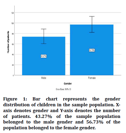
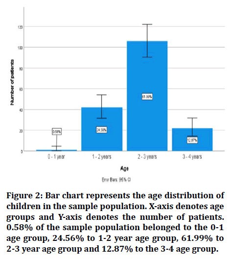
The total study population was 171 children of the age group 0-5 years. In this study population 43.3% belonged to the male gender and 56.7% to the female gender. The demo Figureic data is represented in Figures 1 and Figure 2.

Figure 1: Bar chart represents the gender distribution of children in the sample population. Xaxis denotes gender and Y-axis denotes the number of patients. 43.27% of the sample population belonged to the male gender and 56.73% of the population belonged to the female gender.

Figure 2: Bar chart represents the age distribution of children in the sample population. X-axis denotes age groups and Y-axis denotes the number of patients. 0.58% of the sample population belonged to the 0-1 age group, 24.56% to 1-2 year age group, 61.99% to 2-3 year age group and 12.87% to the 3-4 age group.
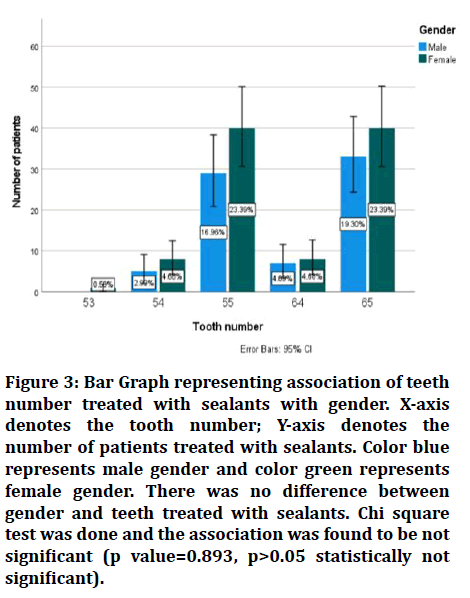
Among the maxillary teeth evaluated, the prevalence of sealant application was 0.6% in 53, 7.6% in 54, 40.4% in 55, 8.8% in 64 and 42.7% in 65. Distribution of sealant application among the male gender was 2.9% in 54, 16.9% in 55, 4.09% in 64 and 19.3% in 65 and among the female gender was 0.58% in 53, 4.68% in 54, 23.39% in 55, 4.68% in 64 and 23.39% in 65.
Discussion
Preventive dental therapy, which includes early and routine dental intervention, fluoride application, and sealants are effective in limiting the disease burden and expenditures. Preventive measures in dentistry are the backbone to improve oral health and dentists play a vital role in achieving this through their attitude and choice of preventive dental procedures and also in the way they motivate their patients regarding preventive treatment 13,15. In the present clinical scenario, non-invasive methods are preferred to invasive treatments. The current study was conducted to evaluate the most commonly treated primary maxillary teeth with sealants.
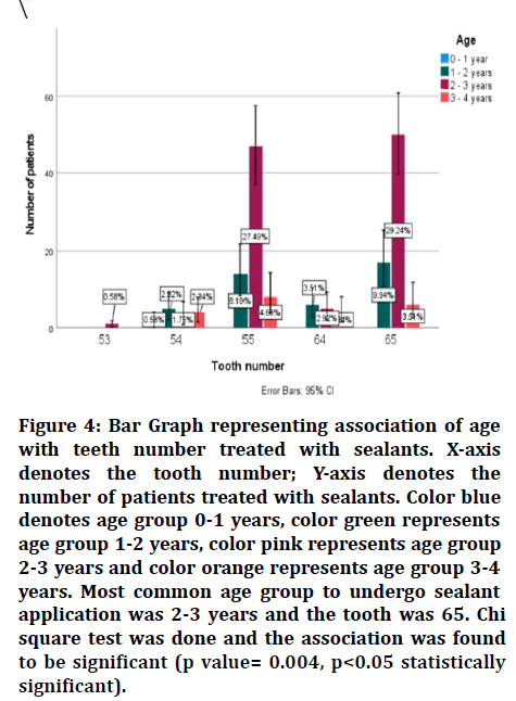
Most commonly treated primary tooth with sealant in the present study was 55 as maxillary right second primary molars were sealed in 40.4% of the sample population. However sealant application in general was more common on the maxillary left side (51.5%). No such significant associations were found to a particular side or arch in previous studies in primary dentition [39]. Most common teeth treated with sealant in males- 65 (19.3%) and in females-55 & 65 (23.39%). There was no significant association between gender and teeth number in the present study (Figure 3). Similar findings were reported with regards to gender in previous studies [40]. There was a significant association between the age group and teeth treated, with 2-3 years being the most commonly treated with sealants and the teeth associated with the maxillary left second molar (Figure 4).

Figure 3: Bar Graph representing association of teeth number treated with sealants with gender. X-axis denotes the tooth number; Y-axis denotes the number of patients treated with sealants. Color blue represents male gender and color green represents female gender. There was no difference between gender and teeth treated with sealants. Chi square test was done and the association was found to be not significant (p value=0.893, p>0.05 statistically not significant).

Figure 4:Bar Graph representing association of age with teeth number treated with sealants. X-axis denotes the tooth number; Y-axis denotes the number of patients treated with sealants. Color blue denotes age group 0-1 years, color green represents age group 1-2 years, color pink represents age group 2-3 years and color orange represents age group 3-4 years. Most common age group to undergo sealant application was 2-3 years and the tooth was 65. Chi square test was done and the association was found to be significant (p value= 0.004, p<0.05 statistically significant).
The limitations of the present study were the small sample size, the treatment plan not being decided by a single operator and the reason for the sealant application (preventive/early caries management) was not included. Further research would enable practitioners to assess the outcome and impact of such preventive interventions.
Higher effort is needed from the professional and government organisations to ensure patients are made aware of the preventive practices available and its impact on the overall health and development of children.
Conclusion
Within the limitations of the present study, we can conclude that the maxillary right second molar is the most commonly sealed primary tooth in the maxillary arch, although sealant application was more frequent in the left side. 2-3 age group was the most frequently treated.
Most commonly sealed teeth in females was the maxillary second molars and among males it was the left maxillary second molar. No significant correlation was found between gender and teeth treated with sealants. Further large scale studies are required to infer these findings as significant.
Acknowledgement
The authors are thankful to the Department of Pediatric Dentistry, Saveetha Dental College & Hospitals, Saveetha Institute of Medical and Technical Sciences, Saveetha University for providing a platform in expressing their knowledge.
Conflict of Interest
The author declares no conflict of interest.
Source of Funding
The present study was supported by the following agencies
Saveetha Dental College & Hospitals, Saveetha Institute of Medical and Technical Sciences, Saveetha University, India
Prompt paper products private LTD.
References
- Lam A. Increase in utilization of dental sealants. J Contemp Dent Pract 2008; 9:81–7.
- Kidd E. The implications of the new paradigm of dental caries. J Dent 2011; 39:3–8.
- Ritter AV. Sturdevant’s Art & science of operative dentistry-E-Book. Elsevier Health Sciences 2017; 544.
- Filstrup SL, Briskie D, da Fonseca M, et al. Early childhood caries and quality of life: Child and parent perspectives. Pediatr Dent 2003; 25:431–40.
- Ten Cate JM. Current concepts on the theories of the mechanism of action of fluoride. Acta Odontol Scand 1999; 57:325–329.
- Abanto J, Carvalho TS, Mendes FM, et al. Impact of oral diseases and disorders on oral health-related quality of life of preschool children. Community Dent Oral Epidemiol 2011; 39:105–14.
- Ayhan H, Suskan E, Yildirim S. The effect of nursing or rampant caries on height, body weight and head circumference. J Clin Pediatr Dent 1996; 20:209–12.
- Kassebaum NJ, Bernabé E, Dahiya M, et al. Global burden of untreated caries: A systematic review and metaregression. J Dent Res 2015; 94:650–658.
- Beauchamp J, Caufield PW, Crall JJ, et al. Evidence-based clinical recommendations for the use of pit-and-fissure sealants: A report of the American dental association council on scientific affairs. Dent Clin North Am 2009; 53:131–47.
- Dye BA, Tan S, Smith V, et al. Trends in oral health status: United States, 1988-1994 and 1999-2004. Vital Health Stat 2007; 1–92.
- Fadm WSE, Hatrick CD. Dental materials: Clinical applications for dental assistants and dental hygienists. Elsevier Health Sciences 2015; 384.
- Low IM, Duraman N, Mahmood U. Mapping the structure, composition and mechanical properties of human teeth. Mater Sci Eng 2008; 28:243–247.
- Wright JT, Tampi MP, Graham L, et al. Sealants for Preventing and arresting Pit-and-fissure occlusal caries in primary and permanent molars. Pediatr Dent 2016; 38:282–308.
- McLean JW. Clinical applications of glass-ionomer cements. Oper Dent 1992; 5:184–90.
- American Academy of Pediatric Dentistry. Guideline on caries-risk assessment and management for infants, children, and adolescents. Pediatr Dent 2013; 35:E157–64.
- Gooch BF, Griffin SO, Gray SK, et al. Preventing dental caries through school-based sealant programs: updated recommendations and reviews of evidence. J Am Dent Assoc 2009; 140:1356–65.
- Horowitz AM, Frazier PJ. Issues in the widespread adoption of pit-and-fissure sealants. J Public Health Dent 1982; 42:312–23.
- Ripa LW. The current status of occlusal sealants. J Prev Dent 1976; 3:6–14.
- Subramanyam D, Gurunathan D, Gaayathri R, et al. Comparative evaluation of salivary malondialdehyde levels as a marker of lipid peroxidation in early childhood caries. Eur J Dent 2018; 12:67–70.
- Ramadurai N, Gurunathan D, Samuel AV, et al. Effectiveness of 2% articaine as an anesthetic agent in children: Randomized controlled trial. Clin Oral Investig 2019; 23:3543–50.
- Ramakrishnan M, Dhanalakshmi R, Subramanian EMG. Survival rate of different fixed posterior space maintainers used in paediatric dentistry – A systematic review. Saudi Dent J 2019; 31:165–72.
- Jeevanandan G, Thomas E. Volumetric analysis of hand, reciprocating and rotary instrumentation techniques in primary molars using spiral computed tomography: An in vitro comparative study. Eur J Dent 2018; 12:21–26.
- Princeton B, Santhakumar P, Prathap L. Awareness on preventive measures taken by health care professionals attending COVID-19 patients among dental students. Eur J Dent 2020; 14:105–109.
- Saravanakumar K, Park S, Mariadoss AVA, et al. Chemical composition, antioxidant, and anti-diabetic activities of ethyl acetate fraction of Stachys riederi var. japonica (Miq.) in streptozotocin-induced type 2 diabetic mice. Food Chem Toxicol 2021; 155:112374.
- Wei W, Li R, Liu Q, et al. Amelioration of oxidative stress, inflammation and tumor promotion by tin oxide-sodium alginate-polyethylene glycol-allyl isothiocyanate nanocomposites on the 1,2-dimethylhydrazine induced colon carcinogenesis in rats. Arabian J Chem 2021; 14:103238.
- Gothandam K, Ganesan VS, Ayyasamy T, et al. Antioxidant potential of theaflavin ameliorates the activities of key enzymes of glucose metabolism in high fat diet and streptozotocin-induced diabetic rats. Redox Rep 2019; 24:41–50.
- Su P, Veeraraghavan VP, Krishna Mohan S, et al. A ginger derivative, zingerone-a phenolic compound-induces ROS-mediated apoptosis in colon cancer cells (HCT-116). J Biochem Mol Toxicol 2019; 33:e22403.
- Mathew MG, Samuel SR, Soni AJ, et al. Evaluation of adhesion of Streptococcus mutans, plaque accumulation on zirconia and stainless steel crowns, and surrounding gingival inflammation in primary molars: randomized controlled trial. Clin Oral Investigat 2020; 24:3275–3280.
- Sekar D, Johnson J, Biruntha M, et al. Biological and clinical relevance of microRNAs in mitochondrial diseases/dysfunctions. DNA Cell Biol 2020; 39:1379–1384.
- Velusamy R, Sakthinathan G, Vignesh R, et al. Tribological and thermal characterization of electron beam physical vapor deposited single layer thin film for TBC application. Surf Topogr Metrol Prop 2021; 9:025043.
- Aldhuwayhi S, Mallineni SK, Sakhamuri S, et al. Covid-19 Knowledge and perceptions among dental specialists: A cross-sectional online questionnaire survey. Risk Manag Healthc Policy 2021; 14:2851–261.
- Sekar D, Nallaswamy D, Lakshmanan G. Decoding the functional role of long noncoding RNAs (lncRNAs) in hypertension progression. Hypertens Res 2020; 43:724–725.
- Bai L, Li J, Panagal M, et al. Methylation dependent microRNA 1285-5p and sterol carrier proteins 2 in type 2 diabetes mellitus. Artif Cells Nanomed Biotechnol 2019; 47:3417–3422.
- Sekar D. Circular RNA: A new biomarker for different types of hypertension. Hypertens Res 2019; 42:1824–1825.
- Sekar D, Mani P, Biruntha M, et al. Dissecting the functional role of microRNA 21 in osteosarcoma. Cancer Gene Ther 2019; 26:179–82.
- Duraisamy R, Krishnan CS, Ramasubramanian H, et al. Compatibility of nonoriginal abutments with implants: Evaluation of microgap at the implant-abutment interface, with original and nonoriginal abutments. Implant Dent 2019; 28:289–295.
- Parimelazhagan R, Umapathy D, Sivakamasundari IR, et al. Association between tumor prognosis marker visfatin and proinflammatory cytokines in hypertensive patients. Biomed Res Int 2021; 2021:8568926.
- Syed MH, Gnanakkan A, Pitchiah S. Exploration of acute toxicity, analgesic, anti-inflammatory, and anti-pyretic activities of the black tunicate, Phallusia nigra (Savigny, 1816) using mice model. Environ Sci Pollut Res Int 2021; 28:5809–5821.
- Ramamurthy P, Rath A, Sidhu P, et al. Sealants for preventing dental caries in primary teeth. Cochrane Database Syst Rev 2018; 2018.
- Lakshmanan L, Ravindran V, Ravindran V, et al. Utilization of sealants and conservative adhesive resin restoration for caries prevention by dental students. Indian J Forensic Med Toxicol 2020; 14:5865.
Indexed at, Google Scholar, Cross Ref
Indexed at, Google Scholar, Cross Ref
Indexed at, Google Scholar, Cross Ref
Indexed at, Google Scholar, Cross Ref
Indexed at, Google Scholar, Cross Ref
Indexed at, Google Scholar, Cross Ref
Indexed at, Google Scholar, Cross Ref
Indexed at, Google Scholar, Cross Ref
Indexed at, Google Scholar, Cross Ref
Indexed at, Google Scholar, Cross Ref
Indexed at, Google Scholar, Cross Ref
Indexed at, Google Scholar, Cross Ref
Indexed at, Google Scholar, Cross Ref
Indexed at, Google Scholar, Cross Ref
Indexed at, Google Scholar, Cross Ref
Indexed at, Google Scholar, Cross Ref
Indexed at, Google Scholar, Cross Ref
Indexed at, Google Scholar, Cross Ref
Indexed at, Google Scholar, Cross Ref
Indexed at, Google Scholar, Cross Ref
Indexed at, Google Scholar, Cross Ref
Indexed at, Google Scholar, Cross Ref
Indexed at, Google Scholar, Cross Ref
Indexed at, Google Scholar, Cross Ref
Indexed at, Google Scholar, Cross Ref
Indexed at, Google Scholar, Cross Ref
Indexed at, Google Scholar, Cross Ref
Indexed at, Google Scholar, Cross Ref
Indexed at, Google Scholar, Cross Ref
Author Info
Sudarsan R and Vignesh Ravindran*
Department of Pedodontics, Saveetha Dental College & Hospitals, Saveetha Institute of Medical and technical Science, Saveetha University, Chennai, IndiaCitation: Sudarsan R, Vignesh Ravindran, Evaluation of Commonly Treated Maxillary Teeth with Preventive Resin Sealant among Children with Primary Dentition, J Res Med Dent Sci, 2022, 10(2): 714-718
Received: 07-Jan-2022, Manuscript No. JRMDS-22-51105; , Pre QC No. JRMDS-22-51105(PQ); Editor assigned: 10-Jan-2022, Pre QC No. JRMDS-22-51105(PQ); Reviewed: 24-Jan-2022, QC No. JRMDS-22-51105; Revised: 27-Jan-2022, Manuscript No. JRMDS-22-51105 (R); Published: 03-Feb-2022
