Research - (2022) Volume 10, Issue 7
Evaluation of Location of the Nutrient Canals using Cone Beam Computed Tomography System in Chennai Population-A Retrospective Study
R Saravanan* and Gurumoorthy Kaarthikeyan
*Correspondence: R Saravanan, Department of Periodontics, Saveetha Dental College and Hospitals, Saveetha Institute of Medical and Technical Sciences (SIMATS), Saveetha University, Chennai, Tamil Nadu, India, Email:
Abstract
Introduction: Nutrient canals are anatomic structures of the alveolar bone through which neurovascular elements transit to supply teeth and supporting structures. Dental identification using the nutrient canal of the mandibular alveolar process as the most compelling anatomic feature placement of implant. Materials and methods: The data were collected form Department of Oral medicine and Radiology patients visiting Saveetha Dental College and Hospital. The retrospective study was carried out among 100 patients in Chennai population. Radiographic Software Galileo's viewer has been used to identify the nutrient canal. Results: The results from the Cone beam computed Tomography system (CBCT). In which Table 1 shows the mean and standard deviation of nutrient canal measurement. Figure 2 shows the nutrient canal distribution among 100 patients. In which 6-10mm (54.55%) shows highest measurement in nutrient canal, 11-12mm(27.27%) of nutrient canal and 12-16mm(18.18%)shows lowest measurement among 100 patients. In which Figure 3 shows the gender distribution among 100 patients. In which 58%where male patients are higher and 42% where female patients (Figure 3), shows the age distribution among patient in which 41-60 (51%)years groups shows higher presence of nutrient canal, 20-40 (41%) years age groups shows and 61-75 (8%) years of age shows lowest presence of nutrient canal. In Figure 1 shows the association between age and nutrient canal in which chi-square test showing P=0.23 is not statistically significant and Figure 4 represents association between age and nutrient canal and chi square test showing 0.721 which is not statistically significant. Conclusion: The results of present study shows that 6-10 mm shows highest measurement of nutrient canal in mandibular anterior, 6-10 mm shows higher measurement at age group of 41-60 years of age compared with other age groups of people.
Keywords
Nutreint canal (NC), Cone beam tomography (CBCT), Implant, Periapical radiograph
Introduction
Nortjé in 1977, where the anatomy of the inferior alveolar nerve path was described based on panoramic radiographs, have led most clinicians for a long time [1,2]. Nutrient canals are derived from the incisive branch of the inferior neurovascular bundle [3], which supplies teeth and gingival tissue in the anterior region of the mandible and are visible in about 5% to 40% of all patients on periapical radiographs [4]. Nutrient canals as the name suggests carry vessels and nerves required for tissue growth and repair [5]. Nutrient canals are frequently observable in dental periapical radiographs, and they are considered to serve as conduits for blood vessels and nerves. Anatomic observations of human skeletons have demonstrated the presence of nutrient canals on the lingual surface of the mandible 80 percent of the time [6]. Nutrient canals have also been called interdental canals, circulatory canals, vascular channels or interdental nutrient canals5.They are more commonly seen in the mandibular anterior segment but are also present elsewhere in the jaws. Nutrient canals carry neurovascular bundles and appear as radiolucent lines of fairly uniform width on periapical radiographs. They are most often seen on mandibular periapical radiographs running vertically from inferior dental canal directly to the apex of a tooth or into the interdental space between mandibular incisors. Nutrient canals are the vertical anatomic structures of the alveolar bone through which neurovascular elements transit to supply teeth and supporting structures. Nutrient canals reportedly are most frequently observed on periapical radiographs of the mandibular central incisor region, where they appear as radiolucent lines. According to literature presence of nutrient canals has been correlated with the presence of pathologic conditions such as periodontal disease, hypertension, diabetes mellitus, tuberculosis, rickets, calcium deficiency, disuse atropy, and coarctation of aorta [7].Various factors govern the appearance of nutrient canals such as above average bone density with small diminutive trabecular spaces, thickness of the alveolar bone, quality of both cortical and cancellous bone, and loss of mandibular teeth. Hence, nutrient canals are commonly seen in patients with periodontal disease, older age people, and edentulous patients [8].The nutrient canal (NC) is a continuation of the mandibular canal and is located within the bone. These canals are derived from the incisive branch of the inferior alveolar neurovascular bundle, which supplies the anterior mandible, and are most frequently found in the anterior region of the mandible, where they appear as linear radiolucent structures between the incisors in dental radiographs. NCs are seen on each side of the midline, branching out from the incisive canal and passing down vertically between the mandibular incisor [9].The anterior region of the mandible is usually considered as a safe region for conducting surgical procedures, such as the placement of endosseous implants in the interforaminal region, and bone harvesting from the chin without the risk of damage to vital anatomical structures, some patients have experienced postoperative neurosensory disturbances [10]. NCs can hold a nerve and blood vessel which can explain the complications. Sensory disturbances of the nutrient canal region or life-threatening severe hemorrhage from nutrient canals may arise [11,12]. Traditional, two-dimensional imaging using panoramic radiographs can offer only little information about the presence of NCs in the anterior region of the mandible due to the superimposition of the cervical vertebrae and orientation of the X-ray beam in relation to the trajectory of the canals. NCs often have a vertical rather than a horizontal course [13]. Panoramic images do not allow for buccal or lingual accessory canals to be diagnosed, while accessory foramina and their content in the body of the mandible are held responsible for problems in achieving efficient mandibular nerve block anesthesia, for intra-operative and post-operative complications when oral surgery, such as implant placement and third molar extractions are performed [14]. However, dental cone beam computed tomography (CBCT) delivers superior image quality than panoramic imaging. In particular, CBCT has been shown to improve the visibility of bony canals that cannot be clearly observed on regular panoramic or other intraoral radiographs [15]. Cone beam computed tomography (CBCT), multislice computed tomography and magnetic resonance imaging are able to show these canals better [7]. CBCT in dentistry, these canals can be detected easily and the information can be used to guide the clinician to perform pre surgical planning for implantology and extractions of impacted teeth. Hence the aim of the present study is to evaluate the location of the nutrient canal using Cone beam computed tomography system.
Material and Methods
The retrospective study was carried out by the analysis of the patients' records who had visited Saveetha dental college and Hospitals from June 2020-March 2021. The study design was reviewed and approved by the ethical committee of Saveetha Institute of Medical and Technical Sciences (SIMATS).Data from 100 patients (58 male and 42 females) were assessed and Cone beam computed tomography (CBCT) analysis were done in department of Oral medicine and radiology Sirona galeilo software were used to analyse the nutrient canals in lower mandible and nutrient canal were analysed by 2 operator to avoid bias in the study. Measurements were analysed and average value taken for the study. Radiographic Software Galileo's viewer has been used to identify the nutrient canal .CBCT involving mandibular region were taken to measure the length from tooth to nutrient canal. The resolution used to see the mandibular region FOV- 8x5 for mandibles. Exclusive criteria patient undergoing history of radiotherapy, severe periodontal disease, systemic disease (eg, blood disorders and diabetes mellitus) with presentation in the mandible, tumor, and cyst with presentation in the mandible.
Statistical analysis
Data collected regarding age, gender and nutrient canal measurement. The data collected were tabulated into excel sheets. It was then transferred to SPSS (23.0) and analysed by Chi - square test.
Results
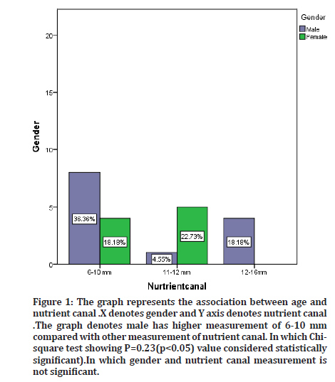
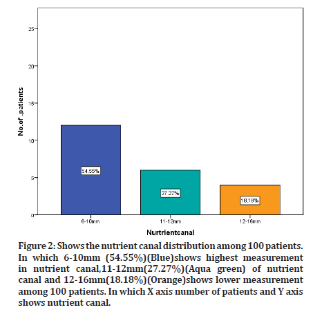
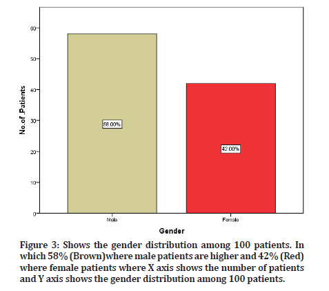
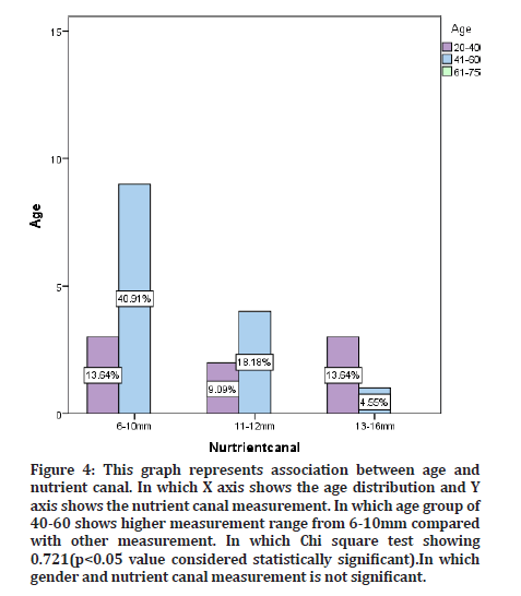
Nutrient canal. In Figure 1 shows the association between age and nutrient canal in which chi-square test showing P=0.23 is not statistically significant and the results from the Cone beam computed Tomography system (CBCT). In which Table 1 shows the mean and standard deviation of nutrient canal measurement. Figure 2 shows the nutrient canal distribution among 100 patients. In which 6-10mm (54.55%) shows highest measurement in nutrient canal, 11-12mm (27.27%) of nutrient canal and 12-16mm (18.18%) shows lowest measurement among 100 patients. In which Figure 3 shows the gender distribution among 100 patients. In which 58%where male patients are higher and 42% where female patients Figure 4 shows the age distribution among patient in which 41-60 (51%) years groups shows higher presence of nutrient canal, 20-40(41%) years age groups shows and 61-75(8%) years of age shows lowest presence of 5 represents association between age and nutrient canal and chi square test showing 0.721 which is not statistically significant.
| N | Mean | Standard Deviation |
|---|---|---|
| 100 | 12.485 | 1.7165 |
Table 1: Shows the mean and standard deviation of nutrient canals present in 100 patients.
Figure 1: The graph represents the association between age and nutrient canal .X denotes gender and Y axis denotes nutrient canal .The graph denotes male has higher measurement of 6-10 mm compared with other measurement of nutrient canal. In which Chisquare test showing P=0.23(p<0.05) value considered statistically significant).In which gender and nutrient canal measurement is not significant.
Figure 2: Shows the nutrient canal distribution among 100 patients. In which 6-10mm (54.55%)(Blue)shows highest measurement in nutrient canal,11-12mm(27.27%)(Aqua green) of nutrient canal and 12-16mm(18.18%)(Orange)shows lower measurement among 100 patients. In which X axis number of patients and Y axis shows nutrient canal.
Figure 3: Shows the gender distribution among 100 patients. In which 58% (Brown)where male patients are higher and 42% (Red) where female patients where X axis shows the number of patients and Y axis shows the gender distribution among 100 patients.
Figure 4: This graph represents association between age and nutrient canal. In which X axis shows the age distribution and Y axis shows the nutrient canal measurement. In which age group of 40-60 shows higher measurement range from 6-10mm compared with other measurement. In which Chi square test showing 0.721(p<0.05 value considered statistically significant).In which gender and nutrient canal measurement is not significant.
Discussion
Nutrient canals as the name suggests carries nutrients. These are the canals that contain nutrient vessels/ blood vessels and nerves which are not detected in all the radiographs, but mandibular anterior radiographs best visualize the nutrient canals [16]. Nutrient canals are the radiolucent spaces in bone which blood vessels and nerves that supply the surrounding structures. Hirschfeld first described nutrient channels and foramina on radiographs which he called “interdental channels [17].They are more commonly seen in the mandibular anterior region followed by premolar and wall of maxillary sinus [18]. Periapical dental radiograph is the best projection to identify the nutrient canals in the anterior mandible. Few authors and others have correlated its presence to be associated with pathologic conditions such as periodontal diseases, hypertension, diabetes, tuberculosis, rickets, calcium deficiency, disuse atrophy, and coarctation of aorta. Incidence of nutrient canals were found to be more in individuals more than 40 years of age and less in people aged less than 30 years and more than 60 years. In which our study also show the same results which age group of 41-60 years shows higher incidence of nutrient canal compared with age group of 20-40 years and 61-75 years. Nutrient canals to be commonly seen in females compared to male are. In our study the results shows that male has higher nutrient canal compared with females. Males had maximum canals in the age group of 21–30‑year whereas females in the age group of 31–40 years had the maximum canals of CT images revealed that nutrient canals were running into the lingual cortical bone which ends at near the alveolar process on CT axial images. Nutrient canals are derived from the incisive branch of the inferior neurovascular bundle and supplies teeth and gingival tissue in the anterior region of the mandible [11,19]. In contrast, CBCT study shows that males had a maximum of 6-10 mm and females had maximum nutrient canal in 11-12mm length of nutrient canals. Clinically, dental implant surgery in the edentulous anterior mandible is considered a routine and relatively safe surgical procedure, but it is not without potential morbidity [13,20]. Complications include hemorrhage from incisive canal and sensory disturbance of incisive canal region [8]. The cause was considered trauma to the incisive canal. These complications are not related to injury of the nutrient canal. However, the nutrient canals carry neurovascular bundles. Morbidity may occur in case the nutrient canals are injured. By preoperative knowledge of the position and anatomy of the nutrient canals, complications such as injury to the nutrient canals can be avoided. Nutrient canals were seen mostly in the anterior region of the mandible. The location of nutrient canals was particularly seen between the central and lateral incisors in the mandible. Patel reported that nutrient canals are seen in the anterior region of the mandible 42.5% on average, Kishi et al reported a prevalence of 1% to 65% in the anterior region [21] and Britt reported nutrient canals were observed in 14% of studied cadavers. These studies evaluated periapical radiographs, but were in the mandible, in the anterior region, and in the molar region. However, there was a significant difference between and females in the mean number of nutrient canals in the premolar region. This may be a result of gender difference of divergent points at which inferior alveolar canal diverges into the incisive canal and the mental nerve near the premolar region8. In which nutrient canals were seen in 94.3% within the mandible. Nutrient canals were most seen in the anterior region of the mandible. Nutrient canals were most commonly seen between the central and lateral incisors. The size of the nutrient foramen varied from 0.4 to 2.0 mm, and the shape are most commonly seen as ovoid .Although anatomic landmarks on reformatted mandibular and maxillary dental CT images and their relation to neurovascular and muscular structures, including NC, have been described by Abrahams this is the first study to our knowledge that describes the incidence and anatomical location of NCs within the anterior mandible using CBCT. In the present study, CBCT enabled the visualization of a well-defined NC and its foramen within the anterior region of the mandible. The presence of NCs within the bone must be taken into consideration during implant placement procedures; the canals can also produce discomfort from an overlying denture if exposed due to resorption of the alveolar process. The anterior mandible has several lingual perforating canals. In which it has variable in number and location and 37 % patients presented with lingual foramina located in lateral incisors to the first premolar area. However, these foramina may include but are not limited to NCs [22]. NCs and their foramina were visible on 17 (16.2 %) images and were bilaterally located in all of them. The results of this study demonstrate that NCs are unusual but not rare. In one radiographic study, NCs were more frequently detected in the anterior region of the mandible in elderly and edentulous patients and in those with advanced periodontal disease9.On the other hand, an increased incidence of NC has also been reported in patients with high bone density, NCs with large diameters could play important roles in osseointegration and in the prevention of postoperative neurosensory disturbances. The size of the canal may also indicate the presence of a well-defined incisive neurovascular bundle coursing through it. The location of the foramen is important to avoid complications that may arise during surgery. For instance, it should be at a distance of three .4 ± 0.7 (1.9–4.7) mm distal to the mandibular midline and three .9 ± 1.4 (1.9–6.6) mm inferior to the alveolar bone crest. The surgical stripping of the periosteum in the mandibles of the aged may noticeably interfere with the healing process because the extra-mandibular portion of the plexus would be damaged16.In unusual case where anastomoses and foramina were detected at the end of NC by means of a periapical radiograph. Based on anatomical studies, they are reported to terminate in the foramina located interdentally on the labial or lingual surfaces of the anterior region of the mandible. NCs are often overlooked during traditional two-dimensional imaging; however, the results of the present study demonstrate that NC is an almost permanent finding on CBCT scans.
Comprehensive information related to the origin of NC from the mandibular canal, bilaterally, is clinically significant. Hirschfeld’ first described nutrient channels and foramina on radiographs which he called interdental channels. Since then there have been a number of reports on these structures. Ennis and associates described the anatomy of nutrient canals by stating that they are derived from the incisive branch of the mandibular artery supplying the region anteror to the mental foramen and that the terminal points of these canals are seen as small nutrient foramina on the superior labial surface of the anterior mandibular area. Shirai reported that the mylohyoid nerve passes through these canals with the sublingual artery [23]. Shimizu observed these foramina anatomically and found them between the right and left central and lateral incisors in approximately 79 percent of the specimens. The diagnostic importance of nutrient canals has been a matter of debate. Stafne stated that these canals are seen more often in the anterior regions of the mandible, particularly in patients in whom the alveolar process is very thin, and considered the canals an anatomic landmark. Worth concluded that there was no evidence to indicate that these shadows had pathologic significance. Nutrient canals tend to be related to advanced age, chronic illness, osteosclerosis, and periodontal disease. Kuroyanagi and associates’ found the radiographic incidence of nutrient canals in the general population to be 46.4 percent, with males showing 49.1 percent and females 44.9 percent incidences. It has been found that nutrient canals were frequently observed where there was no occlusal function in orthodontic patients and stated that variations in anatomic structure were important because these canals were frequently observed in persons with thin ridges in which the medullary spaces appeared to be obliterated [24].
Conclusion
From the present study, it can be concluded that 6-10 mm shows highest measurement of nutrient canal in mandibular anterior, 6-10 mm shows higher measurement at age group of 41-60 years of age compared with other age group of people. Further studies have to be done in larger population to determine the measurements of nutrient canal in different age group and gender of people.
Acknowledgement
The authors are thankful to the Director of academics, Chancellor and Dean of Saveetha Dental College and Hospitals for providing us a platform to do research activities.
References
- Nortjé CJ, Farman AG, Grotepass FW. Variations in the normal anatomy of the inferior dental (mandibular) canal: A retrospective study of panoramic radiographs from 3612 routine dental patients. Br J Oral Sur 1977; 15: 55–63.
- Nortjé CJ, Farman AG, Joubert JD. The radiographic appearance of the inferior dental canal: an additional variation. Br J Oral Sur 1977; 15:171-2.
- Frost D. An anatomical study of mental neurovascular bundle—implants relationships. Br J Oral Maxillofac Surg 1995; 33: 62.
- White SC, Pharoah MJ. Oral radiology-E-Book: Principles and interpretation. Elsevier Health Sciences; 2014.
- Rozylo-Kalinowska I. Imaging techniques in dental radiology: acquisition, anatomic analysis and interpretation of radiographic images. Springer Nature; 2020.
- Omata Y, Sato I, Sato T. An anatomical study of the adult human face: Distribution of elastic fibers and collagen fibers in the skin and subcutaneus tissue. Shigaku = Odontol 1997; 85: 356–375.
- Abdar-Esfahani M, Mehdizade M. Mandibular anterior nutrient canals in periapical radiography in relation to hypertension. Nephro-Urol Monthly. 2014.
- Kishi K, Nagaoka T, Gotoh T, et al. Radiographic study of mandibular nutrient canals. Oral Surg Oral Med Oral Pathol 1982; 54: 118–122.
- Wang PD, Serman NJ, Kaufman E. Continuous radiographic visualization of the mandibular nutrient canals. Dentomaxillofac Radiol 2001; 30: 131–132.
- Abarca M, van Steenberghe D, Malevez C, et al. Neurosensory disturbances after immediate loading of implants in the anterior mandible: an initial questionnaire approach followed by a psychophysical assessment. Clin Oral Investig 2006; 10: 269–277.
- Wismeijer D, van Waas MA, Vermeeren JI, et al. Patients’ perception of sensory disturbances of the mental nerve before and after implant surgery: a prospective study of 110 patients. Br J Oral Maxillofac Surg 1997; 35: 254–259.
- Lee CYS, Craig Yanagihara L, Suzuki JB. Brisk, pulsatile bleeding from the anterior mandibular incisive canal during implant surgery. Implant Dent 2012; 21: 368–373.
- Patel JR, Wuehrmann AH. A radiographic study of nutrient canals. Oral Surg Oral Med Oral Pathol 1976; 42: 693–701.
- Mraiwa N, Jacobs R, Steenberghe D, et al. Clinical assessment and surgical implications of anatomic challenges in the anterior mandible. Clin Implant Dent Relat Res 2003; 5: 219–225.
- Juodzbalys G, Wang H-L, Sabalys G. Anatomy of mandibular vital structures. part ii: mandibular incisive canal, mental foramen and associated neurovascular bundles in relation with dental implantology. J Oral Maxillofac Res 2010; 1: e3.
- Hirschfeld I. A study of skulls in the american museum of natural history in relation to periodontal disease. J Dental Res 1923; 5: 241–265.
- Guruprasad Y, Kumar V, Naik R, et al. Incidence of nutrient canals in hypertensive patients: A radiographic study. J Nat Sci Bio Med 2014; 5: 164.
- Jaju PP, Suvarna PV, Parikh NJ. Incidence of mandibular nutrient canals in hypertensive patients: a radiographic study. Indian J Dent Res 2007; 18: 181–185.
- Laboda G. Life-threatening hemorrhage after placement of an endosseous implant: report of case. J Am Dental Ass 1990; 121: 599–600.
- Hirsch J-M, Brånemark P-I. Fixture stability and nerve function after transposition and lateralization of the inferior alveolar nerve and fixture installation. Br J Oral Maxillofac Surg 1995; 33: 276–281.
- Yildirim YD, Güncü GN, Galindo-Moreno P, et al. Evaluation of mandibular lingual foramina related to dental implant treatment with computerized tomography. Implant Dent 2014; 23: 57–63.
- Bradley JC. Age changes in the vascular supply of the mandible. Br Dental J 1972; 132: 142–144.
- https://ur.booksc.me/book/42272014/ecedcb
- Lovett DW. Nutrient Canals: A roentgenographic study. J Am Dental Ass 1948; 37: 671–675.
Indexed at, Google Scholar, Cross Ref
Indexed at, Google Scholar, Cross Ref
Indexed at, Google Scholar, Cross Ref
Indexed at, Google Scholar, Cross Ref
Indexed at, Google Scholar, Cross Ref
Indexed at, Google Scholar, Cross Ref
Indexed at, Google Scholar, Cross Ref
Indexed at, Google Scholar, Cross Ref
Indexed at, Google Scholar, Cross Ref
Indexed at, Google Scholar, Cross Ref
Indexed at, Google Scholar, Cross Ref
Indexed at, Google Scholar, Cross Ref
Indexed at, Google Scholar, Cross Ref
Indexed at, Google Scholar, Cross Ref
Indexed at, Google Scholar, Cross Ref
Indexed at, Google Scholar, Cross Ref
Indexed at, Google Scholar, Cross Ref
Author Info
R Saravanan* and Gurumoorthy Kaarthikeyan
Department of Periodontics, Saveetha Dental College and Hospitals, Saveetha Institute of Medical and Technical Sciences (SIMATS), Saveetha University, Chennai, Tamil Nadu, IndiaReceived: 21-Jun-2022, Manuscript No. JRMDS-22-45720; , Pre QC No. JRMDS-22-45720 (PQ; Editor assigned: 23-Jun-2022, Pre QC No. JRMDS-22-45720 (PQ; Reviewed: 08-Jul-2022, QC No. JRMDS-22-45720; Revised: 13-Jul-2022, Manuscript No. JRMDS-22-45720 (R); Published: 20-Jul-2022


