Research - (2022) Volume 10, Issue 9
Evaluation of Smear Layer Removal during Canal Preparation Using TruNatomy, ProTaper Next, and Dia-X File Systems: In Vitro Comparative Study
Khanda Ali Kareem* and Shakhawan Kadir Kadir
*Correspondence: Khanda Ali Kareem, Department of Conservative Dentistry and Endodontics, College of Dentistry, Hawler Medical University, Iraq, Email:
Abstract
Background: To guarantee effective endodontic therapy, any smear layer that forms as a result of the debridement of dentinal walls must be eliminated, as it may reduce the overall efficacy of therapy. Aims: This study aims to evaluate the smear layer removal of TruNatomy (TRN), Protaper Next (PTN), and Dia-X systems. Thirty extracted mandibular premolars with a single canal were chosen. The teeth were randomly divided into three groups of 10 specimens based on the type of rotary file used for instrumentation: (1) TruNatomy (TRN) system, (2) Protaper Next (PTN) system, and (3) Dia-X system. Samples were irrigated with 5.25 percent NaOCl and Normal saline. Then samples were analyzed using a Scanning Electron Microscope (SEM) at a magnification of 2000x at the center of the coronal, middle, and apical areas. Statistical analysis of the data was conducted using the Kruskal-Wallis and Mann-Whitney U tests. The apical area had the highest mean values in all three groups [mean(SD)=3.00(0.0)] than the coronal and middle. In the middle region, no significant differences could be seen between the three groups (P>0.05). The coronal region had a lower mean smear layer level across all groups TRN 2.300(0.67), PTN 2.600(0.51), and Dia-X 2.800(0.42). TRN eliminated the smear layer better than PTN and Dia-X in the coronal region, with no statistically significant differences (P>0.05). All tested groups exhibited greater cleanliness at the coronal and middle thirds comparing to the apical region. TruNatomy files demonstrated superior cleaning capacity in the coronal third compared to other groups.
Keywords
Smear layer, Root canal preparation, Protaper next, TruNatomy, Dia-X
Introduction
Root canal preparation includes adequate shaping of the restrictive dentin which facilitates the elimination of pulpal tissue, bacteria, and endotoxins. Furthermore, it creates space for the extra volume of irrigants to flow through the canal to reach the goal of a successful cleaning and prepares the root canal to accommodate the filling material for an efficient apical seal treatment [1].
However, canal preparation with instruments results in the formation of an amorphous, granular, and uneven layer covering the radicular dentin called the smear layer, which was initially reported by McComb and Smith [2]. This layer is composed of inorganic dentin debris and organic elements such as pulp tissue, odontoblastic process, necrotic debris, bacteria, and their metabolic products [3]. According to studies, the existence of the smear layer on the dentinal walls restricts the entry of the endodontic sealers into the dentinal tubule openings and prevents a close adaptation of the obturating materials with the prepared canal walls [4].
The development of nickel-titanium (NiTi) rotary instruments into dental practice has significantly altered the root canal preparation procedure. They gained increased popularity in the last two decades due to their ability of faster canal preparation, superior cutting efficiency, less operator fatigue, maintenance canal curvature, and reduction in procedural mishaps like ledging and transportation [5].
TruNatomy (Dentsply Sirona Maillefer, Ballaigues, Switzerland) consists of five files: two initial instruments, a TRN orifice modifier (20/.08 taper), and a TRN Glider file (17/.02) in addition to three shaping files as follows: Small (20/.04), Prime (26/.04), and Medium (36/.03), with the manufacturer claiming that the Prime shaping file is appropriate for most cases [6]. These files have a variable regressive taper, with just two cutting edges, and an off-center square parallelogram cross-section. The TRN is constructed with post-grinding thermal treatments from a unique narrow NiTi wire design of 0.8 mm as opposed to the standard 1.2 mm design utilized commonly by other rotary systems and operates at higher speeds of 500 rpm [7].
ProTaper Next (Dentsply Sirona, Ballaigues, Switzerland) this system is manufactured with a pre-manufacturing heat treatment technique utilizing M-wire. It consists of five files with various sizes as follows: X1(17.04), X2 (25.06), X3 (30.07), X4 (40.06), and X5 (50.06). PTN shaping files have a variable progressive taper and an off-center rectangular cross-section design. The manufacturers claim that the off-center cross-sectional design provides a snake-like, swaggering movement of the instrument within the canal, which generates a mechanical wave-like oscillation that accelerates the movement of irrigating solution within the canal [8].
Dia-X Rotary Files from (DiaDent Group International, Korea) are NiTi instruments that have been gold-heattreated. Dia-X files have a convex triangular crosssection with a gradually regressive taper, which lowers contact with canal walls and, consequently, rotational friction. They have a non-cutting tip that efficiently removes debris and soft tissue with increased safety. Three shaping and three finishing files are included in this multi-file system. The shaping files DX (16.04), D1 (18.02), and D2 (18.02) have increasingly larger tapers along the length of the cutting blades, whereas the finishing files D3 (20.06), D4 (25.07), and D5 (35.08) are designed with decreasing taper to increase flexibility and reduce the possibility of taper lock [9].
Many studies evaluated the smear layer removal for ProTaper Next files. However, to the best of our knowledge, limited studies are available for the TruNatomy system and no available information exists for the Dia-X system regarding their cleaning ability. The researcher could not find a study that comparatively examined these three systems together in a single experiment, thus, this study aimed to evaluate the amount of smear layer removed when the canals are prepared with: TruNatomy (TRN), Dia-X, and Protaper Next (PTN) systems.
Materials and Methods
Sample selection
The thirty extracted human mandibular premolars with a single canal were used in this study. The teeth that were included were those extracted due to orthodontic reasons and periodontal problems. Radiographs from both the mesiodistal and buccolingual views were taken with digital X-ray (FONA XDG, Assago, Italy) so that only single canaled teeth be selected to exclude the teeth that are with open apex, curved roots, resorption, and endodontically treated. The teeth were cleaned with an ultrasonic scaler to remove soft tissue and hard deposit from the root surface then they were stored in purified distilled water at room temperature before and during the experiment.
Root canal instrumentation
The teeth were divided randomly into three groups each with 10 specimens prepared according to the file systems used in this study. To reach a standard root length of 12 mm, the specimens were de-coronated at the cement enamel junction using a diamond disc on a low-speed handpiece and water cooling. The canal patency was determined by inserting a stainless steel ISO #10 K-file (Dentsply Sirona, Ballaigues, Switzerland) through the apical foramen, and working length (WL) was determined by subtracting 1 mm from the file length when the file tip was visible at the apical foramen. A K-file ISO #15 was used in a watch-winding motion to standardize the size of the apex and to create a glide path [10].
Mechanical preparation performed using endodontic rotary device ENDO-MATE DT Endo-motor (NSK, JAPAN). An important point to be noted is that in each system only two files will be used based on the standardization of file number, tip size, and taper. Not following the usual sequence has been recommended by the manufacturer.
Group 1: Root canal preparation with TRN system
The canal preparation with TruNatomy rotary system (TRN; Dentsply Sirona, Ballaigues, Switzerland) started by adjusting the setting of the endodontic rotary device at 500 rpm and 1.5 Ncm following the manufacturer's recommendations. The sequence of instrumentation was as follows: TRN Glider (17/.02) was used to enhance the efficacy of root canal preparation with a reproducible glide path, worked until the working length is reached, then canals were shaped to the working length with TRN Prime (26/.04) file.
Group 2: Root canal preparation with PTN system
Canal preparation was done using the PTN X1 instrument (taper 0.04, Size 17) until the working length is reached, then the PTN X2 file (taper 0.06, Size 25) was used to shape the canal to the working length. Following the manufacturer’s recommendations at a speed of 300 rpm /2 Ncm torque.
Group 3: Root canal preparation with the DIA-X system
In this group the file sequence was as followed: D2 file (taper 0.04, Size 17) was used until the working length is reached, then D4 file # 25/07 was used to shape the canal to the working length, at 300 rpm speed with a torque of 2.4 Ncm following the manufacturer’s recommendations.
In all the experimental groups, files were used in a brushing motion with the crown-down technique in a continuous rotation as recommended by the manufacturers. Samples were irrigated with 5.2% of NaOCl solution after each file with a total of 5 ml using a 30-gauge side vented needle placed 2 mm short from WL after that canals were flushed with 5 ml of normal saline then canals were dried with absorbent points.
Scanning Electron Microscope (SEM) evaluations
For evaluation of the smear layer removal with the Scanning Electron Microscope, samples were split along the long axis of the teeth by making grooves on the lingual and buccal sides of the roots using the diamond disc with a low-speed handpiece. Then, the root was separated into two halves using a chisel and mallet, and the root half containing the most visible part of the canal was taken.
The specimens were dehydrated with ethyl alcohol in an increasing concentration, mounted on the metal stubs, and then gold sputtered. For each specimen, the canal was divided into the apical, middle, and coronal thirds with equal lengths of four mm, and microscopic images were taken from one point at the center of each third at 2000x magnifications. Then, the scanned images were coded randomly and two investigators observed separate blind evaluations for the presence of a smear layer on the surface of the root canal based on the criteria used by Torabinejad et al [11]:
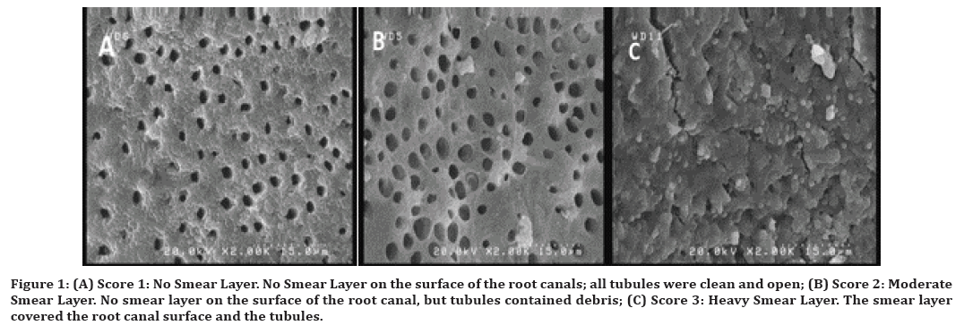
“Score 1: No Smear Layer. No Smear Layer on the surface of the root canals; all tubules were clean and open, Score 2: Moderate Smear Layer. No smear layer on the surface of the root canal, but tubules contained debris, Score 3: Heavy Smear Layer. The smear layer covered the root canal surface and the tubules” (Figure 1).

Figure 1. (A) Score 1: No Smear Layer. No Smear Layer on the surface of the root canals; all tubules were clean and open; (B) Score 2: Moderate Smear Layer. No smear layer on the surface of the root canal, but tubules contained debris; (C) Score 3: Heavy Smear Layer. The smear layer covered the root canal surface and the tubules.
The Weighted Kappa test showed a very good interexaminer agreement in all three regions as it recorded a high Kappa value (0.760 and 0.862) and a statistically significant result (P-value 0.05) at the middle and coronal parts, respectively. On the other hand, no result was computed for the apical area due to exact consistency in the dataset.
Statistical analysis
The SPSS Version 26 was used for the data analysis. Descriptive analysis for the sample, mean values, standard deviation, and mode were calculated for the statistical examination of the data gained from the SEM images. Then the Kruskal-Wallis Test and Mann-Whitney U Test used for the between-groups and within-groups comparisons at different root regions. The level of statistical significance for all tests was set at p<0.05.
Results
By using a variety of descriptive statistics (Mean, Mode, Standard deviation), the amounts of smear layer removal in the apical, middle, and coronal levels of the canal are shown in Table 1.
| Groups | Areas | Mean ± SD | Mode | Minimum | Maximum |
|---|---|---|---|---|---|
| TRN | Apical | 3.000 ± 0.000 | 3 | 3 | 3 |
| Middle | 2.600 ± 0.699 | 3 | 1 | 3 | |
| Coronal | 2.300 ± 0.675 | 2 | 1 | 3 | |
| PTN | Apical | 3.000 ± 0.000 | 3 | 3 | 3 |
| Middle | 2.600 ± 0.699 | 3 | 1 | 3 | |
| Coronal | 2.600 ± 0.516 | 3 | 2 | 3 | |
| Dia-X | Apical | 3.000 ± 0.000 | 3 | 3 | 3 |
| Middle | 2.800 ± 0.422 | 3 | 2 | 3 | |
| Coronal | 2.800 ± 0.422 | 3 | 2 | 3 |
Table 1: Descriptive statistics for all groups and areas.
According to Table 1, the apical region in all groups showed a heavy smear layer as per their mean values scoring 3. Regarding the middle third, all groups recorded lower mean values than the apical third, but TRN and PTN groups (both with Mean ± SD 2.6 ± 0.699) showed improved cleaning efficiency than the Dia-X group (Mean ± SD 2.800 ± 0.422).
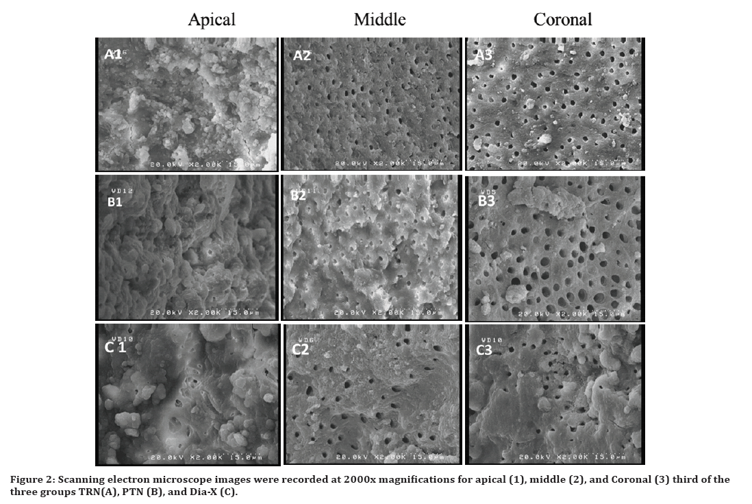
The coronal region was considered to be a more cleaned area regarding smear layer removal compared to other areas and lower scores were recorded based on the mean values shown in Table 1. TRN group showed the lowest means of smear layer among the other two groups. In Table 2, the Kruskal-Wallis test reported no significant differences between the three experimented groups at all root thirds apical, middle, and coronal (P>0.05). Thus, no further investigation was needed with the implementation of the Mann-Whitney test. However, it was vital to determine within-group comparisons for each file system using the Friedman test for three canal regions and the Wilcoxon signed rank test for pairwise comparisons Table 3. It's notable from Friedman’s Test that TRN had a higher effective influence as its p-value was (0.034) (P<0.05), additionally, Wilcoxon signed Ranks Test showed that only apical and coronal regions in TRN had a statistically significant difference regarding smear layer scoring. Meanwhile, the other groups revealed the same influence in terms of smear layer removal at different canal regions. (Figure 2) shows SEM images for groups at different root thirds.
| Areas | Groups | Mean Rank | Kruskal Wallis (P-value) |
|---|---|---|---|
| Apical | TRN | 15 | 0 (1.000) |
| PTN | 15 | ||
| Dia-X | 15 | ||
| Middle | TRN | 14.9 | 0.466 (0.792) |
| PTN | 14.9 | ||
| Dia-X | 16.7 | ||
| Coronal | TRN | 12.2 | 3.604 (0.165) |
| PTN | 15.7 | ||
| Dia-X | 18.6 |
Table 2: Non-parametric between-group comparison test at different root areas.
| Groups | Apical Mean Rank | Middle Mean Rank | Coronal Mean Rank | Friedman Test (P-value) | Wilcoxon Signed Ranks Test |
|---|---|---|---|---|---|
| TRN | 2.45 | 2 | 1.55 | 6.750 (0.034) | (A-C)* |
| PTN | 2.35 | 1.85 | 1.8 | 5.692 (0.058) | |
| Dia-X | 2.2 | 1.9 | 1.9 | 4.000 (0135) | |
| * Statistically significant with<0.05 | |||||
Table 3: Within-group comparison test between root canal areas for each file system.

Figure 2. Scanning electron microscope images were recorded at 2000x magnifications for apical (1), middle (2), and Coronal (3) third of the three groups TRN(A), PTN (B), and Dia-X (C).
Discussion
Chemo-mechanical preparation is a crucial aspect of root canal therapy. For this goal, a variety of materials and instruments are utilized. However, during cleaning and shaping, root dentin is cut and shaped using hand or rotary files to remove necrotic tissue and microorganisms, resulting in the production of a smear layer that covers the whole root canal wall [12]. Numerous authors have studied the detrimental effect of the smear layer presence in the dentinal walls on the penetration of intra-canal disinfectants and sealers into the dentinal tubules, which may fail to seal the canals [13,14].
If it is impossible to entirely avoid the production of debris and smear layer, it is imperative to select a file system that removes the maximum amount of smear layer [15]. This study, therefore, investigated the cleaning efficacy of several nickel-titanium rotary tools by evaluating the degree of the smear layer that remains on the root canals following mechanical instrumentation.
Regarding the evaluation of the three canal sections (coronal, middle, and apical), the descriptive statistics from Table 1 show that all three systems failed to remove the smear layer. Table 2, shows the Kruskal- Wallis test of between-group comparison and illustrates that on average, all three systems recorded more smear layer removal from coronal to apical region (coronal 0.165, middle 0.792, and apical 1.000) regardless of the instrument used, although the results were not statistically significant. This observation is consistent with previous studies that found all rotary files were more effective at cleaning the coronal third of the root canal than the middle and apical thirds, and that the apical third of the canal was less clean overall [16–18]. Such studies underline those wider coronal preparations, which improve irrigant solution flow and promote more effective fluid dynamics and turbulence, are the cause of increased smear layer removal at coronal areas. However, the variations between the mean value of this study and those of earlier investigations may be contributed to the use of EDTA in the above-mentioned studies, as in this study only sodium hypochlorite NaOCl of 5.25% was used as the final irrigation solution to avoid any influence of various irrigation solutions [18,19].
Results revealed that all three studied files failed to remove the smear layer at the apical third (Mean ± SD 3.000 ± 0.00). This can be due to the use of a needle and syringe as a method of irrigation that study findings by Htun concluded that using a needle and syringe was not an adequate irrigation method to produce hydrodynamic shear stresses enough to dislodge the materials adhered to the canal walls [20]. Also, Nangia, et al. [21], claimed that it would be challenging for a needle and syringe to effectively remove the debris and smear layer from this area as more sclerotic dentine in the apical portion may make it harder to remove the smear layer. However, Akcay, et al. [22] reported that manual needle irrigation considerably increased the quantity of smear layer removal on the apical and middle thirds of the root canals.
Another factor can be due to the small apical size of experimented files (TRN Prime #26, PTN X2 #25, Dia-X D4 #25) which may limit the ability of irrigant solution to reach the apical third due to the size of apical enlargement or the depth of penetration of the needle which can be supported by study findings of Andreani, et al. [23] on the use of Conventional needle irrigation who reported that larger taper and apical preparation sizes will result in higher irrigant flow and effective removal of smear layer. Contradicting the findings of Nangia [21], who examined both #30 and #40 final apical preparation sizes and discovered that larger apical preparation did not significantly reduce smear layer and debris.
Various taper percentages were investigated in this study (TRN Prime 4%, PTN X2 6%, and Dia-X D4 7%), however an increase in taper from 4 to 7 percent did not appreciably lessen the smear layer in the apical third. The results from Zarei, et al. [24] utilizing RaCe files with two distinct tapers (30/02 and 30/04) are in agreement with these findings. they stated that the amount of smear layer in the apical third was unaffected by an increase in taper. On the other hand, the SEM results of Reddy et al. [1] have demonstrated that lower smear layer scores of ProTaper Gold at the 9 mm level, may be due to a 9% taper of the F3 file, which may have decreased the number of untouched areas. While the decreased taper of 4% of XP Endo Shaper might not have made contact with all canal walls, is the cause of the higher smear layer scores.
With no significant differences, Table 2 demonstrates that TRN removes the smear layer better than PTN and Dia-X (12.200, 15.700, and 18.600, respectively) at the coronal third. These results are consistent with Waleed and Selivany [18], which found that TruNatomy removes the smear layer significantly better than PTN. The TRN files have off-centered parallelogram cross-sections, active cutting flutes with only two cutting edges, a unique slim NiTi wire design, and variable taper that ensures the shank ends up with a maximum flute diameter of 0.8 mm. This design allows for more debridement space and minimizes the accumulation of smear and dentinal chips [7]. Another reason may be related to the higher speed of TRN instruments (500 rpm) than the other two systems which operate at speeds of 300-350 rpm. This finding is consistent with that of Mohammed et al. [25] who suggested that the high rotational speed (800 rpm) of the XP may contribute to its superior cleaning performance when compared to other experimental systems.
According to the Kruskal-Wallis test, the PTN demonstrated better canal cleaning than the Dia-X in the middle (PTN 14.900, Dia-X 16.700) and coronal thirds (PTN 15.700, D.X 18.600) with no significant differences. This is in line with the conclusions reached by Girgis et al. [17], and Al-Khafaji and Al-Huwaizi [19], who found that PTN displayed less amount of smear layer than other systems used in their study. This difference might be caused by the file's off-center rectangular cross-section, which creates a special mechanical wave of motion that goes along the file's active length and helps to reduce the file's engagement with the dentin by ensuring that the file always hits the canal walls twice. As a result, there is less lateral debris compaction and greater room for coronal debris clearance [8]. While, a study claims that the PTN file's swaggering motion which is caused by its off-center rectangular cross-section, demonstrates the lesser cutting capability and higher smear layer buildup of PTN files than WOG [18].
Tables 1 and 2 demonstrate that Dia-X recorded the most smear layer in the coronal and middle areas. This outcome could be influenced by the file´s convex triangular crosssectional design that gives the instrument a bigger core area. This finding can be supported by Girgis et al. [17] who found that the M-Pro file's higher amount of smear layer may be related to its convex triangular crosssection, which provides it with a thick core and increases cutting capacity in comparison to the ProTaper Next's smaller core. An increase in the instrument's ability to cut will increase its ability to produce a smear layer [16]. However, more research is required to fully understand this system's cleaning capacity.
Conclusion
In conclusion, none of the examined files could completely clean root canals. There were no statistically significant differences between the three systems, however, the coronal and middle thirds of each showed superior cleanliness to the apical third. TruNatomy group showed better cleaning capacity at the coronal third than the other groups, with no statistically significant differences.
References
- Mankeliya S, Singhal RK, Gupta A, et al. A comparative evaluation of smear layer removal by using four different irrigation solutions like root canal irrigants: An in vitro SEM study. J Contemp Dent Pract 2021; 22:527-531.
- McComb D, Smith DC. A preliminary scanning electron microscopic study of root canals after endodontic procedures. J Endod 1975; 1:238-242.
- Torabinejad M, Handysides R, Khademi AA, et al. Clinical implications of the smear layer in endodontics: A review. Oral Surg Oral Med Oral Pathol Oral Radiol Endodont 2002; 94:658-666.
- Shaikh M, Shetty P, Tekwani D, et al. Comparative evaluation of smear layer removal using three chelating agents and their effect on the penetrability of epoxy resin-based sealer into dentinal tubules using SEM and CLSM-in vitro study. J Adv Med Dent Sci Res 2021; 9:44-50.
- Lall AG, Saha SG, Alageshan V, et al. A comparative evaluation of cyclic fatigue resistance of reciproc blue, wave one gold and 2shape nickel–titanium rotary files in different artificial canals. Endodontology 2021; 33:1.
- Riyahi AM, Bashiri A, Alshahrani K, et al. Cyclic fatigue comparison of TruNatomy, twisted file, and ProTaper next rotary systems. Int J Dent 2020; 2020.
- Van der Vyver PJ, Vorster M, Peters OA. Minimally invasive endodontics using a new single-file rotary system. Int DentAfrican 2019; 9:6-20.
- Ruddle CJ, West JD, Machtou P. Fifth-generation technology in endodontic: The shaping movement. Roots 2014; 1:22-8.
- Micoogullari Kurt S, Kaval ME, Serefoglu B, et al. Cyclic fatigue resistance and energy dispersive X‐ray spectroscopy analysis of novel heat‐treated nickel–titanium instruments at body temperature. Microsc Res Tech 2020; 83:790-794.
- Mahdi SF, Bakr DK. Evaluation of the effect of smear OFF on smear layer removal and erosion of root canal dentin: An in vitro study. Erbil Dent J 2019; 2:269-277.
- Torabinejad M, Khademi AA, Babagoli J, et al. A new solution for the removal of the smear layer. J Endod 2003; 29:170-175.
- Kaushal R, Bansal R, Malhan S. A comparative evaluation of smear layer removal by using ethylenediamine tetraacetic acid, citric acid, and maleic acid as root canal irrigants: An in vitro scanning electron microscopic study. J Conserv Dent 2020; 23:71.
- Sonu KR, Girish TN, Ponnappa KC, et al. Comparative evaluation of dentinal penetration of three different endodontic sealers with and without smear layer removal-Scanning electron microscopic study. Saudi Endod J 2016; 6:16.
- Machado R, Garcia LD, da Silva Neto UX, et al. Evaluation of 17% EDTA and 10% citric acid in smear layer removal and tubular dentin sealer penetration. Microsc Res Tech 2018; 81:275-282.
- Chatterjee S, Desai PD, Mukherjee S, et al. Evaluation of debris and smear layer formation using three different NI-TI rotary instrument systems: An in vitro scanning electron microscope study. J Conserv Dent 2021; 24:568.
- Machado R, Comparin D, Back ED, et al. Residual smear layer after root canal instrumentation by using Niti, M-Wire and CM-Wire instruments: A scanning electron microscopy analysis. Eur J Dent 2018; 12:403-409.
- Girgis D, Roshdy N, Sadek H. Comparative assessment of the shaping and cleaning abilities of M-Pro and Revo-S versus ProTaper Next Rotary Ni-Ti systems: An in vitro study. Adv Dent J 2020; 2:162-176.
- Waleed D, Selivany BJ. Debridement ability of trunatomy, S-One Plus, and other single file systems. Open Access Macedonian J Med Sci 2022; 10:91-7.
- Al-Khafaji HA, Al-Huwaizi HF. Cleaning efficiency of root canals using different rotary instrumentation systems: A comparative in vitro study. IJMRHS 2019; 8:89-93.
- Htun PH, Ebihara A, Maki K, et al. Cleaning and shaping ability of Gentlefile, HyFlex EDM, and ProTaper Next instruments: a combined micro–computed tomographic and scanning electron microscopic study. J Endod 2020; 46:973-979.
- Nangia D, Nawal RR, Yadav S, et al. Influence of final apical width on smear layer removal efficacy of Xp Endo finisher and endodontic needle: An ex vivo study. Eur Endod J 2020; 5:18.
- Akçay A, Gorduysus M, Aydin B, et al. Evaluation of different irrigation techniques on dentin erosion and smear layer removal: A scanning electron microscopy study. J Conserv Dent 2022; 25:311.
- Andreani Y, Gad BT, Cocks TC, et al. Comparison of irrigant activation devices and conventional needle irrigation on smear layer and debris removal in curved canals. (Smear layer removal from irrigant activation using SEM). Aust Endod J 2021; 47:143-149.
- Zarei M, Javidi M, Afkhami F, et al. Influence of root canal tapering on smear layer removal. NY State Dent J 2016; 82:35-38.
- Mohammed MN, Kamel WH, Rokaya ME. Comparison of the cleaning effectiveness and incidence of dentinal defects after biomechanical preparation using different ni ti rotary instruments in root canals. Al-Azhar Dent J Girls 2020; 7:165-170.
Indexed at, Google Scholar, Cross Ref
Indexed at, Google Scholar, Cross Ref
Indexed at, Google Scholar, Cross Ref
Indexed at, Google Scholar, Cross Ref
Indexed at, Google Scholar, Cross Ref
Indexed at, Google Scholar, Cross Ref
Indexed at, Google Scholar, Cross Ref
Indexed at, Google Scholar, Cross Ref
Indexed at, Google Scholar, Cross Ref
Indexed at, Google Scholar, Cross Ref
Indexed at, Google Scholar, Cross Ref
Indexed at, Google Scholar, Cross Ref
Indexed at, Google Scholar, Cross Ref
Indexed at, Google Scholar, Cross Ref
Indexed at, Google Scholar, Cross Ref
Indexed at, Google Scholar, Cross Ref
Indexed at, Google Scholar, Cross Ref
Author Info
Khanda Ali Kareem* and Shakhawan Kadir Kadir
Department of Conservative Dentistry and Endodontics, College of Dentistry, Hawler Medical University, Erbil, IraqReceived: 03-Sep-2022, Manuscript No. jrmds-22-73667; , Pre QC No. jrmds-22-73667(PQ); Editor assigned: 05-Sep-2022, Pre QC No. jrmds-22-73667(PQ); Reviewed: 19-Sep-2022, QC No. jrmds-22-73667(Q); Revised: 23-Feb-2022, Manuscript No. jrmds-22-73667(R); Published: 30-Sep-2022
