Research - (2021) Volume 9, Issue 6
Green Preparation of Gold Nano Particles Procured from Neem and Ginger Plant Extract to Evaluate the Antimicrobial Activity Against Oral Aerobic Pathogens
Harsh Kasabwala1*, Dhanraj Ganapathy1 and Rajeshkumar Shanmugam2
*Correspondence: Harsh Kasabwala, Department of Prosthodontics, Saveetha Dental College and Hospitals, Saveetha Institute of Medical And Technical Sciences, Saveetha University, Chennai, India, Email:
Abstract
Gold has been used therapeutically in Chinese medicine since 2500 BC. Swarna Bhasma, or red colloidal gold, is still used in Indian Ayurvedic medicine for rejuvenation and revitalization in old age. Green metal nanoparticle synthesis is an important technique for developing more environmentally friendly nanoparticle production methods. The biomedically valid gold nanoparticle was synthesised in this study using Neem and Ginger plant extract. The transition of the extract from brown to pinkish red confirmed the synthesis of gold nanoparticles. Scanning Electron Microscope revealed triangle, rectangle, and square shaped gold nanoparticles with an average size of 60 nm (SEM). Energy dispersive analysis (EDS) was used to investigate the nature of elemental gold. Finally, the antibacterial activity of gold nanoparticles was tested; it was discovered that E faecalis had the highest level of inhibition, while S mutans, S Aureus and Candida Albicans and had a medium range of inhibition.
Keywords
Gold nanoparticles, Green synthesis, Neem, Ginger, Antibacterial activity
Introduction
In recent years, there have been significant advancements in the field of nanotechnology, with numerous methodologies developed to synthesise nanoparticles of specific shape and size based on specific requirements. There is a growing need to develop environmentally friendly nanoparticle synthesis processes that do not involve the use of toxic chemicals [1,2]. Magneto tactic bacteria (which synthesise magnetite nanoparticles) [3], diatoms (which synthesise siliceous materials), and Slayer bacteria are all examples of microorganisms that synthesise inorganic materials [4].
While microorganisms such as bacteria, actinomycetes, and fungi are still being studied in metal nanoparticle synthesis, using parts of whole plants in similar nanoparticle synthesis methodologies is an exciting and underexplored possibility [5]. Even though gold nanoparticles are biocompatible, chemical synthesis methods may result in the presence of toxic chemical species adsorbed on the surface, which could cause problems in medical applications [6]. The use of microorganisms or plants to synthesise nanoparticles could potentially solve this problem by making the nanoparticles more biocompatible. Plant-based nanoparticle synthesis could be more environmentally friendly than other biological processes because it eliminates the time-consuming process of maintaining cell cultures. It can also be scaled up for nanoparticle synthesis on a large scale.
Jose-Yacaman, et al. recently demonstrated the synthesis of gold and silver nanoparticles within live alfalfa plants using gold and silver ion uptake from solid media, respectively [3,7,8]. The synthesis of pure metallic nanoparticles of gold by reduction of aqueous Ag+ and AuCl4 ions, as well as the synthesis of bimetallic core–shell nanoparticles of gold by simultaneous reduction of aqueous Ag+ and AuCl4 ions with the broth of Neem (Azadirachta indica). and Ginger leaves (Zingiber officinale), are described in this paper.
Materials and Methods
Visual observation
The pink colour of a reaction mixture of 1 mM aqueous gold chloride and Neem and Ginger extract indicates that gold ions have been reduced to gold nanoparticles. The reaction mixture initially turned brownish pink. After that, as the incubation time was increased from 30 minutes to 48 hours, the colour of the solution changed rapidly to pink. After that, the colour of the solution remained stable with no change in intensity, indicating that the reduction was complete. The excitations of surface plasmon resonance in the nanoparticles caused a colour change in the reaction mixture. This significant finding points to the reduction of Au+ ions and gold nanoparticle biosynthesis. Singaravelu et al. [9] previously reported that the gold nanoparticle synthesis process took 15 hours to complete. In the current study, however, the gold nanoparticle synthesis process is started quickly at 50 minutes and completed after 48 hours of incubation (Figure 1).
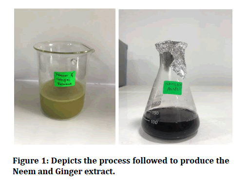
Figure 1: Depicts the process followed to produce the Neem and Ginger extract.
Scanning electron microscopy
Figure 2 shows a SEM image obtained at room temperature using Neem and Ginger extracts and a 1 mm gold chloride solution. The formation of square, rectangle, cubic, and triangle shaped nanoparticles with an average diameter of 60 nm and some divergence is demonstrated. Similar results were obtained in the studies by Venkatraman et al [10] and Daisy et al [10,11], where gold nano particles were obtained using Turbinaria conoides and Cassia fistula, respectively.
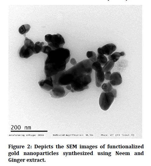
Figure 2: Depicts the SEM images of functionalized gold nanoparticles synthesized using Neem and Ginger extract.
Energy dispersive analysis
Figure 3 depicts the EDS spectra obtained from AuNps. The EDS profile shows a strong gold signal, as well as weak oxygen, carbon, magnesium, and silica peaks, which could have come from biological and chemical molecules bound to the gold nanoparticle's surface.
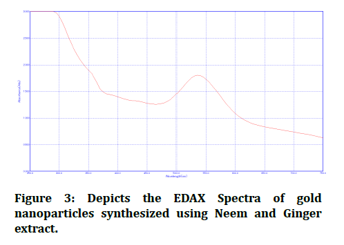
Figure 3: Depicts the EDAX Spectra of gold nanoparticles synthesized using Neem and Ginger extract.
Antibacterial activity
The antibacterial activity of gold nanoparticles was tested using four pathogenic bacteria isolates: E. faecalis, S mutaNS, S Aureus, and C. Albicans. The disease-causing bacteria E. faecalis exhibits the greatest inhibition zone in this study (22 mm). S mutans and S Aureus, the opportunistic bacteria, had a moderate range of inhibition (13mm). The reason behind this could be the addition of 100 µl of colloidal gold nanoparticles. However, C Albicans had the smallest inhibition zone (10mm). Nazari et al. investigated the antimicrobial properties of gold nanoparticles against P. aeruginosa, Staphylococcus aureus, and Escherichia coli using the disc diffusion method in 2012 [12]. However, the current study illustrates good zone of inhibition against all the pathogenic bacterial strains. This research also found that high concentrations of gold nanoparticles resulted in a larger zone of inhibition. As a result, the inhibition zone is proportional to the concentration of gold nanoparticles (Figure 4 and Figure 5).
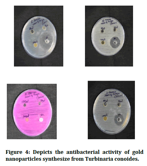
Figure 4: Depicts the antibacterial activity of gold nanoparticles synthesize from Turbinaria conoides.
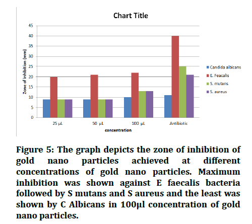
Figure 5: The graph depicts the zone of inhibition of gold nano particles achieved at different concentrations of gold nano particles. Maximum inhibition was shown against E faecalis bacteria followed by S mutans and S aureus and the least was shown by C Albicans in 100μl concentration of gold nano particles.
Results and Discussion
Visual observation
The pink colour of a reaction mixture of 1 mM aqueous gold chloride and Neem and Ginger extract indicates that gold ions have been reduced to gold nanoparticles. The reaction mixture initially turned brownish pink. After that, as the incubation time was increased from 30 minutes to 48 hours, the colour of the solution changed rapidly to pink. After that, the colour of the solution remained stable with no change in intensity, indicating that the reduction was complete. The excitations of surface plasmon resonance in the nanoparticles caused a colour change in the reaction mixture. This significant finding points to the reduction of Au+ ions and gold nanoparticle biosynthesis. Singaravelu et al. [9] previously reported that the gold nanoparticle synthesis process took 15 hours to complete. In the current study, however, the gold nanoparticle synthesis process is started quickly at 50 minutes and completed after 48 hours of incubation (Figure 1).
Scanning electron microscopy
Figure 2 shows a SEM image obtained at room temperature using Neem and Ginger extracts and a 1 mm gold chloride solution. The formation of square, rectangle, cubic, and triangle shaped nanoparticles with an average diameter of 60 nm and some divergence is demonstrated. Similar results were obtained in the studies by Venkatraman et al [10] and Daisy et al [10,11], where gold nano particles were obtained using Turbinaria conoides and Cassia fistula, respectively.
Energy dispersive analysis
Figure 3 depicts the EDS spectra obtained from AuNps. The EDS profile shows a strong gold signal, as well as weak oxygen, carbon, magnesium, and silica peaks, which could have come from biological and chemical molecules bound to the gold nanoparticle's surface.
Antibacterial activity
The antibacterial activity of gold nanoparticles was tested using four pathogenic bacteria isolates: E. faecalis, S mutaNS, S Aureus, and C. Albicans. The disease-causing bacteria E. faecalis exhibits the greatest inhibition zone in this study (22 mm). S mutans and S Aureus, the opportunistic bacteria, had a moderate range of inhibition (13mm). The reason behind this could be the addition of 100 µl of colloidal gold nanoparticles. However, C Albicans had the smallest inhibition zone (10mm). Nazari et al. investigated the antimicrobial properties of gold nanoparticles against P. aeruginosa, Staphylococcus aureus, and Escherichia coli using the disc diffusion method in 2012 [12]. However, the current study illustrates good zone of inhibition against all the pathogenic bacterial strains. This research also found that high concentrations of gold nanoparticles resulted in a larger zone of inhibition. As a result, the inhibition zone is proportional to the concentration of gold nanoparticles (Figure 4 and Figure 5).
Conclusion
Because of their functionally active properties, gold nanoparticles have a lot of potential in the fields of physics, chemistry, and biomedicine. Gold nanoparticles have been used in drug delivery, cancer therapy, and antimicrobial agents in biomedicine. We concluded that green synthesis of gold nanoparticles using Neem and ginger leaf extracts was possible. Additionally, the nano particles did display their antibacterial activity against pathogenic bacteria. The synthesis of gold nanoparticles took 48 hours to form completely. As a result, the gold nanoparticles that were created were stable for several months.
References
- Simkiss K, Wilbur KM. Biomineralization. Elsevier 2012; 337.
- Mann S. Biomineralization and biomimetic materials chemistry. J Mater Chem 1995; 5:935.
- Philipse AP, Maas D. Magnetic colloids from magnetotactic bacteria: Chain formation and colloidal stability. Langmuir 2002; 18:9977–84.
- Lovley DR, Stolz JF, Nord GL, et al. Anaerobic production of magnetite by a dissimilatory iron-reducing microorganism. Nature 1987; 330:252–4.
- Shaikh SM, Shaikh TJ. Biogenic synthesis of silver and gold nano particles using fungal species. Plantae Scientia 2018; 1:25–30.
- Sharon M. A Peep into what was behind the incorporation of gold nano particles in nano medicine. J Nanomed Res 2015; 2.
- Gardea-Torresdey JL, Parsons JG, Gomez E, et al. Formation and growth of au nanoparticles inside live alfalfa plants. Nano Letters 2002; 2:397–401.
- Gardea-Torresdey JL, Gomez E, Peralta-Videa JR, et al. Alfalfa sprouts: A natural source for the synthesis of silver nanoparticles. Langmuir 2003; 19:1357–61.
- Singaravelu G, Arockiamary JS, Ganesh Kumar V, et al. A novel extracellular synthesis of monodisperse gold nanoparticles using marine alga, Sargassum wightii Greville. Colloids Surfaces Biointerfaces 2007; 57:97–101.
- Venkatraman A, Yahoob SAM, Nagarajan Y, et al. Pharmacological activity of biosynthesized gold nanoparticles from brown algae- seaweed turbinaria conoides. Nano World J 2018; 4.
- Daisy P, Saipriya. Biochemical analysis of Cassia fistula aqueous extract and phytochemically synthesized gold nanoparticles as hypoglycemic treatment for diabetes mellitus. Int J Nanomed 2012; 1189.
- Nazari ZE, Banoee M, Sepahi AA, et al. The combination effects of trivalent gold ions and gold nanoparticles with different antibiotics against resistant Pseudomonas aeruginosa. Gold Bulletin 2012; 53–59.
Author Info
Harsh Kasabwala1*, Dhanraj Ganapathy1 and Rajeshkumar Shanmugam2
1Department of Prosthodontics, Saveetha Dental College and Hospitals, Saveetha Institute of Medical And Technical Sciences, Saveetha University, Chennai, India2Department of Pharmacology, Saveetha Dental College and Hospitals, Saveetha Institute of Medical and Technical Sciences, Saveetha University, Chennai, India
Citation: Harsh Kasabwala, Dhanraj Ganapathy, Rajeshkumar Shanmugam,Green Preparation of Gold Nano Particles Procured from Neem and Ginger Plant Extract to Evaluate the Antimicrobial Activity Against Oral Aerobic Pathogens, J Res Med Dent Sci, 2021, 9(6): 259-262
Received: 04-May-2021 Accepted: 21-Jun-2021
