Research Article - (2022) Volume 10, Issue 7
Incidence of Congenitally Missing Mandibular Incisors among Patients with Orthodontic Treatment
Revathi B and Remmiya Mary Varghese*
*Correspondence: Remmiya Mary Varghese, Department of Orthodontics, Saveetha Dental College, Saveetha Institute of Medical and Technical Sciences, Poonamallee High Road, Chennai, India, Email:
Abstract
Aim: To assess the incidence of congenitally missing mandibular incisors in patients reporting for orthodontic treatment.
Introduction: Hypodontia is the most common condition reported among people. Missing mandibular incisors plays an important role in planning the treatment in the field of orthodontics. The knowledge on the mandibular teeth agenesis and other different tooth positions aids in analyzing the etiological basis of the orthodontic treatment.
Materials and methods: A total of 962 patients' case sheets were taken in the present study. From which, 155 patients were categorized under the orthodontic population and 807 patients under the general population. The prevalence and average missing of the mandibular incisors were analyzed in both the groups using intraoral images and panoramic radiograph.
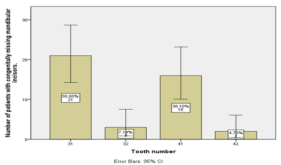
Results: The prevalence of missing mandibular incisors among orthodontic and general population were 8.38% and 1.11% respectively. Tooth number 31 (50%) is found to be the most commonly missing teeth followed by 41, 32 and 42. Females show a high incidence of missing lower incisors compared to males. And also, females have more missing teeth than males.
Conclusion: Hypodontia is found considerably more frequent in mandibular incisors in orthodontic patients. Early detection of missing teeth can obtain a satisfactory permanent dentition and to reduce the complications of hypodontia.
Keywords
Incidence, Innovative approach, Hypodontia, Mandibular incisors, Panoramic radiographsIntroduction
Hypodontia is the one of the most common dental anomalies encountered by the dentist in both primary and permanent dentition. Numerous researches were carried out assessing the hypodontia with overall prevalence rate from 0.1 to 10% [1]. The prevalence of hypodontia varies based on geographical locations and races. The incidence of tooth agenesis, excluding third molars in both genders, is reported to be 3.5% among American population, 11.3% among Irish and Solvenian population and 0.3% among Israeli population [2-4]. The different findings could be explained by the variety in the samples examined in terms of age range and ethnicity used for evaluation. Multiple factors are employed for the emergence of hypodontia. A study conducted by Boruchov and Green, Moller et al. and Townsend et al proved that environmental factors may be important in the expression of the trait of hypodontia such as infection, trauma and drugs [5-7]. Another study by Parkin stated that occurrence of hypodontia is not completely dependent on genetic factors but is supported by the variable expression of hypodontia [8]. Furthermore, it is also suggested that anterior hypodontia may depend more on genetics while posterior missing might be sporadic [9]. The different findings could be explained by the variety in the samples examined in terms of age range and ethnicity used for evaluation.
Assessment of other congenital dental anomalies might be established easily, whereas congenitally missing teeth are not commonly seen through the naked eyes of a dentist due to minimal discrepancy in the other teeth in the arch [10]. Thorough clinical and radiographic examination, care and conservatism must be exercised to arrive at decisions that concerned congenitally missing teeth. The treatment plan is established on the basis of presence of even one congenital missing tooth as it influences the complete profile, masticatory efficiency, and aesthetics of the patient undergoing orthodontic treatment [11].
Many literature analyzed the incidence rates of missing maxillary lateral incisors, maxillary and mandibular premolars [12,13]. So far, studies focused only on either normal individuals or orthodontic patients. Hence in contrast, this study aims to determine the incidence of missing mandibular incisors among the orthodontic population against a comparable sample of the general Indian population.
Materials and Methods
Study design
This was a retrospective study for a period of 2 months from December 2020 to February 2021. A total of 962 patients were included in this study that visited the Department of Orthodontics between the 18-40 years of age group and were divided into two groups. Group A (N=155) patients undergoing orthodontic treatment. Group B (N=807) patients without any requirement of orthodontic treatment. Informed consent was obtained from all the patients.
Data collection
The case sheets of those patients were verified and recorded the demographic details such as name, age and gender. The number of missing mandibular incisors was analyzed using intraoral images and panoramic radiograph which were uploaded in the case history of each patient along with the tooth number.
Inclusion criteria
- Patients of more than 18 years of age.
Exclusion criteria
- Missing mandibular incisors due to trauma, extraction and orthodontic camouflage.
- Case sheets with improper/missing intraoral images and radiographs.
Statistical analysis
Collected data was tabulated and imported to Statistical Package for Social Science (SPSS, version 22, SPSS Inc. Chicago, IL, USA) for statistical analysis. Association of missing mandibular incisors at each position in the right and left quadrant of the mouth for all the patients was analyzed using the chi-square test. The differences in number of missing teeth and teeth number between gender in orthodontic and general population were compared.
Results
Figure 1: Graph shows the frequency of congenitally missing mandibular incisors. Tooth number 31 is reported to be most commonly missing mandibular incisors with 50% followed by 41 (38.1%), 32(7.1%) and 42 with the least percentage of 4.7%.
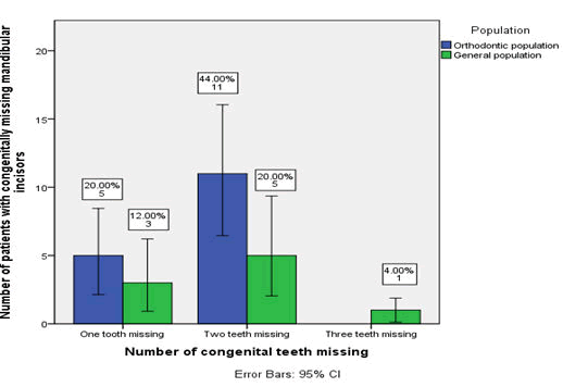
Figure 2: Graph shows the distribution of number of congenitally missing mandibular incisors among orthodontic and general population. In the orthodontic population, 20% of the individuals had lost one tooth, 44% had two lost teeth and no individuals with three missing teeth, whereas in the general population, 12% had lost one tooth, 20% had lost two teeth and 4% with three missing teeth. The correlation is found to be statistically significant with P<0.05.
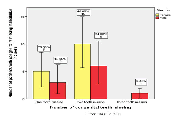
Figure 3: Graph shows the distribution of number of congenitally missing mandibular incisors among males and females. Females showed a high incidence of one and two missing teeth (20% and 40%) compared to males (12% and 24%) with no statistical significance of P>0.05.
Discussion
From the study, the incidence of missing mandibular incisors among the orthodontic population is found to be in 16 patients (8.38%) and the general population 9 (1.11%). Females showed a higher incidence of congenitally missing mandibular incisors than males with 6.54% and 4.59% respectively.
Our results suggest that the congenitally missing mandibular incisors are found to be higher among the orthodontic population compared to the general population. The prevalence in the present study is found to be 8.38%. Similar results were obtained in a study conducted by Sterzik et al., with a prevalence rate of 8.1% among German orthodontic population [14]. In contrast, a study conducted by reported 0.3% prevalence in Jewish population [15] and a study conducted by concluded with 10.1% prevalence rate among Norwegian population which is slightly higher compared to the present study [3]. This wide range of variation can be due to the factors such as age, gender, sampling methods and clinical examination.
Females showed a higher incidence than males with 6.54% and 4.59% respectively. The result is in line compared to the other previous studies. A study conducted by at UK population at Iceland population and Nik-Hussein at Malaysian population reported higher prevalence of hypodontia in females than males [16-18]. However, some studies reported no differences were found when comparing the total prevalence of hypodontia between males and females [19-22]. The higher rates observed in females might be associated with biological differences such as smaller jaws which might trigger environmental factors. This might be confirmed by the suggestion that teeth might be absent also when the development of tooth germs is delayed and thus the required space has been compromised by the surrounding tissues [23]. Another factor that females show higher prevalence of congenitally missing mandibular incisors is due to higher requirement of orthodontic treatment and the higher values that society gives for aesthetics, particularly for females [24]. However the latter is not acceptable as most of the studies conducted among female school children have shown higher rates [25].
The number of missing teeth is found to be higher among the orthodontic population with predominantly one or two teeth missing compared to the general population. Tooth number 31 is found to be a highly predominant missing tooth followed by 41, 32 and 42. Mandibular central incisor [31-41] is the most common congenitally missing tooth in the present study. Interestingly, mandibular lateral incisor agenesis has a higher prevalence rate in Japanese orthodontic patients. Higher incidence of lateral incisor prevalence in samples of orthodontic patients could be explained by missing tooth’s localization. Compromised esthetics may provoke anxiety among patients with missing anterior teeth that leads to an undeniable need for orthodontic treatment. Hence future studies must concentrate more on genetic traits causing congenital missing teeth. The prevalence should be estimated with larger samples to arrive at a probability of getting affected with congenitally missing teeth among the related familial members.
Our team has extensive knowledge and research experience that has been translated into high quality publications [26-45].
Conclusion
Hypodontia is found considerably more frequent in mandibular incisors in orthodontic patients. The most frequent missing tooth is mandibular central incisors followed by lateral incisors. The majority of the patients had one or two missing teeth, seldom three. By early detection of congenitally missing mandibular incisors, the alternative treatment modalities can be planned accordingly in order to obtain a satisfactory permanent dentition and to reduce the complications of hypodontia.
Acknowledgements
This research was done under the supervision of the Department of Orthodontics, Saveetha dental College and Hospital. We sincerely show gratitude to the corresponding guide who provided insight and expertise that greatly assisted the research.
Conflict of Interest
There was no potential conflict of interest.
Source of Funding
The present project is supported by
- Saveetha Institute of Medical and Technical Sciences
- Saveetha Dental College & Hospitals
- Saveetha University
- Sunder Palayakat and Co. Chennai.
References
- Sisman Y, Uysal T, Gelgor IE. Does the prevalence and distribution pattern differ in orthodontic patients? Eur J Dent 2007; 1:167-173.
[Cross Ref] [Google Scholar] [Indexed]
- Rosenzweig KA, Garbarski D. Numerical aberrations in the permanent teeth of grade school children in Jerusalem. Am J Phys Anthropol 1965; 23:277-283.
[Cross Ref] [Google Scholar] [Indexed]
- Dowling IB, McNamara TG. Congenital absence of permanent teeth among Irish school-children. J Ir Dent Assoc 1990; 36:136-138.
- Fekonja A. Hypodontia in orthodontically treated children. Eur J Orthod 2005; 27:457-460.
[Cross Ref] [Google Scholar] [Indexed]
- Boruchov MJ, Green LJ. Hypodontia in human twins and families. Am J Orthod 1971; 60:165â??174.
[Cross Ref] [Google Scholar] [Indexed]
- Moller P, Ruud AF, Kvien TK. Variable experession of familial hypodontia in monozygotic triplets. Eur J Oral Sci 1981; 89:16-18.
[Cross Ref] [Google Scholar] [Indexed]
- Townsend G, Rogers J, Richards L, et al. Agenesis of permanent maxillary lateral incisors in South Australian twins. Aust Dent J 1995; 40:186-192.
[Cross Ref] [Google Scholar] [Indexed]
- Parkin N, Elcock C, Smith RN, et al. The aetiology of hypodontia: the prevalence, severity and location of hypodontia within families. Arch Oral Biol 2009; 54:52â??56.
[Cross Ref] [Google Scholar] [Indexed]
- Galluccio G, Pilotto A. Genetics of dental agenesis: anterior and posterior area of the arch. Eur Arch Paediatr Dent 2008; 9:41-45.
[Cross Ref] [Google Scholar] [Indexed]
- Niswander JD, Sujaku C. Congenital anomalies of teeth in japanese children. Am J Phys Anthropol 1963; 21:569-574.
- Muller TP, Hill IN, Peterson AC, et al. A survey of congenitally missing permanent teeth. J Am Dent Assoc 1970; 81:101-107.
[Cross Ref] [Google Scholar] [Indexed]
- Zachrisson BU, Rosa M, Toreskog S. Congenitally missing maxillary lateral incisors: Canine substitution. Am J Orthod Dentofacial Orthop 2011; 139:436.
[Cross Ref] [Google Scholar] [Indexed]
- Park JH, Okadakage S, Sato Y, et al. Orthodontic treatment of a congenitally missing maxillary lateral incisor. J Esthet Restor Dent 2010; 22:297-312.
[Cross Ref] [Google Scholar] [Indexed]
- Silva Meza R. Radiographic assessment of congenitally missing teeth in orthodontic patients. Int J Paediatr Dent 2003; 13:112-116.
[Cross Ref] [Google Scholar] [Indexed]
- Eidelman E, Chosack A, Rosenzweig KA. Hypodontia: prevalence amongst Jewish populations of different origin. Am J Phys Anthropol 1973; 39:129-133.
[Cross Ref] [Google Scholar] [Indexed]
- Ng ang a RN, Ng ang a PM. Hypodontia of permanent teeth in a Kenyan population. East Afr Med J 2001; 78:200â??203.
[Cross Ref] [Google Scholar] [Indexed]
- Magnusson TE. Hypodontia, hyperodontia, and double formation of primary teeth in Iceland An epidemiological study. Acta Odontol Scand 1984; 42:137â??139.
[Cross Ref] [Google Scholar] [Indexed]
- Hussein NNN. Hypodontia in the Permanent Dentition: A Study of Its Prevalence in Malaysian Children. 1989.
- Rolling S. Hypodontia of permanent teeth in Danish schoolchildren. Scand J Dent Res 1980; 88:365â??369.
[Cross Ref] [Google Scholar] [Indexed]
- Lynham A. Panoramic radiographic survey of hypodontia in Australian Defence Force recruits. Aust Dent J 1990; 35:19â??22.
[Cross Ref] [Google Scholar] [Indexed]
- Gaitan-Cepeda LA, Rodriguez-Alonso B, Cevallos-Salobrena A, et al. Non systemic multiple hypodontia. Boletin medico-hospital infantil de mexico 2002; 59:166â??173.
- Behr M, Proff P, Leitzmann M, et al. Survey of congenitally missing teeth in orthodontic patients in Eastern Bavaria. Eur J Orthod 2011; 33:32â??36.
[Cross Ref] [Google Scholar] [Indexed]
- Yanagida I, Mori S. Statistical studies on numerical anomalies of teeth in children using orthopantomograms-congenital hypodontia. Osaka Daigaku Shigaku Zasshi 1990; 35:580-593.
- Varghese RM, Subramanian AK, Sreenivasagan S, et al. Comparison of dentoskeletal changes in skeletal class II cases using two different fixed functional appliances: Forsus fatigue resistant device and powerscope class II corrector A clinical study. Int J Oral Health Dent 2021; 13:234.
- Varela M, Arrieta P, Ventureira C. Non-syndromic concomitant hypodontia and supernumerary teeth in an orthodontic population. Eur J Orthod. 2009; 31:632-637.
[Cross Ref] [Google Scholar] [Indexed]
- Felicita AS, Sumathi Felicita A. Orthodontic extrusion of Ellis Class VIII fracture of maxillary lateral incisor. The sling shot method. Saudi Dent J 2018; 30:265-269.
[Cross Ref] [Google Scholar] [Indexed]
- Chandrasekar R, Chandrasekhar S, Shantha Sundari KK, et al. Development and validation of a formula for objective assessment of cervical vertebral bone age. Progress in Orthodontics 2020; 21.
[Cross Ref] [Google Scholar] [Indexed]
- Arvind P, Jain RK. Skeletally anchored forsus fatigue resistant device for correction of Class II malocclusions A systematic review and metaâ?analysis. Orthod Craniofac Res 2021; 24:52-61.
[Cross Ref] [Google Scholar] [Indexed]
- Khan A, Verpoort F, Asiri AM, et al. Metal Organic Frameworks for Chemical Reactions: From Organic Transformations to Energy Applications. Elsevier 2021; 500.
- Alam MK, Alfawzan AA, Haque S, et al. Sagittal Jaw Relationship of Different Types of Cleft and Non-cleft Individuals. Front Pediatr 2021; 9:651951.
[Cross Ref] [Google Scholar] [Indexed]
- Marya A, Venugopal A. The Use of Technology in the Management of Orthodontic Treatment-Related Pain. Pain Res Manag 2021; 5512031.
- Adel S, Zaher A, El Harouni N, et al. Robotic Applications in Orthodontics: Changing the Face of Contemporary Clinical Care. Biomed Res Int 2021; 9954615.
[Cross Ref] [Google Scholar] [Indexed]
- Sivakumar A, Nalabothu P, Thanh HN, et al. A Comparison of Craniofacial Characteristics between Two Different Adult Populations with Class II Malocclusion-A Cross Sectional Retrospective Study. Biology 2021; 10.
[Cross Ref] [Google Scholar] [Indexed]
- Venugopal A, Vaid N, Jay Bowman S. Outstanding, yet redundant. After all, you may be another Choluteca Bridge. Seminars in Orthodontics. 2021; 27:53-56.
- Gopalakrishnan U, Sumathi Felicita A, Mahendra L, et al. Assessing the Potential Association Between Microbes and Corrosion of Intra-Oral Metallic Alloy-Based Dental Appliances Through a Systematic Review of the Literature. Front Bioeng Biotechnol 2021; 9.
[Cross Ref] [Google Scholar] [Indexed]
- Venugopal A, Vaid N, Bowman SJ. The quagmire of collegiality vs competitiveness. Am J Orthod Dentofacial Orthop 2021; 159:553-555.
[Cross Ref] [Google Scholar] [Indexed]
- Marya A, Karobari MI, Selvaraj S, et al. Risk Perception of SARS-CoV-2 Infection and Implementation of Various Protective Measures by Dentists Across Various Countries. Int J Environ Res Public Health 2021; 18.
[Cross Ref] [Google Scholar] [Indexed]
- Ramesh A, Varghese S, Jayakumar ND, et al. Comparative estimation of sulfiredoxin levels between chronic periodontitis and healthy patients-A case control study. J Periodontol 2018; 89:1241-1248.
[Cross Ref] [Google Scholar] [Indexed]
- Arumugam P, George R, Jayaseelan VP. Aberrations of m6A regulators are associated with tumorigenesis and metastasis in head and neck squamous cell carcinoma. Arch Oral Biol 2021; 122:105030.
[Cross Ref] [Google Scholar] [Indexed]
- Joseph B, Prasanth CS. Is photodynamic therapy a viable antiviral weapon against COVID-19 in dentistry. Oral Surg Oral Med Oral Pathol Oral Radiol 2021; 132:118-119.
[Cross Ref] [Google Scholar] [Indexed]
- Ezhilarasan D, Apoorva VS, Vardhan NA. Syzygium cumini extract induced reactive oxygen speciesâ?mediated apoptosis in human oral squamous carcinoma cells. J Oral Pathol Med
- Duraisamy R, Krishnan CS, Ramasubramanian H, et al. Compatibility of Nonoriginal Abutments With Implants. Implant Dent 2019; 28:289-295.
[Cross Ref] [Google Scholar] [Indexed]
- Gothandam K, Ganesan VS, Ayyasamy T, et al. Antioxidant potential of theaflavin ameliorates the activities of key enzymes of glucose metabolism in high fat diet and streptozotocin-induced diabetic rats. Redox Report 2019; 24:41-50.
[Cross Ref] [Google Scholar] [Indexed]
- Ezhilarasan D. Hepatotoxic potentials of methotrexate: Understanding the possible toxicological molecular mechanisms. Toxicology 2021; 458:152840.
[Cross Ref] [Google Scholar] [Indexed]
- Preethi KA, Sekar D. Dietary microRNAs: Current status and perspective in food science. J Food Biochem 2021; 45:13827.
[Cross Ref] [Google Scholar] [Indexed]
Author Info
Revathi B and Remmiya Mary Varghese*
Department of Orthodontics, Saveetha Dental College, Saveetha Institute of Medical and Technical Sciences, Poonamallee High Road, Chennai, IndiaCitation: Revathi B, Remmiya Mary Varghese, Incidence of congenitally missing mandibular incisors among patients with orthodontic treatment, J Res Med Dent Sci, 2022, 10 (7): 001-005.
Received: 25-Apr-2022, Manuscript No. JRMDS-22-46908; , Pre QC No. JRMDS-22-46908; Editor assigned: 27-Apr-2022, Pre QC No. JRMDS-22-46908; Reviewed: 11-May-2022, QC No. JRMDS-22-46908; Revised: 25-Jun-2022, Manuscript No. JRMDS-22-46908; Published: 02-Jul-2022


