Research - (2021) Volume 9, Issue 2
Incidental Findings in Orthodontic Patients Studied Using Two Dimensional Images
*Correspondence: Maragathavalli G, Department of Oral Medicine and Radiology, Saveetha Dental College and Hospitals, Saveetha Institute of Medical and Technical Sciences (SIMATS), Saveetha University Tamilnadu, India, Email:
Abstract
Aim: This study aims to analyze the pre-treatment and post-treatment incidental findings that can be elicited through orthopantomogram in individuals who underwent orthodontic treatment. Materials and method: A total of 155 patients who attended the orthodontics clinic between May 2016 and December 2019 at Saveetha Dental College and Hospital were taken in the study. Two researchers from the same institution were involved in the study. Participants were selected randomly in order to overcome any bias. Results: The result of the study reveals all the subjects involved had at least a minimal change post orthodontic treatment. Of which, the pulpal anomalies (53.5%) were commonly seen in number. Conclusion: The examiners observed changes on comparing the two sets of panoramic radiographs of the patients and concluded that it is fundamental for a dentist to rule out the current changes. Clinicians should be knowledgeable to appreciate the evident changes.
Keywords
OPG, Panoramic imaging, Orthodontic treatment, Incidental findings
Introduction
Panoramic radiographs are two-dimensional imaging modalities employed widely in dentistry for diagnosis and treatment planning. Observations made from orthopantomogram shows the dental and its surrounding structures in a two-dimensional pattern for evaluation of various anomalies. Skeletal growth and malformations in children and teenagers can also be appraised using 2-dimensional (2D) images like lateral cephalogram. Assessment of the radiographic images in surgical procedures have also been testified widely in the field of dentistry. Any alterations in the bone or trabecular pattern, anatomical deformities, or complex structures can be appreciated in the 2D radiographs [1].
In orthodontics, the purpose of panoramic radiographs is to visualise the dentofacial hard and soft tissue morphology, the eruption of teeth and its pattern, or malocclusion of dental or skeletal origin which may require orthodontic mode of management [2]. In a study by Atchison et al. (1992), he inferred that the most common indication for radiography in orthodontics was to study the skeletal relationship of the jaws, followed by root formation/length and molar position or development [3]. However, they largely utilize panoramic imaging for studying the subjects, plan simple or complex treatment and even to perform orthognathic surgeries. Previous studies conducted by various researchers reveals 90% of the orthodontists subject the patients for panoramic images like orthopantomogram and/or lateral cephalogram for diagnosis and treatment planning [4].
Handful of studies have analysed the prevalence of numerous pathologic or abnormal findings in radiographs advised primarily for orthodontic purposes [5]. Few other studies have shown that 80% of the patients undergoing orthodontic treatment are found to endure age transition between mixed dentition to permanent dentition, a period where there are plenty of dental anomalies observed in the radiographs [6]. The findings in the radiographs are of special interest to the assessing dentists as they may indicate pathologies that can be incidental, which might require additional dental therapies.
The aim of this study is to analyze the incidental findings evident in panoramic radiographs such as orthopantomogram of orthodontic patients prior and post treatment. Our recent research portfolio slides numerous articles in reputed journals [7–11]. Based on this experience we planned to pursue the incidental findings in orthodontic patients studied using 2-dimensional images.
Materials and Method
Study population
This was a retrospective study conducted among the orthodontic patients who attended the Orthodontic clinic at Saveetha Dental College and Hospital in Chennai between May 2016 to December 2019.
Ethical clearance
As this is a retrospective study performed using radiographs, there was no direct involvement of the patients. Hence patients' consent and ethical clearance was not required.
Inclusion criteria
A. Patients with fixed orthodontic appliances alone. B. Patients with both pre-treatment and post-treatment radiographs. C. Both extraction and non-extraction cases were also included in the study.
Exclusion criteria
A. Debonded against medical advice. B. Without pre-treatment and post- treatment radiographs. C. With underlying systemic illness reflecting changes in oral cavity. D. Patients exposed to surgical management were all excluded from the study.
Description of the sample collection
Panoramic radiographs of patients treated with fixed orthodontic appliances from May 2016 to December 2019 were selected from the archives of the Department of Oral radiology and Department of Orthodontics at Saveetha Dental College and Hospital in Chennai. 155 dental records of patients with complete orthodontic documentation, prior and post to orthodontic treatment were taken and examined.
Study method
Two researchers - a post graduate resident and a specialist in the field of oral and maxillofacial radiology for 40 years were involved in the study. Prior to analysing the radiographs, they were asked to analyse 20 randomly selected radiographs for inter-observer reliability. Interobserver differences, if present, were reconciled by discussion of each radiograph to reach a consensus. The orthopantomograph involved in the study were captured with orthophos XG 3D machine at 65 Kvp and 6 mAs. The images were assessed with Planmeca Romexis viewer, Ver 5.0, Planmeca Oy, Finland in the native format. The radiographs were manipulated to obtain good subjective density and contrast. Analysis of each preoperative and postoperative radiographs were done separately and the findings were stored in Microsoft excel 2019 version 16 Redmond, WA, USA. Sample data were classified into four groups. The first were the postoperative radiographs of female and male patients which were tabulated as Group A (female) and Group B (male) and studied. The second was the analysis between the preoperative and postoperative radiographs tabulated as Group C and Group D respectively. From simple pulpal anomalies to complex cystic lesions, all the pathologies observed were recorded.
Statistical analysis
Statistical analysis was carried out using SPSS v20.0 software. Descriptive and inferential statistical analysis has been carried out. Frequency distribution and percentage of the demographic data of the study were calculated. The Wilcoxon test was performed to compare the findings in the pre-treatment and post-treatment radiographs, adopting the significance level at 5%. Continuous variables were reported as means ± standard deviation (SD). Cohen’s kappa statistics was performed to evaluate the interobserver reliability which was corresponding to kappa index 0.71 denoting substantial agreement.
Results
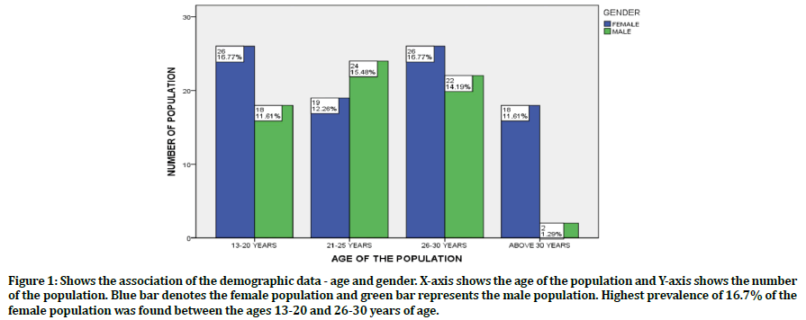
Of 155 participants, 89 of them were female and 66 were male. Between the age group 13- 20 years 44 patients, between 21-25 years 43 patients, between 26-30 years 48 patients and 20 patients above 30 years of age were present. Graph 1 denotes the frequency distribution of the demographic data of the population - age and gender.
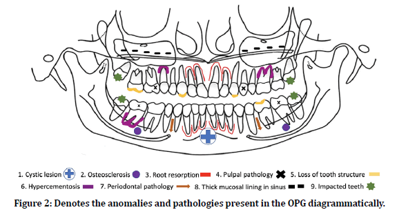
Criteria based on which the findings were calculated: 1. Periodontal pathology/anomaly comprises - bone loss, periodontal ligament space widening, loss of lamina dura. 2. Pulpal pathology/anomaly - dental caries with or without pulpal involvement, periapical lesion due to dental caries, chronic hyperplastic pulpitis. 3. Osteosclerosis - idiopathic and condensing osteosclerosis. 4. Hypercementosislocalised or generalized. 5. Root resorption - internal and external. 6. Loss of tooth structure - attrition, fracture. 7. Cystic lesions (∼10 to 25 mm diameter) - periodontal cyst, simple bone cyst, paradental cyst. 8. Thickening of the mucosal lining in the maxillary sinus. 9. Impacted teeth. (Till criteria 9 represented in figure 1) 10. Dilaceration. 11. Hypodontia.

Figure 1. Shows the association of the demographic data - age and gender. X-axis shows the age of the population and Y-axis shows the number of the population. Blue bar denotes the female population and green bar represents the male population. Highest prevalence of 16.7% of the female population was found between the ages 13-20 and 26-30 years of age.
Comparing Group A and Group B, it is observed that on an average female patient were largely affected. 43.2% of periodontal and 53.5% of pulpal anomalies, 18.06% of osteosclerosis, and 33.5% of hypercementosis conditions was seen. On further examination, 44.5% of root resorption, 25.1% of loss of tooth structures like attrition, 13.5% of mucosal thickening in maxillary sinus, 29.6% of impacted teeth, 18.06% of dilaceration and 27.09% of hypodontia were observed. Only 3.8% of cystic lesions were evident during the study period (Table 1).
| Findings | Group A (Female) | Group B (Male) | Total | Mean | SD | P (=0.05) |
|---|---|---|---|---|---|---|
| Periodontal pathology/anomaly | 52 (33.5%) | 15 (9.6%) | 67 (43.2%) | 1.42 | 0.422 | 0.198 |
| Pulpal pathology/anomaly | 69 (44.5%) | 14 (9.03%) | 83 (53.5%) | 1.37 | 0.394 | 0.076 |
| Osteosclerosis | 14 (9.03%) | 14 (9.03%) | 28 (18.06%) | 1.84 | 0.412 | 0.085 |
| Hypercementosis | 22 (14.1%) | 30 (19.3%) | 52 (33.5%) | 1.55 | 0.503 | 0.357 |
| Root resorption | 31 (20%) | 38 (24.5%) | 69 (44.5%) | 1.41 | 0.495 | 0.148 |
| Loss of tooth structure | 27 (17.4%) | 12 (7.7%) | 39 (25.1%) | 1.82 | 0.389 | 0.078 |
| Cystic lesion | 5 (3.2%) | 1 (0.64%) | 6 (3.8%) | 1.94 | 0.232 | 0.08 |
| Thickened mucosal lining in sinus | 18 (11.6%) | 3 (1.9%) | 21 (13.5%) | 1.8 | 0.404 | 0.025 |
| Impacted teeth | 30 (19.3%) | 16 (10.3%) | 46 (29.6%) | 1.76 | 0.432 | 0.205 |
| Dilaceration | 13 (8.3%) | 15 (9.6%) | 28 (18.06%) | 1.73 | 0.355 | 0.011 |
| Hypodontia | 24 (15.4%) | 18 (11.6%) | 42 (27.09%) | 1.77 | 0.446 | 0.933 |
Table 1: Reveals the association between Group A and Group B.
While examining the preoperative and postoperative radiographs of the orthodontic patients, it is observed that there was a decrease in the number of impacted teeth from 58 (11%) to 46 (29.6%). This can be attributed due to the impacted third molars in young patients. However, there was no evidence of any underlying cystic lesions or thickened mucosal lining in maxillary sinus was seen in the preoperative radiographs. There was a drastic increase in the pulpal anomalies 53.5% postoperatively was seen (Table 2 and Figure 2).
| Findings | Group C (PRE-OP) | Group D (POST-OP) |
|---|---|---|
| Periodontal pathology/anomaly | 28 (5.3%) | 67 (43.2%) |
| Pulpal pathology/anomaly | 25 (4.7%) | 83 (53.5%) |
| Osteosclerosis | - | 28 (18.06%) |
| Hypercementosis | 4 (0.8%) | 52 (33.5%) |
| Root resorption | 6 (1.1%) | 69 (44.5%) |
| Loss of tooth structure | - | 39 (25.1%) |
| Cystic lesion | - | 6 (3.8%) |
| Thickened mucosal lining in sinus | - | 21 (13.5%) |
| Impacted teeth | 58 (11%) | 46 (29.6%) |
| Dilaceration | 10 (1.9%) | 10 (6.4%) |
| Hypodontia | 24 (4.5%) | 24 (15.4%) |
| STATISTICAL ANALYSIS | PRE-OP | POST-OP |
| Mean ± SD | 2.99 ± 2.24 | 1.97 ± 0.751 |
Table 2: Reveals the association between Group C and Group D.

Figure 2. Denotes the anomalies and pathologies present in the OPG diagrammatically.
Discussion
In this study, the female participants were higher in number compared to their male counter population. Of the abnormalities present, the pulpal anomalies such as dental caries with irreversible pulpitis, chronic hyperplastic pulpitis or, dental caries with periapical lesion were highly evident (53.5%) which requires an immediate endodontists attention. Periodontal pathologies were found to be 43.2% whereas root resorption and loss of tooth structure was found to be about 44.5% and 25.1% respectively.
Cral et al. conducted a similar study in Brazilian population and found out the correlation between pre-treatment and post-treatment incidental findings in OPGs. He reported changes like supernumerary root, dilaceration, pulp stones, root resorption, apical endodontic lesions, osteosclerosis and many others in his study [6]. Findings from the present study are reported to be in unanimity with the previous study.
Levander et al. studied about the root resorption in their respective studies. Levander also stated that the root resorption rate is less in patients undergoing the treatment with pause thus paving way for the cementum to recover [12]. Apajalahti et al. did a study in orthodontic patients to check for apical root resorption using radiographs (OPG and IOPA). He concluded that the degree of root resorption increases with increase in the duration of the orthodontic treatment and suggested IOPA is the method used to determine them accurately [13].
Over a period of time, the dental pulp undergoes physiological changes due to various factors like aging. Deposition of the mineralized tissues in the form of nodules in the interior of the pulp cavity may occur with influence of factors like caries, periodontal diseases [14]. Our study reported higher pulpal changes in female patients (44.5%) than male patients.
The presence of hypercementosis may not impede much in orthodontic treatment however, the dentist is aware of the evolution of each specific case [15]. In our study 52 (33.5%) patients were present with hypercementosis which was evident in postoperative radiographs. However, the study did not report any changes pertaining to temporomandibular joint (TMJ) or area associated.
This article reports retrospective investigations done only with the information obtained in the radiographs. Clinical evaluation of the population was not done to evaluate the presence of any periodontal findings, or swelling for cystic lesions. Examination of swelling clinically can be helpful in diagnosing the cysts present which may require medical or surgical management. Examination of periodontal pocket or attachment loss clinically can help in ruling out the periodontal pathologies and can be directed towards necessary periodontal management.
Maintenance of good oral hygiene is advised to patients during and after the orthodontic treatment. Excellent oral care was emphasized as it consisted of brushing the teeth twice daily using a soft bristle brush, flossing on a daily basis, and advised scaling at accurate intervals in having satisfying oral hygiene [16].
As the ionizing radiation exposed is minimal and safe, the 2-dimensional radiographs are advised largely in orthodontic therapy [17]. It is reported that these panoramic radiographs are equivalent to four intraoral periapical radiographs [18]. Since the evaluation of the patients were based completely on the radiographs, clinical assessment of the patients post orthodontic therapy were uncertain. Radiographs also showed image distortion of 15-20%. Few radiographs even had artifacts and other errors like spine shadow or soft tissue shadow which made interpretation of the radiographs, challenging. Few authors consider OPG is not an accurate radiographic modality to identify the root morphology because of a resultant two-dimensional image and overlapping of the structures and recommend for 3-dimensional imaging modalities [19]. In case of analyzing root resorption, intraoral periapical radiographs are ideal for measuring the length of the root resorption as there are possibilities of positioning errors which can be of limitation.
Avoiding any error or false positive observation, the study was analyzed repeatedly for a few times and the reliability between the two examiners were in the range of acceptance given by the kappa indices. While the study was carried out in an orthodontic population, it is necessary for a dentist to picture the possible changes that can be expected post treatment and precautions to be carried out to protect from causing abnormalities which can help patients undergoing or willing to undergo orthodontic treatment. Watchful observations to be made not just by orthodontists but by all dentists, in orthodontic patients is recommended. Since the population of the study is small, further anomalies like odontome, enamel pearls etc. couldn’t be appreciated which is of limited information.
Conclusion
Clinicians should be knowledgeable to evaluate the pathologies and anomalies present in the radiographs in order to avoid any further radical changes in the oral cavity. This study showed higher incidence of pulpal abnormalities (53.5%) while the cystic lesions being the least (3.8%). The anomalies can be overcome with proper necessary treatments prior or during the orthodontic treatment and maintenance of oral hygiene. Comparison between the pre-treatment and post-treatment panoramic image should be analyzed for ruling out abnormalities and clinicians should pay attention to these in each follow up.
Clinical Significance
Changes experienced in the oral cavity after orthodontic treatment should be identified by the dentist and necessary treatment should be planned, as the findings might require special oral care. Such changes, when ignored, might aggravate to further pathologies.
Acknowledgement
We thank the Department of Oral and Maxillofacial Radiology and Department of Orthodontics at Saveetha Dental College and Hospital, Chennai for providing us with the necessary materials, time and opportunity to prepare this manuscript. There are no conflicts of interest between the examiners. No financial support obtained.
References
- Kurol J, Owman-Moll P. Hyalinization and root resorption during early orthodontic tooth movement in adolescents. Angle Orthod 1998; 68:161–165.
- Bondemark L, Jeppsson M, Lindh-Ingildsen L. et al. Incidental findings of pathology and abnormality in pretreatment orthodontic panoramic radiographs. Angle Orthod 2006; 76:98–102.
- Atchison KA, Luke LS, White SC. An algorithm for ordering pretreatment orthodontic radiographs. Am J Orthod Dentofacial Orthop 1992; 102:29–44.
- Atchison KA. Radiographic examinations of orthodontic educators and practitioners. J Dent Educ 1986; 50:651–655.
- Kuhlberg AJ, Norton LA. Pathologic findings in orthodontic radiographic images. Am J Orthod Dentofacial Orthop 2003; 123:182–184.
- Cral WG, Silveira MQ, Rubira-Bullen IRF, et al. Incidental findings in pretreatment and post-treatment orthodontic panoramic radiographs. Int J Radiol Radiat Ther 2018; 5:00132.
- Subramaniam N, Muthukrishnan A. Oral mucositis and microbial colonization in oral cancer patients undergoing radiotherapy and chemotherapy: A prospective analysis in a tertiary care dental hospital. J Investig Clin Dent 2019; 10:12454.
- Vadivel JK, Govindarajan M, Somasundaram E, et al. Mast cell expression in oral lichen planus: A systematic review. J Investig Clin Dent 2019; 10:12457.
- Patil SR, Maragathavalli G, Ramesh DNSV, et al. Assessment of maximum bite force in oral submucous fibrosis patients: A preliminary study. Pesqui Bras Odontopediatria Clin Integr 2020; 20:482.
- Patil SR, Maragathavalli G, Araki K, et al. Three rooted mandibular first molars in a Saudi Arabian population: A CBCT Study. Pesqui Bras Odontop Clin Integr 2018; 18:4133.
- Patil SR, Yadav N, Al-Zoubi IA, et al. Comparative study of the efficacy of newer antioxitands lycopene and oxitard in the treatment of oral submucous fibrosis. Pesqui Bras Odontopediatria Clin Integr 2018; 18:1–7.
- Levander E, Bajka R, Malmgren O. Early radiographic diagnosis of apical root resorption during orthodontic treatment: A study of maxillary incisors. Eur J Orthod 1998; 20:57–63.
- Apajalahti S, Peltola JS. Apical root resorption after orthodontic treatment a retrospective study. Eur J Orthod 2007; 29:408–412.
- Nixon CE, Saviano JA, King GJ, et al. Histomorphometric study of dental pulp during orthodontic tooth movement. J Endod 1993; 19:13–16.
- Humerfelt A, Reitan K. Effects of hypercementosis on the movability of teeth during orthodontic treatment. Angle Orthod 1966; 36:179–189.
- Subashri A, Uma Maheshwari TN. Knowledge and attitude of oral hygiene practice among dental students. Res J Pharm Technol 2016; 9:1840.
- Granlund CM, Lith A, Molander B, et al. Frequency of errors and pathology in panoramic images of young orthodontic patients. Eur J Orthod 2012; 34:452–457.
- Freeman JP, Brand JW. Radiation doses of commonly used dental radiographic surveys. Oral Surg Oral Med Oral Pathol 1994; 77:285–289.
- Patil SR, Maragathavalli G, Araki K, et al. Three rooted mandibular first molars in a Saudi Arabian population: A CBCT study. Pesquisa Br Odontop Clín Integrada 2018; 18:4133.
Author Info
Department of Oral Medicine and Radiology, Saveetha Dental College and Hospitals, Saveetha Institute of Medical and Technical Sciences (SIMATS), Saveetha University Tamilnadu, Chennai, IndiaCitation: Indra G, Maragathavalli G, Incidental Findings in Orthodontic Patients Studied Using 2 Dimensional Images, J Res Med Dent Sci, 2021, 9 (2): 330-335.
Received: 23-Sep-2020 Accepted: 15-Feb-2021
