Research - (2022) Volume 10, Issue 12
Jaleel hernia in nine month of male patients
Jaleel Hussien Hammoodi Al Obaidi*
*Correspondence: Jaleel Hussien Hammoodi Al Obaidi, Department of surgery, Alshafah private Hospital Diyala Govermente Medical Directer, Iraq, Email:
Abstract
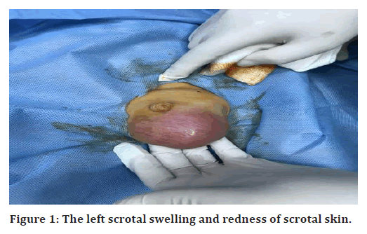
Congenital inguinal hernias occur when the abdominal cavity extends as a sac-like projection in the groin on one or both sides toward the labia or scrotum. The aim of the present study was to operate rare type of left inguinal hernia with the surgical procedure. A nine month old male patient was admitted at Alshafah private Hospital during Feb 2019 and diagnosed with left scrotal swelling. Congenital malrotation was diagnosed by various clinical tests such as Ultrasound (to determine the normal position of the abdomen), X-ray and CT scans. In the X-rays, abdominal X-ray (intestinal obstructions); Barium enema X-ray (barium is inserted into the intestine through the anus) and X-ray were performed for the prognosis. CT scans contrast was performed by the harmless dye. It was injected to see the sight of obstruction. The patient was kept for 48 hours under clinical examination. The left scrotal swelling was red and irreducible inguinal swelling was observed for two days (Figure 1). The patients also had tender fever. A professional diagnoses acute epididymo orchitis or torsion of left testis. The ultrasound revealed indirect inguinal hernia, defect measuring 27mm with irreducible herniation of bowel loops. It showed diminish vascularity at time of examination with normal left testis displaced inferiorly by hernia sac, smooth outline, and homogenous in texture. There was no focal lesion could be seen. It shows normal parenchymal in color Doppler flow and LT epididymis with a right mild hydrocele. Roentgen equivalent man (REM) for an ultrasound of testis revealed that the bowel loops on the side of inguinal hernia sac, showing diminished vascularity of bowel loops. The patient was discharged in a good general condition. The sutures were removed after 7 days. The postoperative chest X-ray showed the normal location of the heart. The ultrasound showed malrotation of the bowel only. The study can be concluded as the left side inguinal hernia can be treated successfully, through its prevalence is very rare. The family history should be taken into consideration while diagnosing the hernial condition.
Keywords
Appendex, Cecum, Jaleel hernia
Introduction
Amyands hernia, an intestine appendix in an inguinal hernia sac that may or may not be associated with appendicitis. A French surgeon at St Georges and Westminster Hospital (London, 1735) accomplished the first successful appendectomy on 11 year-old kid who arrived with an appendix in his inguinal hernia sac [1]. Hernia in children is about 3% of childhood more in the right side. The inter-abdominal structure may pass from a week internal ring in childhood. The incidence of an appendix in the left side inguinal hernia is extremely uncommon, occurring in only 0.08 percent of cases [2]. The primordial bowel protruded from the abdominal cavity during embryonic development. The large bowel generally rotates counterclockwise when it returns, with the cecum resting in the right lower quadrant [3-5]. Mallrotation occurs when the bowel does not return to its normal position in the right quadrant during intrauterine life [6]. With this background, the aim of the present study was to operate rare type of left inguinal hernia with the surgical procedure.
Case Report
A 9 months old male patient was admitted at Alshafah private Hospital during Feb 2019 and diagnosed with left scrotal swelling. The patient was kept for 48 hour under clinical examination. The left scrotal swelling was red and irreducible inguinal swelling was observed for two days (Figure 1). The patients also had tender fever.

Figure 1. The left scrotal swelling and redness of scrotal skin.
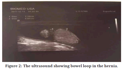
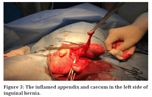
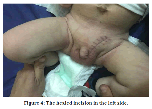
A professional diagnoses acute epididymo orchitis or torsion of left testis. The ultrasound reveals the indirect inguinal hernia, defect measuring 27mm irreducible herniation of bowel loops. It showed diminished vascularity at time of examination with normal left testis displaced inferiorly by hernia sac, smooth outline, and homogenous in texture. There was no focal lesion could be seen. It shows normal parenchymal in color Doppler flow and LT epididymis with a right mild hydrocele. Roentgen equivalent man (REM) for ultrasound of testis revealed that the bowel loops in the side of inguinal hernia sac, showing diminished vascularity of bowel loop. Figure 2 showed the ultrasound image showing bowel loop in the hernia. Figure 3 represents the inflamed appendix and caecum in the left side of inguinal hernia. Similarly, Figure 4 showed healed incision in the left side of the patient. The operation was done through the left iniguanal incision, there were caecum and inflamed appendix in the left inguinal sac. Appendectomy for the inflamed appendix and reduction of the caecum, along with herniotomy were done. The wound is closed in layers. The patient was discharged in a good general condition. The sutures were removed after 7 days. The postoperative chest X-ray showed the normal location of the heart. The ultrasound showed malrotation of the bowel only.

Figure 2. The ultrasound showing bowel loop in the hernia.

Figure 3. The inflamed appendix and caecum in the left side of inguinal hernia.

Figure 4. The healed incision in the left side.
Discussion
The appendix occurrence within the hernia sac is extremely uncommon, especially on the left side [7]. In the present study, such type of rare case (left inguinal hernia) is successfully operated. The majority of hernias occur on the right side, most likely due to the appendix's usual anatomical position as well as the point that the right-sided hernias seem to be more prevalent than leftsided hernias. Although a left-sided Amyand hernia has been documented, it is rare and may be caused by situs inversus, intestinal malrotation, or a movable cecum [8].
The appendix existence in the left side inguinal hernia this occur only in malrotation of bowel. It occurs during the first trimester [9]. The presence of an appendix and caecum on the left side is extremely uncommon. Hence, it is extremely necessary to investigate the child postoperatively to govern whether there are congenital anomalies or molestation [10]. Balamaddaiah, et al. [11] reported that the right side hernia is more common than the left side hernia. Due to testis late fall down and more recurrent closure of right processus vaginalis failure, right side hernia predominance observed [12-14].
Abnormal intestinal positioning, often including
On the abdomen’s right side, small intestine is found.
The cecum emigrant from its normal position in the right hypochondrium or right lower quadrant into the epigastrium.
Displaced or absent or ligament treitz.
Fibrous peritoneal bands known as Ladd bands that runs vertically across the duodenum.
Congenital mallrotation was diagnosed by various clinical tests such as Ultrasound (to determine the normal position of the abdomen), X-ray and CT scan. In the X-rays, abdominal X-ray (intestinal obstructions); Barium enema X-ray (barium is inserted into the intestine through the anus) and X-ray were performed for the prognosis. CT scan +contrast were performed by the harmless dye. It was injected to see the site of obstruction.
Conclusion
A hernia surgeon may encounter unexpected intraoperative finding such as an Amyands hernia, appendectomy must be done at the same time to prevent inflammation of appendix occurring in the future, and investigates the patients for other congenital abnormalities and follow up of the patient.
References
- Sharma H, Gupta A, Shekhawat NS, et al. Amyand’s hernia: A report of 18 consecutive patients over a 15-year period. Hernia 2007; 11:31-35.
- Psarras K, Lalountas M, Baltatzis M, et al. Amyand's hernia-a vermiform appendix presenting in an inguinal hernia: A case series. J Med Case Rep 2011; 5:1-3.
- Berrocal T, Lamas M, Gutiérrez J, et al. Congenital anomalies of the small intestine, colon, and rectum. Radiographics 1999; 19:1219-1236.
- Kadhim M. Total oxidants, lipid peroxidation and antioxidant capacity in the serum of rheumatoid arthritis patients. J Pharma Negative Results 2022; 13.
- Hameed RY, Nathir I, Abdulsahib WK, et al. Study the effect of biosynthesized gold nanoparticles on the enzymatic activity of alpha-Amylase. Res J Pharm Technol 2022; 15:3459-3465.
- Gamblin TC, Stephens RE, Johnson RK, et al. Adult malrotation: A case report and review of the literature. Curr Surg 2003; 60:517-520.
- Ghafouri A, Anbara T, Foroutankia R. A rare case report of appendix and cecum in the sac of left inguinal hernia (left Amyand's hernia). Med J Islamic Republic of Iran 2012; 26:94.
- Johari HG, Paydar S, Davani SZ, et al. Left-sided Amyand hernia. Ann Saudi Med 2009; 29:321.
- Morales-Cárdenas A, Ploneda-Valencia CF, Sainz-Escárrega VH, et al. Amyand hernia: Case report and review of the literature. Ann Med Surg 2015; 4:113-115.
- Dietz DW, Walsh RM, Grundfest-Broniatowski S, et al. Intestinal malrotation. Dis Colon Rectum 2002; 45:1381-1386.
- Balamaddaiah G, Reddy SR. Prevalence and risk factors of inguinal hernia: a study in a semi-urban area in Rayalaseema, Andhra Pradesh, India. Int Surg J 2016; 3:1310-1313.
- Mohammed AK, Al-Shaheeb S, Fawzi OF, et al. Evaluation of Interlukein-6 and Vitamin D in Patients with COVID-19. Res J Biotechnol 2022; 17.
- Garba ES. The pattern of adult external abdominal hernias in Zaria. Nigerian J Sur Res 2000; 2:12-15.
- Mbah N. Morbidity and mortality associated with inguinal hernia in northwestern Nigeria. West African J Med 2007; 26:288-292.
Indexed at, Google scholar, Cross Ref
Indexed at , Google scholar, Cross Ref
Indexed at, Google scholar, Cross Ref
Indexed at, Google scholar, Cross Ref
Indexed at, Google scholar, Cross Ref
Indexed at, Google scholar, Cross Ref
Indexed at, Google scholar, Cross Ref
Author Info
Jaleel Hussien Hammoodi Al Obaidi*
Department of surgery, Alshafah private Hospital Diyala Govermente Medical Directer, IraqReceived: 25-Nov-2022, Manuscript No. jrmds-22-78196; , Pre QC No. jrmds-22-78196(PQ); Editor assigned: 28-Nov-2022, Pre QC No. jrmds-22-78196(PQ); Reviewed: 13-Dec-2022, QC No. jrmds-22-78196(Q); Revised: 19-Dec-2022, Manuscript No. jrmds-22-78196(R); Published: 26-Dec-2022
