Research - (2021) Volume 9, Issue 1
Knowledge and Awareness About Periapical Lesions Among Clinical Students and General Practitioners: A Survey
Dhinesh Kumar S and S Delphine Priscilla Anthony*
*Correspondence: S Delphine Priscilla Anthony, Department of Conservative Dentistry and Endodontics, Saveetha Dental College and Hospital, Saveetha Institute of Medical and Technical Sciences, Saveetha University Tamilnadu, India, Email:
Abstract
Periapical lesions of endodontic origin are common pathological conditions affecting periradicular tissues. Microbial infection of pulpal tissues is primarily responsible for initiation and progression of periapical lesions. The primary objective of endodontic therapy should be to restore involved teeth to a state of normalcy nonsurgically. The aim of this survey was to assess knowledge and awareness about periapical lesion among clinical students including third year, final year, interns, postgraduate students and general practitioners. This cross-sectional study was done among clinical students and general practitioners from various institutions in Chennai. A total of 120 participants took part in this survey study. Results were analysed and compared using the SPSS Statistical Software by doing both the frequency tests and correlation tests. Chi-square test shows P=0, P<0.05, a significant value, proving that there is significant association between having skills and knowledge to handle a patient with periapical lesions and occupation and between gender and awareness about symptoms of periapical lesions. Regarding their knowledge, respondents of the survey showed an acceptable level of knowledge and awareness about periapical lesions.
Keywords
Endodontic treatment, Periapical lesion, Root canal treatment, Survey
Introduction
The infection caused by bacteria to the dental pulp can cause periapical lesions [1]. Periapical lesions are usually diagnosed during radiographic examination or when there is an acute pain the patient’s tooth [2]. One of the most common pathological conditions affecting periradicular tissues are periapical lesions. It is usually involved with one or more non-vital teeth with size greater than 200 mm2 and it is seen radiographically as a well circumscribed, well-defined radiolucent area bounded by a thin radiopaque line. Upon aspiration or during drainage, periapical lesion produces a strawcolored fluid. Periapical lesions are usually the sequelae of inflammation of the pulp or necrosis with inflammatory mediators spreading through the apical foramen to start a periapical lesion. Periapical lesions may also be seen in rare instances not related to pulpal inflammation and present as a neoplasm [3]. Histologically, periapical lesions of endodontic origin consist mainly of inflammatory cysts, granulomas, abscesses or apical scar tissue.
Imaging plays an especially important role in diagnosis of various lesions. Periapical lesions secondary to endodontic infection are usually diagnosed and treated based on the initial radiological findings. The final confirmatory diagnosis is performed by histopathological examination of the tissues. Several radiographic features, such as the size and shape of the lesion and the presence of sclerotic border demarcating the lesion, support the diagnosis of periapical lesions. Traditionally, the size of a periapical radiolucent lesion has been thought to give an indication as to the nature of the underlying disease process.
The goal of endodontic therapy should be to return the affected teeth to a state of health and function without surgical intervention due to the drawbacks, which limit its use in the management of periapical lesions. Initially, all inflammatory periapical lesions should be treated with conservative nonsurgical procedures. When non-surgical techniques have failed, then surgical intervention is preferred [4].
For successful outcomes of root canal therapy, microorganisms within the canal should be eradicated. Different techniques can be used in the nonsurgical management of periapical pathologies, for example method using calcium hydroxide, aspiration-irrigation technique, orthograde root canal therapy decompression technique, lesion sterilization and repair therapy, active nonsurgical decompression technique and the apexum procedure. Surgical intervention has many disadvantages, and hence should be considered as an option only in the case of failure of nonsurgical techniques [5].
Bacteria present in the periradicular area will not be accessible to disinfection procedures. Canals with negative cultures for bacteria are said to have higher success rates as opposed to those canals which test positive [6]. There are higher chances of treatment failure in these teeth with pretreatment periradicular rarefactions. Other than improper cleaning and shaping of the canal, a leaky apical seal is also a contributory factor in endodontic failure due to microbiological activity which can lead to periapical lesions. Seepage of fluids is likely to occur if there is no proper apical seal establishment. This can also perpetuate periradicular inflammation anytime. The chances of a favorable outcome are invariably higher when a proper cleaning of the canal has been undertaken. Thus, the importance of thorough disinfection procedure cannot be over emphasized.
We have numerous highly cited publications on well-designed clinical trials and lab studies [7- 24]. Now we are focusing on epidemiological surveys. The idea for this survey stemmed from the current interest in our community.
Materials and Methods
Study setting
This survey was done among clinical students and practitioners from various institutions in Chennai. A total of 120 participants took part in this survey study.
Study subjects
A total of 120 dental interns and practitioners took part in this survey
Methodology
A survey was conducted through an online standard questionnaire with 16 yes or no questions sent via a Google Form application. The questionnaire consisted of questions about the knowledge, awareness, and practice of periapical lesions among Saveetha Dental College Clinical students and general practitioners. Adequate time was provided to fill the questionnaire. The responses of the students were recorded, analyzed for flaws, checked for completeness and were taken up for assessment.
Statistical analysis
After data was collected and coded, the statistical analysis was done using the IBM SPSS Statistical Software package (Version 20.0). All the frequency tests were carried out and the Chisquare test was done at a significance level.
Results and Discussion
A total of 120 dental practitioners participated in the study, where all these dental practitioners which includes 58% males and 43% females (Figure 1) with third year and final year students, dental Intern, postgraduate students, and general practitioners. Most of the respondents are from the third year and final year student category (Figure 2).
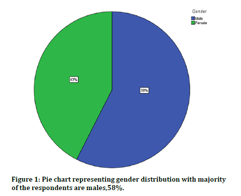
Figure 1: Pie chart representing gender distribution with majority of the respondents are males,58%.
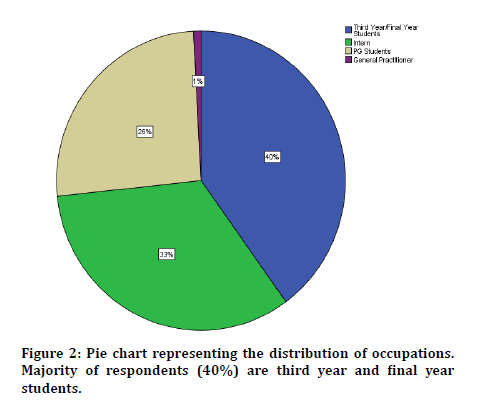
Figure 2: Pie chart representing the distribution of occupations. Majority of respondents (40%) are third year and final year students.
When questioned on their awareness of symptoms of periapical lesions, majority of respondents (84%) reported that they are aware about the symptoms of periapical lesions (Figure 3).
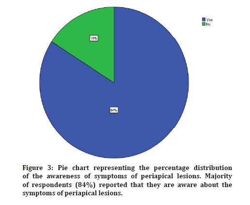
Figure 3: Pie chart representing the percentage distribution of the awareness of symptoms of periapical lesions. Majority of respondents (84%) reported that they are aware about the symptoms of periapical lesions.
When inquired about awareness about etiological factors of periapical lesions. Majority of respondents (80%) reported that they are aware about the etiological factors of periapical lesions by saying ‘Yes’ (Figure 4).
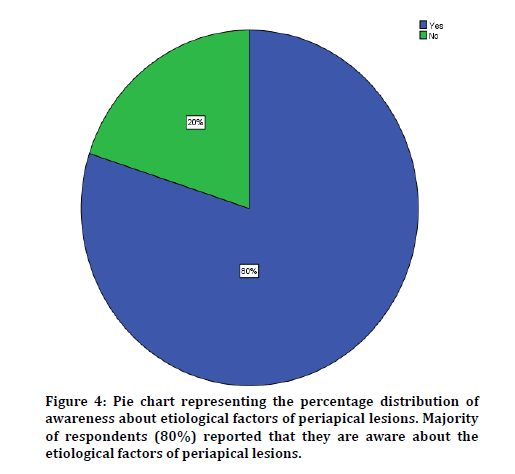
Figure 4: Pie chart representing the percentage distribution of awareness about etiological factors of periapical lesions. Majority of respondents (80%) reported that they are aware about the etiological factors of periapical lesions.
When the respondents were questioned if the respondents ever encountered patients with periapical lesions in their dental practice, most respondents (84%) reported that they have encountered patients with periapical lesions before in their dental practice (Figure 5).
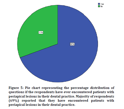
Figure 5: Pie chart representing the percentage distribution of questions if the respondents have ever encountered patients with periapical lesions in their dental practice. Majority of respondents (69%) reported that they have encountered patients with periapical lesions in their dental practice.
On a question about the knowledge about periapical lesion being asymptomatic sometimes among the participants of the survey, most respondents (71%) reported that they are aware about periapical lesion being asymptomatic sometimes by saying ‘Yes’ (Figure 6).
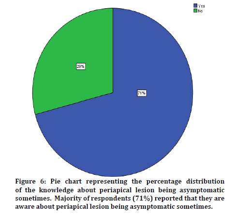
Figure 6: Pie chart representing the percentage distribution of the knowledge about periapical lesion being asymptomatic sometimes. Majority of respondents (71%) reported that they are aware about periapical lesion being asymptomatic sometimes.
On assessment on awareness about the methods of diagnosing periapical lesions among the participants of most respondents (79%) reported that they are aware about the methods of diagnosing periapical lesions (Figure 7).
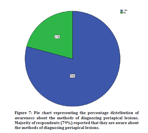
Figure 7: Pie chart representing the percentage distribution of awareness about the methods of diagnosing periapical lesions. Majority of respondents (79%) reported that they are aware about the methods of diagnosing periapical lesions.
When inquired about the ability to manage if they ever encounter a patient with periapical lesion, majority of respondents ,75% reported that they are aware about the ability to manage if they ever encounter a patient with periapical lesion by saying ‘Yes’ and 25% of the respondents said ‘No’ (Figure 8).
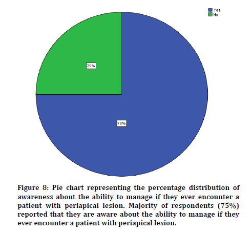
Figure 8: Pie chart representing the percentage distribution of awareness about the ability to manage if they ever encounter a patient with periapical lesion. Majority of respondents (75%) reported that they are aware about the ability to manage if they ever encounter a patient with periapical lesion.
When questioned about the antibiotics prescribed for periapical lesions majority of respondents ,79% reported that they are aware about the antibiotics prescribed for periapical lesions by saying ‘Yes’ and the remaining 21% responded by saying ‘No’ (Figure 9).
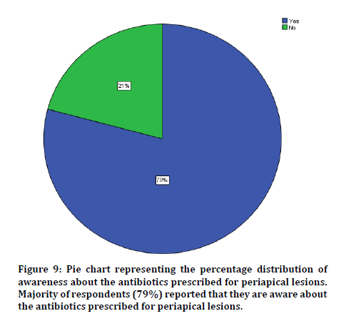
Figure 9: Pie chart representing the percentage distribution of awareness about the antibiotics prescribed for periapical lesions. Majority of respondents (79%) reported that they are aware about the antibiotics prescribed for periapical lesions.
When inquired about about intracanal medicament in management of periapical lesions majority of respondents answering ‘Yes’, 70% which reports that they are aware about intracanal medicament in management of periapical lesions and the remaining of the respondents, 30% answering ‘No’ (Figure 10).
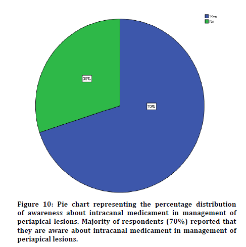
Figure 10: Pie chart representing the percentage distribution of awareness about intracanal medicament in management of periapical lesions. Majority of respondents (70%) reported that they are aware about intracanal medicament in management of periapical lesions.
When questioned on awareness about the consequences if periapical lesions are left untreated majority of respondents, 71% reported that they are aware about the consequences if periapical lesions are left untreated and 29% answer ‘No’ (Figure 11).
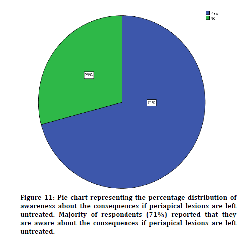
Figure 11: Pie chart representing the percentage distribution of awareness about the consequences if periapical lesions are left untreated. Majority of respondents (71%) reported that they are aware about the consequences if periapical lesions are left untreated.
When inquired about awareness about the sequelae of periapical lesion majority of respondents which is 75% answered ‘Yes’ which implies that they are aware about the sequelae of periapical lesion (Figure 12).
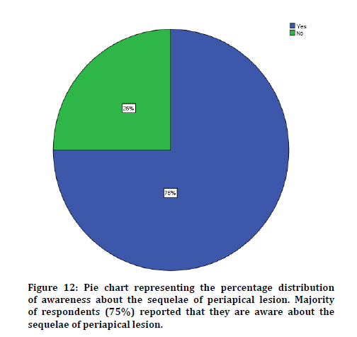
Figure 12: Pie chart representing the percentage distribution of awareness about the sequelae of periapical lesion. Majority of respondents (75%) reported that they are aware about the sequelae of periapical lesion.
When assessed about the clinical signs of periapical lesions, majority of respondents, 84% reported that they are aware about the symptoms of periapical lesions and the remaining 16% are unaware (Figure 13).
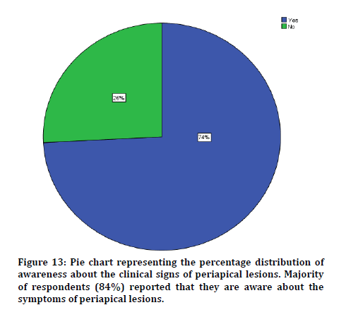
Figure 13: Pie chart representing the percentage distribution of awareness about the clinical signs of periapical lesions. Majority of respondents (84%) reported that they are aware about the symptoms of periapical lesions.
When questioned about having enough skills and knowledge to handle a patient periapical lesion majority of respondents which is 63% reported that they are having enough skills and knowledge to handle a patient with periapical lesion (Figure 14).
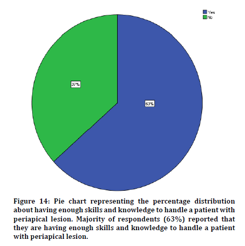
Figure 14: Pie chart representing the percentage distribution about having enough skills and knowledge to handle a patient with periapical lesion. Majority of respondents (63%) reported that they are having enough skills and knowledge to handle a patient with periapical lesion.
When assessed about the knowledge that the Weine’s classification is used in obturation of periapical lesions majority of respondents, 68% reported having knowledge that Weine’s classification is used in obturation of periapical lesions (Figure 15).
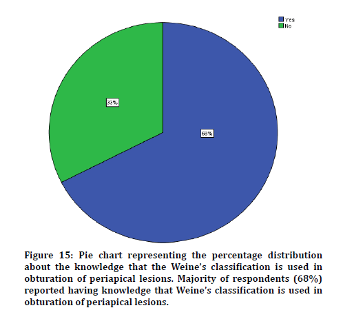
Figure 15: Pie chart representing the percentage distribution about the knowledge that the Weine’s classification is used in obturation of periapical lesions. Majority of respondents (68%) reported having knowledge that Weine’s classification is used in obturation of periapical lesions.
When inquired about having awareness about the term lesion sterilization and tissue repair, majority of respondents, 84% reported that they are aware about the term lesion sterilisation and tissue repair (Figure 16).
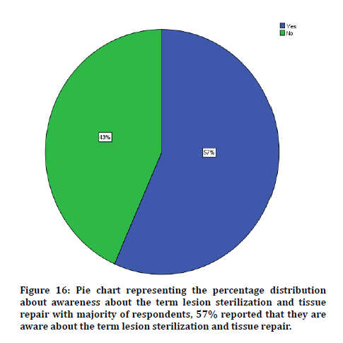
Figure 16: Pie chart representing the percentage distribution about awareness about the term lesion sterilization and tissue repair with majority of respondents, 57% reported that they are aware about the term lesion sterilization and tissue repair.
When questioned about having awareness about the awareness if a non-surgical treatment would workout in treating periapical lesion cases, majority of respondents, 56% reported that they are aware that non-surgical treatment would workout in treating periapical lesion cases (Figure 17).
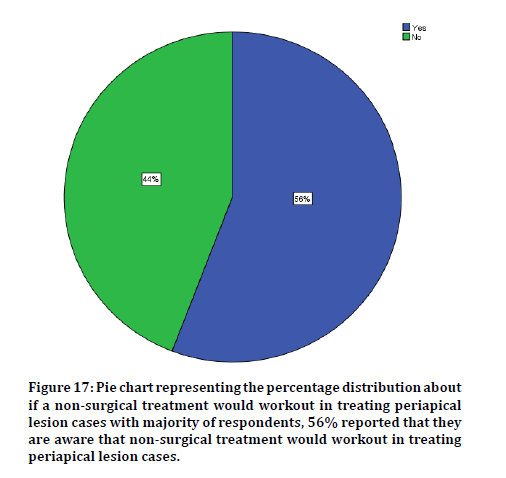
Figure 17: Pie chart representing the percentage distribution about if a non-surgical treatment would workout in treating periapical lesion cases with majority of respondents, 56% reported that they are aware that non-surgical treatment would workout in treating periapical lesion cases.
When inquired about awareness on how to treat periapical lesions in endodontically treated tooth and majority of the respondents, 53% reported that they know how to treat periapical lesions in endodontically treated tooth (Figure 18).
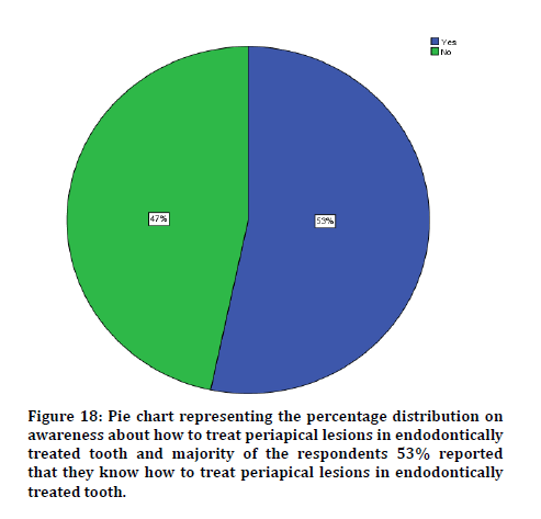
Figure 18: Pie chart representing the percentage distribution on awareness about how to treat periapical lesions in endodontically treated tooth and majority of the respondents 53% reported that they know how to treat periapical lesions in endodontically treated tooth.
Figure 19 shows association between having skills and knowledge to handle a patient with periapical lesion and occupation with majority of the respondents in each category answered ‘Yes’ with postgraduate students being the highest with 28%.
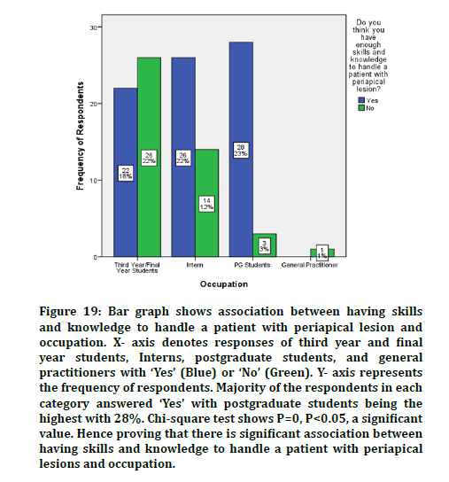
Figure 19: Bar graph shows association between having skills and knowledge to handle a patient with periapical lesion and occupation. X- axis denotes responses of third year and final year students, Interns, postgraduate students, and general practitioners with ‘Yes’ (Blue) or ‘No’ (Green). Y- axis represents the frequency of respondents. Majority of the respondents in each category answered ‘Yes’ with postgraduate students being the highest with 28%. Chi-square test shows P=0, P<0.05, a significant value. Hence proving that there is significant association between having skills and knowledge to handle a patient with periapical lesions and occupation.
Figure 20 shows association between gender and awareness about symptoms of periapical lesions. Majority of the respondents in each category answered ‘Yes’ with male respondents being the highest, 66%.
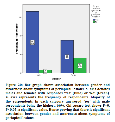
Figure 20: Bar graph shows association between gender and awareness about symptoms of periapical lesions. X- axis denotes males and females with responses ‘Yes’ (Blue) or ‘No’ (Green). Y- axis represents the frequency of respondents. Majority of the respondents in each category answered ‘Yes’ with male respondents being the highest, 66%. Chi-square test shows P=0, P<0.05, a significant value. Hence proving that there is significant association between gender and awareness about symptoms of periapical lesions.
Conclusion
Knowledge and awareness about periapical lesion are important in diagnosis and treatment of the disease. Dental practitioners including third year, final year, interns, postgraduate students, and also general practitioners should always be updated with all the information about periapical lesions. Within our study limits, regarding their knowledge, they showed an acceptable level of knowledge and awareness about periapical lesions.
References
- Möller ÅJ, Fabricius L, Dahlen G, et al. Influence on periapical tissues of indigenous oral bacteria and necrotic pulp tissue in monkeys. Eur J Oral Sci 1981; 89:475–484.
- Fernandes M, de Ataide I. Nonsurgical management of periapical lesions. J Conservative Dent 2010; 13:240–245.
- Abbott PV. Unusual periapical pathosis-Adenoid cystic carcinoma. Australian Endodont J2001; 27:73–75.
- Chan V. When should infective endocarditis be treated with early surgical intervention? Canadian J Cardiol 2018; 34:1110–1111.
- Sood N, Maheshwari N, Gothi R, et al. Treatment of large periapical cyst like lesion: A noninvasive approach: A report of two cases. Int J Clin Pediatr Dent 2015; 8:133–137.
- Sritharan A. Discuss that the coronal seal is more important than the apical seal for endodontic success. Australian Endodont J 2002; 28:112–115.
- Govindaraju L, Neelakantan P, Gutmann JL. Effect of root canal irrigating solutions on the compressive strength of tricalcium silicate cements. Clin Oral Investigations 2017; 21:567–571.
- Azeem RA, Sureshbabu NM. Clinical performance of direct versus indirect composite restorations in posterior teeth: A systematic review. J Conservative Dent 2018; 21:2–9.
- Jenarthanan S, Subbarao C. Comparative evaluation of the efficacy of diclofenac sodium administered using different delivery routes in the management of endodontic pain: A randomized controlled clinical trial. J Conservative Dent 2018; 21:297–301.
- Manohar MP, Sharma S. A survey of the knowledge, attitude, and awareness about the principal choice of intracanal medicaments among the general dental practitioners and nonendodontic specialists. Indian J Dent Res 2018; 29:716–720.
- Nandakumar M, Nasim I. Comparative evaluation of grape seed and cranberry extracts in preventing enamel erosion: An optical emission spectrometric analysis. J Conservative Dent 2018; 21:516–520.
- Teja KV, Ramesh S, Priya V. Regulation of matrix metalloproteinase-3 gene expression in inflammation: A molecular study. J Conservative Dent 2018; 21:592–596.
- Janani K, Sandhya R. A survey on skills for cone beam computed tomography interpretation among endodontists for endodontic treatment procedure. Indian J Dent Res 2019; 30:834–838.
- Khandelwal A, Palanivelu A. Correlation between dental caries and salivary albumin in adult population in Chennai: An In Vivo Study. Br Dent Sci 2019; 22:228–233.
- Malli Sureshbabu N, Selvarasu K, Nandakumar M, et al. Concentrated growth factors as an ingenious biomaterial in regeneration of bony defects after periapical surgery: A report of two cases. Case Reports Dent 2019; 2019:7046203.
- Poorni S, Srinivasan MR, Nivedhitha MS. Probiotic strains in caries prevention: A systematic review. J Conservative Dent 2019; 22:123–128.
- Rajakeerthi R, Ms N. Natural product as the storage medium for an avulsed tooth–A systematic review. Cumhuriyet Dent J 2019; 22:249–256.
- Rajendran R, Kunjusankaran RN, Sandhya R, et al. Comparative evaluation of remineralizing potential of a paste containing bioactive glass and a topical cream containing casein phosphopeptide-Amorphous calcium phosphate: An in Vitro study. Pesquisa Br Odontopediatr Clin Integrada 2019; 19:1–10.
- Ramarao S, Sathyanarayanan U. CRA grid-A preliminary development and calibration of a paper-based objectivization of caries risk assessment in undergraduate dental education. J Conservative Dent 2019; 22:185–190.
- Siddique R, Nivedhitha MS. Effectiveness of rotary and reciprocating systems on microbial reduction: A systematic review. J Conservative Dent 2019a; 22:114–122.
- Siddique R, Sureshbabu NM, Somasundaram J, et al. Qualitative and quantitative analysis of precipitate formation following interaction of chlorhexidine with sodium hypochlorite, neem, and tulsi. J Conservative Dent 2019; 22:40–47.
- Siddique R, Nivedhitha MS, Jacob B. Quantitative analysis for detection of toxic elements in various irrigants, their combination (precipitate), and para-chloroaniline: An inductively coupled plasma mass spectrometry study. J Conservative Dent 2019; 22:344–350.
- Antony DP, Thomas T, Nivedhitha MS. Two-dimensional periapical, panoramic radiography versus three-dimensional cone-beam computed tomography in the detection of periapical lesion after endodontic treatment: A systematic review. Cureus 2020; 12:e7736.
- Kumar D, Delphine Priscilla Antony S. Calcified canal and negotiation-A review. Res J Pharm Technol 2018; 11:3727-3730.
Author Info
Dhinesh Kumar S and S Delphine Priscilla Anthony*
Department of Conservative Dentistry and Endodontics, Saveetha Dental College and Hospital, Saveetha Institute of Medical and Technical Sciences, Saveetha University Tamilnadu, Chennai, Tamil Nadu, IndiaCitation: Dhinesh Kumar S, S Delphine Priscilla Anthony, Knowledge and Awareness About Periapical Lesions Among Clinical Students and General Practitioners: A Survey, J Res Med Dent Sci, 2021, 9 (1): 97-104.
Received: 29-Sep-2020 Accepted: 14-Dec-2020
