Research - (2023) Volume 11, Issue 5
Laser: A Ray of Light in the Field of Dentistry
Farzeen Tanwir1, Bushra Ijaz1, Nabeel Hafeez3, Tauqeer Bibi1, Saima Mazhar1*, Ahmed Bin Khalid Khan1 and Anum Baqar2
*Correspondence: Saima Mazhar, Pakistan, Email:
Abstract
The term Laser stands for “Light Amplification by the Stimulated Emission of Radiation”. It was introduced in dentistry in 1960s. Laser dentistry offers an effective method for treating a variety of dental condition; it can be used on dental hard and soft tissues. Lasers of different wavelength are recommended for various clinical procedures. Laser technology has become a valuable tool in many procedures, including caries removal, cavity preparation, soft tissue surgeries, caries detection, peri implantitis treatment, maxillofacial surgery, bonded restoration, and endodontic procedure. Laser treatment in dentistry can be quicker and more efficient with the benefits that include less pain, lack of bleeding, and less postoperative discomfort. It is making great inroads into lot of areas of dentistry, but possible hazard allied with lasers should also be considered. This paper gives an insight on laser in dentistry.
Keywords
Clinical application, Dentistry, Laser, Laser technology, Photo-bio modulation, Wavelength
Introduction
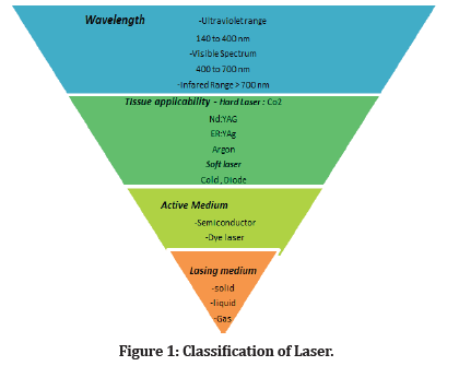
Science has revolutionized the modern era of technology, one of its innovations was laser that has brought much advancement in General Dentistry and other fields of medicine. LASER stands for ‘Light Amplification by the Stimulated Emission of Radiation’. In the 1960s, it was introduced by Theodore Maiman. She has led continuous research in the various applications of lasers in dental practice [1]. Laser is a unique instrument that disperses the light of several frequencies into chromatic emissions in the ultraviolet, visible spectrum and infrared areas. Glare formed by the laser are precise form of electromagnetic radiation. Energy wave emitted by electromagnetic spectrum pass over gamma rays with 10 to 12 m to radio waves having wavelengths of thousands of meters [2]. In addition, Lasers can be classified in different ways: Based on lasing medium, wavelengths, tissue applicability and type of active medium is used accordingly [3, 4]. As shown in Figure 1. Laser dentistry offers an effective method for treating a variety of dental condition; it can be used on dental hard and soft tissues. Lasers that are used on hard tissue are considered as a better alternate as compared to conventional lasers, it renders clinicians a better field of work that aid in better treatment outcomes and affirmative results. This paper aims at over viewing the application/trends of laser technology in the field of dentistry.

Figure 1: Classification of Laser.
Different Laser Type
Erbium: yttrium aluminum garnet (Er: YAG), Diode laser, Erbium chromium: yttrium scandium gallium garnet, Argon, Carbon dioxide (Co2) and Neodymium Yttrium Aluminum Garnet (Nd: YAG) laser are commonly used in the field of dentistry as summarized in (Table 1). These lasers can be resourceful for the following procedure like periodontal surgery, cavity preparation, detection of caries, peri implantitis treatment, maxillofacial surgery, bonded restoration, and endodontic procedure [5, 6].
| Laser | Type | Wavelength | Construction | Use |
|---|---|---|---|---|
| Argon | 488, 515 | Gas state | Ablation and soft-tissue incision, curing resins, bleaching | |
| Carbon dioxide CO | 966, 10600 | De epithelialization of gingival during periodontal regenerative procedures | ||
| Helium-Neon | 633 | caries detection, diagnosis, wound healing, pain reduction | ||
| Diode | 635, 670, 810, 830, 980 | Semiconductor | Soft-tissue incision and ablation | |
| Nd:YAG | 1064 | Solid state | soft-tissue incision and ablation; incipient caries removal | |
| Er:YAG | 2940 | Restorative procedure and root canal preparation | ||
| Er,Cr:YSGG | 2780 | root canal preparation caries removal, cavity preparation |
Table 1: Common Type of Dental Laser Along With Wavelength and Their Application.
Clinical Application of Laser
Oral Medicine
Premalignant lesion like oral lichen planus can be treated by different low-level laser therapy that includes ultraviolet, diode and Helium neon. Low- Level Laser Therapy (LLLT) has especially designed to treat erosive lichen planus and has minimal side effect [7, 8]. Oral leukoplakia has shown lapse with the laser, for its management CO2 lasers is quite effective but it has minimal side effect that is pain and swelling. Photodynamic therapy with 5-aminolevulinic acid and a pulse dye laser is used as an alternate for regression of leukoplakia [9]. Combination of Low intensity and high intensity lasers are beneficial for the excision of Fordyce granule and have swift healing and minimal post-operative pain [10].
Restorative Dentistry
In restorative procedure, High intensity lasers such as Er: YAG and Er, Cr, YSGG have been effectively used for caries removal, and cavity preparation, their use is based on ablation mechanism in which hard dental tissue are excised via mechanical and thermal effect [11-13].
Moreover, laser device works based on fluorescence principal. It can remove faulty restorations and the pulsed laser is also helpful to achieve automated removal of caries [14, 15]. Furthermore, in direct pulp capping CO2 laser can be used as it restraint the hemorrhage and eases the placement of calcium hydroxide paste at exposure site and provide better clinical results [16].
Endodontic Procedure
Laser beam has capability to destroy microbes and eliminate mullock and smear layer from root canal. For the canal debridement and cleaning Erbium and Nd: YAG wavelength can be utilized [17]. Nd: YAG laser has been used for the evaporation and contraction of the smear layer whereas Erbium lasers used for the laser activated irrigation and photon-initiated photo acoustic streaming. Additionally, the use of laser is recommended as an adjunct to debridement protocol and traditional disinfection rather than as an alternative to NaOCl. Further clinical studies should be done to assess endodontic treatment outcome following the use of laser [18, 19].
Periodontology
Laser technology has impacted the field of Periodontology in many ways they help to manage periodontal disease. In Periodontics treatment Er: YAG laser is beneficial for the removal of calculus, destroying tooth substances and calculus of sub gingival root surface [20]. Erbium group of lasers have been found to be effective in removing lipopolysaccharides and root surface endotoxins has shown notable bactericidal effect against Actinobacillus actinomycetemcomitans and Porphyromonas gingivalis [21]. In addition, diode, C02 and Nd: YAG is also useful in periodontal management as they exhibit good soft tissue ablation and homeostatic quality. However, according to the research when this laser technology was used on bone or root surface thermal damage and carbonization was observed and reported. Hence, it is limited to soft tissue procedures like removal of melanin pigmentation of gingiva, frenectomy and gingivectomy [22]. Laser devise is used as an adjunct to treatment procedure like scaling and root planning.
LANAP
In LANAP (Laser Assisted New Attachment Procedure) was introduced by Gregg and McCarthy in 1990 [23]. According to the research it’s an invasive procedure which uses special laser that is Periolase MVP-7 and Nd: YAG 1064nm (neodymium yttrium aluminum garnet) [24, 25]. This device works on the principal of regeneration. It is used to treat periodontal pocket by removing diseased inflamed epithelium lining from the connective tissue. Nd: YAG has been shown to decrease microbial loading from the pocket [26]. Furthermore Periolase MVP-7 has FDA clearance for regeneration and recreation of bone around the teeth. It can be used in biopsies, frenectomy, and removing leukoplakia and excellent for hemostasis.
Oral Surgery
In Oral and maxillofacial surgery different laser wavelengths have been used. It has changed the practice to such an extent that now a days it has become a valuable tool in dentistry and cosmetic surgeries. Common lasers that are used in oral surgeries are Diode, C02 and Er: YAG. Low level lasers are used in the procedure of healing and disinfection. Multiple oral diseases, biopsies, benign and malignant tumor and post-traumatic facial scarring have improved notably with the advent of laser surgery are treated by laser technology [27, 28].
Prosthodontics
Uses of laser therapy are not confined to anyone filed in dental or medical domain, like other fields its use in prosthetic dentistry and oral Implantology are appreciable. Many surgical and minimally invasive procedures like crown lengthening and procedures involving soft tissue managements are dealt using laser technology for better outcomes. Making implant site, related gingival modification, and associated soft tissue managements are most likable work in oral Implantology. As far as the use of laser technology in non-fixed domain of prosthetic is concerned, it is ideal for pre-prosthetic surgeries before definitive prosthesis. For such prophylactically performed surgeries laser technique are highly recommended as they exhibit clear operation site and give most favorable access due to lesser bleeding and distress to nearby soft tissue structures. Taking these substantial advantages into consideration, this article was designed to elaborate the most recent advancements and applications of laser in the field of prosthodontics [29, 30] As shown in Figure 2.

Figure 2:Depicts Benefit, Hazard and Limitations of Hazard.
Conclusion
Laser technology has opened many doors in the field of dentistry. Laser of different wavelength and their capability to be used for diverse applications in dentistry may influence the management and treatment planning of dental patients. As laser therapy is famous for its minimal invasive approach with increased efficacy it will become important tool of contemporary dental practice in the upcoming year.
Future Advocacy for Laser technology
In fact, there is currently promising evidence for the use of light therapy across a spectrum of oral hard and soft tissues and in a variety of important dental subspecialties, such as endodontic, Periodontics, orthodontics, and maxillofacial surgery.
The merging of diagnostic and therapeutic light procedures is also seen as a promising area of future development. In the next decade, several light technologies are predicted to become integral components of the practice of modern dentistry.
To conclude, photobiology plays a crucial role in changing paradigms regarding optimizing or implementing new clinical protocols in contemporary dental medicine.
This research recommends proficient outcome of laser in reducing pain sensation, improving the healing index postoperatively and increased patient comfort during and after the procedure, reducing chair site time of clinician, as a matter of fact, currently plenty of potential evidence is present in dental literature in favor of biophoto modulation therapy (PBMT).
References
- Gheorghe I, Priya S. Lasers and Its Applications in Dentistry: A Brief Review.
- Verma SK, Maheshwari S, Singh RK, et al. Laser in dentistry: An innovative tool in modern dental practice. Natl J Maxillofac Surg 2012; 3:124–132.
- Srivastava VK, Srivastava SM, Bhatt A, et al. Lasers classification revisited. Famdent Practical Dentistry Handbook. 2013; 13:1-5.
- Coluzzi DJ. An overview of laser wavelengths used in dentistry. Dent Clin N Am 2000; 44:753-65.
- Frame JW. Carbon dioxide laser surgery for benign oral lesions. Br Dent J 1985; 158:125-8.
- Walsh LJ. The current status of laser applications in dentistry. Aust Dent 2003; 48:146-55.
- Mishra MB, Mishra S. Lasers and its Clinical Applications in Dentistry. Int J Dent Clin 2011; 3:35-38.
- Cauwels RG, Martens LC. Low level laser therapy in oral mucositis: A pilot study. Eur Arch Paediatr Dent 2011; 12: 34-78.
- Montebugnoli l, Frini F, Gissi DB, et al. Histological and immunohistochemical evaluation of new epithelium after removal of oral leukoplakia with Nd: YAG laser treatment. Laser Med Sci 2012; 27:205-10.
- Baeder FM, Pelino JE, De Almeida ER, et al. High-power diode laser use on Fordyce granule excision: A case report. J Cosmet Dermatol 2010; 9:321-4.
- Ana PA, Blay A, Miyakawa W, et al. Thermal analysis of teeth irradiated with Er, Cr: YSGG at low fluences. Laser Phys Lett 2007; 4:827-834.
- De Moor RJ, Delme KI. Laser-assisted cavity preparation and adhesion to erbium-lased tooth structure: part 2. present-day adhesion to erbium-lased tooth structure in permanent teeth. J Adhes Dent 2010; 12:91-102.
- Matos AB, de Azevedo CS, da Ana PA, et al. Laser technology for caries removal. Cont Appr Dental Caries 2012; 1.
- Glockner K, Rumpler J, Ebeleseder K, et al. Intrapulpal temperature during preparation with the Er: YAG laser compared to the conventional burr: an in vitro study. J Lasers Med Sci 1998; 16:153-7.
- Louw NP, Pameijer CH, Ackermann WD, et al. Pulp histology after Er: YAG laser cavity preparation in subhuman primates – A pilot study. SADJ 2002; 57:313-7.
- Miserendino LJ. The laser apicoectomy: Endodontic application of the CO2 laser for the periapical surgery. Oral Surg Oral Med Oral Pathol 1988; 66: 615-19.
- Bhatia S, Kohli S. Lasers in root canal sterilization-a review. Int J Sci Study 2013; 1:107-11.
- Jurič IB, Anić I. The use of lasers in disinfection and cleanliness of root canals: A review. Acta Stomatol Croat 2014; 48:6-15.
- Sabari M. Effectiveness of newer tip design of laser in removing the smear layer at the apical third of curved root canals: An ex vivo study. 2012.
- Pandarathodiyil AK, Anil S. Lasers and their applications in the dental practice. Int J Oral Health Dent 2020; 7:936-43.
- Gheorghe I, Priya S. Lasers and Its Applications in Dentistry: A Brief Review.
- Rossmann JA, Cobb CM. Lasers in periodontal therapy. Periodontology 1995; 9:150-64.
- https://en.wikipedia.org/wiki/Laser-assisted_new_attachment_procedure
- Shafei RM, Jefri AA, Alhindi MA. Laser-Assisted New Attachment Procedure–LANAP. Egypt J Hosp Med 2017; 69:1641-5.
- Jha A, Gupta V, Adinarayan R. LANAP, periodontics and beyond: A review. J Lasers Med Sci 2018; 9:76.
- Cobb CM, McCawley TK, Killoy WJ. A preliminary study on the effects of the Nd: YAG laser on root surfaces and subgingival microflora in vivo. J Periodontol 1992; 63:701-7.
- Asnaashari M, Zadsirjan S. Application of laser in oral surgery. J Lasers Med Sci. 2014;5(3):97–107.
- Wörle B, Bayerl C. Aesthetic Dermatology. Braun-Falco´ s Dermatology. 2020:1-24.
- Yadav I, Gupta S, Bhaumik K, et al. Latest advancements and applications of lasers in the field of prosthodontics: A review. J Pharm Negat Results 2022; 4044-8.
- Sikri A, Sikri J. LASERS: A Boon in Prosthodontics. J Dent Oral Sci 2020; 2:1-3.
Indexed at, Google Scholar, Cross Ref
Indexed at, Google Scholar, Cross Ref
Indexed at, Google Scholar, Cross Ref
Indexed at, Google Scholar, Cross Ref
Indexed at, Google Scholar, Cross Ref
Indexed at, Google Scholar, Cross Ref
Indexed at, Google Scholar, Cross Ref
Indexed at, Google Scholar, Cross Ref
Indexed at, Google Scholar, Cross Ref
Indexed at, Google Scholar, Cross Ref
Indexed at, Google Scholar, Cross Ref
Indexed at, Google Scholar, Cross Ref
Indexed at, Google Scholar, Cross Ref
Indexed at, Google Scholar, Cross Ref
Indexed at, Google Scholar, Cross Ref
Indexed at, Google Scholar, Cross Ref
Indexed at, Google Scholar, Cross Ref
Indexed at, Google Scholar, Cross Ref
Author Info
Farzeen Tanwir1, Bushra Ijaz1, Nabeel Hafeez3, Tauqeer Bibi1, Saima Mazhar1*, Ahmed Bin Khalid Khan1 and Anum Baqar2
1Department of Periodontology, Bahria University Health Sciences, Karachi, Pakistan2Department of prosthodontics, Bahria University Health Sciences, Karachi, Pakistan
3Department of Oral & Maxillofacial Surgery, PNS Shifa Hospital, Pakistan
Citation: Farzeen Tanwir, Bushra Ijaz, Nabeel Hafeez, Tauqeer Bibi, Saima Mazhar, Ahmed Bin Khalid Khan, Anum Baqar, Laser: A Ray of Light in the Field of Dentistry, J Res Med Dent Sci, 2023, 11(5):18-21.
Received: 26-Apr-2023, Manuscript No. jrmds-23-97635; Accepted: 28-Apr-2023, Pre QC No. jrmds-23-97635; Editor assigned: 28-Apr-2023, Pre QC No. jrmds-23-97635; Reviewed: 12-May-2023, QC No. jrmds-23-97635; Revised: 17-May-2023, Manuscript No. jrmds-23-97635; Published: 24-May-2023
