Research Article - (2021) Volume 9, Issue 12
Maxillary soft tissue lesions (An analysis of 348 cases)
Shahad Abbas Waheed* and Taghreed F. Zaidan
*Correspondence: Shahad Abbas Waheed, Department of Oral diagnosis, College of dentistry, Baghdad University, Baghdad, Iraq, Email:
Abstract
Background: oral cavity soft tissue lesions represent a wide range of entities with the majority of them being reactive in nature. This study aims to describe the clinicopathological features and prevalence of different soft tissue lesions that affect the upper jaw. Materials and methods: data for this study was collected from the histopathology files for patients with a diagnosis of soft tissue lesions affecting the maxilla from 2010 to 2020. Data were collected with respect to gender, age, origin, and site. Result: 348 cases were identified. 260 cases were non neoplastic, of which reactive conditions were the most common disease subcategory 216 (83%) and pyogenic granuloma (110, 42.3%) followed by peripheral giant cell granuloma (45, 17.3%) were the most common diagnose. 88 cases were neoplasms, most cases were malignant in nature 48 (54.5%) and the most common diagnosis was squamous cell carcinoma (38, 43.2%) while the most common benign tumors were giant cell fibroma (9, 10.2%) and neurofibroma (9, 10.2%). Conclusion: the demographic characteristics of soft tissue lesions in maxilla are similar to that seen in other parts of oral cavity; reactive conditions are the most common lesions and squamous cell carcinoma is the most common malignancy. True mesenchymal tumors are generally rare in oral cavity however they showed equal frequency to epithelial tumors in maxilla which pose a challenge and require careful assessment when approaching the diagnosis in this location.
Keywords
Maxilla, Soft tissue, maxillary Squamous cell carcinoma, pyogenic granuloma, Iraq, reactive
Introduction
Maxillae are bones that join to form the upper jaw. Each maxilla consists of a body and four processes, namely: the frontal, zygomatic, alveolar and palatine processes [1]. A wide variety of diseases can affect the maxilla ranging from reactive to neoplastic conditions. Bone pathologies are more commonly encountered than soft tissue [2]. In general the majority of soft tissue lesions of oral cavity are reactive in response to local chronic trauma. Most soft tissue neoplasms are epithelial in origin with squamous cell carcinoma (SCC) accounting for the majority of cases [3]. On the other hand tumors of mesenchymal origin are rare [4]. This study aims to describe the clinicopathological features and prevalence of various soft tissue lesions affecting the upper jaw.
Material and Method
The data was collected from the histopathology archives of nine hospitals in the city of Baghdad during the period (2010 – 2020). The inclusion criteria were any histopathology diagnosis of a soft tissue lesion affecting the maxilla.
The following variables were recorded: (diagnosis, age, gender and site). In relation to the anatomic site, lesions were divided into three sites based on the clinical findings; anterior from right canine to left canine, posterior from first premolar to maxillary tuberosity and palate. The side was also divided into right, left, crossing the midline and bilateral (affecting both sides).
The lesions were classified into two main groups; non neoplastic and tumors or tumors like lesions which include both benign and malignant tumors and according to origin into epithelial and mesenchymal. Further subcategories were assigned such as reactive, developmental, infections, peripheral nerve sheath tumors, vascular tumors, smooth muscle tumors, skeletal muscle tumors, adipose tissue tumors, etc.
Histopathology examination for available slides was performed. The data was analyzed using Statistical Package for Social Sciences (SPSS) version 23. Descriptive statistics were performed.
Results
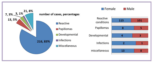
Non neoplastic lesions; this group consists of 260 cases. 142 (54.6%) cases reported in female patients and 118 (45.4%) in male patients. The mean age was 33.5 years with a SD of 18.27 years. The most common diagnosis was pyogenic granuloma (110, 42.3%) followed by peripheral giant cell granuloma (45, 17.3%) and fibrous hyperplasia (36, 13.8%). Table (1) demonstrates the frequency and distribution of non neoplastic soft tissue lesions according to age and gender. The exact site was reported in 181 cases as following: 72 (39.77%) cases in anterior maxilla, 41 (22.65%) cases in posterior maxilla and 68 (37.56%) cases in palate. The side was reported in 106 cases as following: 52 (49.1%) in the right side, 50 (47.2%) in the left side, three cases in midline (2.8%) and one case bilaterally (0.9%). In regard to diseases subcategories, Reactive condition was the most common group 216 (83%) followed by papilloma 13 (5%). All subcategories showed higher frequency in females than males. Figure (1) shows the frequency and distribution of soft tissue non neoplastic diseases subcategories and according to gender.
| Frequency | Gender | Age (years) | |||||||
|---|---|---|---|---|---|---|---|---|---|
Diagnosis |
N | % | M | F | M:F | Mean | SD | Min | Max |
| pyogenic granuloma | 110 | 42.3 | 47 | 63 | 0.7 | 31.69 | 17.5 | 2 M | 80 |
| Peripheral giant cell granuloma | 45 | 17.3 | 23 | 22 | 1.04 | 33.4 | 19.18 | 3 | 72 |
| fibrous hyperplasia | 36 | 13.8 | 19 | 17 | 1.1 | 38.38 | 19.04 | 9 | 70 |
| nonspecific inflammation | 17 | 6.5 | 8 | 9 | 0.8 | 38 | 20.26 | 6 | 67 |
| Peripheral ossifying fibroma | 16 | 6.2 | 8 | 8 | 1 | 32.56 | 13.08 | 13 | 55 |
| squamous papilloma | 13 | 5 | 5 | 8 | 0.6 | 37.3 | 18.95 | 7 | 65 |
| chronic hyperplastic gingivitis | 6 | 2.3 | 2 | 4 | 0.5 | 40.66 | 24.19 | 1 | 68 |
| vascular malformation | 5 | 1.9 | 2 | 3 | 0.6 | 34.8 | 24.36 | 12 D | 61 |
| Others* | 5 | 1.9 | 3 | 2 | 1.5 | 29 | 6.2 | 24 | 38 |
| Gingival fibromatosis | 4 | 1.5 | 0 | 4 | 13.5 | 7.5 | 4 | 22 | |
| Infections* | 3 | 1.2 | 1 | 2 | 0.5 | 23 | 21.5 | 2 | 45 |
| Total | 260 | 100 | 118 | 142 | 0.8 | 33.5 | 18.27 | 12 D | 80 |
Infections: actinomycosis, noma, tuberculosis (one case of each)
Others: denture induced fibrous hyperplasia, intramucosal nevus, plasma cell gingivitis, verruciform xanthoma and lymphangioma (one case of each)
Table1: The frequency and distribution of non neoplastic maxillary soft tissue lesions according to gender and age.

Figure1: the frequency and distribution of non neoplastic disease subcategories and according to gender
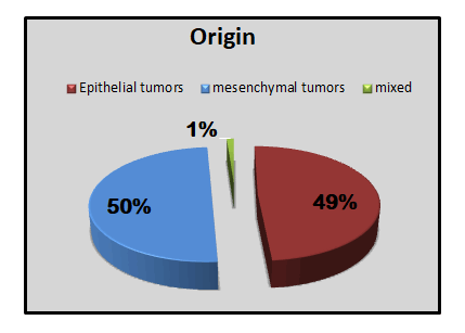
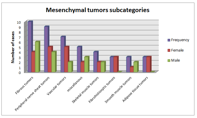
Maxillary tumors and tumors like lesions; this group consists of 88 cases. Female patients were affected more 47 (53.4%) than males 41 (46.6%) with a male to female ratio of 0.87:1. The mean age was 44.96 years with a SD of 23.17 years. Most cases were malignant in nature 48 (54.5%), while benign tumors were 37 (42%) cases and three cases of dysplasia (3.4%). The most common diagnosis was squamous cell carcinoma (38, 43.2%) followed by giant cell fibroma (9, 10.2%) and neurofibroma (9, 10.2%). Table (2) demonstrates the frequency and distribution of maxillary soft tissue tumors and tumors like lesions according to gender and age. In regard to origin, there were almost an equal percentage of mesenchymal tumors (50%) and epithelial tumors (49%) with one case being of mixed origin (mature teratoma) as shown in figure (2). In regard to mesenchymal tumors subcategories, fibrous tumors were the most common 10 (22.7%), followed by peripheral nerve sheath tumors 9 (20.45%) and vascular tumors 7 (15.9%) as shown in figure (3). In regard to site, 33 cases reported in general as maxillary lesions, 12 cases reported in anterior maxilla, 9 cases in posterior maxilla, 31 cases in the palate, and 3 cases involving both maxilla and maxillary sinus. The Side was reported in 35 cases and as following: left side 18 (51.4%), right side 16 (45.7%) and one case in the midline (2.9%).
| Frequency | Gender (N) | Age (years) | |||||||
|---|---|---|---|---|---|---|---|---|---|
| Diagnosis | N | % | M | F | M:F | Mean | SD | Min | Max |
| Benign tumors | 37 | 42 | 15 | 22 | 0.68 | 29.7 | 18.15 | 48 D | 74 |
| Giant cell fibroma | 9 | 10.2 | 6 | 3 | 2 | 20.55 | 10.17 | 10 | 36 |
| Neurofibroma | 9 | 10.2 | 4 | 5 | 0.8 | 40.1 | 16 | 29 | 74 |
| Hemangioma | 5 | 5.7 | 1 | 4 | 0.25 | 36.8 | 20.5 | 11 | 60 |
| Other benign tumors* | 4 | 4.5 | 1 | 3 | 0.3 | 13.5 | 27 | 48 D | 54 |
| Fibrous histiocytoma | 3 | 3.4 | 0 | 3 | 18.66 | 1.15 | 18 | 20 | |
| Lipoma | 3 | 3.4 | 0 | 3 | 44.3 | 18 | 31 | 65 | |
| Hobnail hemangioma | 2 | 2.3 | 1 | 1 | 1 | 22.5 | 3.5 | 20 | 25 |
| Leiomyoma | 2 | 2.3 | 2 | 0 | 41 | 5.65 | 37 | 45 | |
| Dysplasia | 3 | 3.4 | 2 | 1 | 2 | 65.66 | 16 | 50 | 82 |
| Malignant tumors | 48 | 54.5 | 24 | 24 | 1 | 55.4 | 20.2 | 4 | 82 |
| Oral SCC | 38 | 43.2 | 18 | 20 | 0.9 | 62.4 | 11.8 | 39 | 82 |
| Rhabdomyosarcoma | 4 | 4.5 | 2 | 2 | 1 | 8.5 | 5.06 | 4 | 15 |
| Undifferentiated pleomorphic sarcoma | 2 | 2.3 | 2 | 0 | 45.5 | 20.5 | 31 | 60 | |
| Fibrosarcoma | 1 | 1.1 | 0 | 1 | 33 | . | 33 | 33 | |
| leiomyosarcoma | 1 | 1.1 | 0 | 1 | 20 | . | 20 | 20 | |
| melanoma | 1 | 1.1 | 1 | 0 | 33 | . | 33 | 33 | |
| metastatic carcinoma | 1 | 1.1 | 1 | 0 | 76 | . | 76 | 76 | |
| Total | 88 | 100 | 41 | 47 | 0.87 | 44.96 | 23.17 | 48 D | 82 |
Other benign tumors: congenital granular cell epulis, mature teratoma, melanotic neuroectodermal tumor of infancy, oral focal mucinosis (one case of each).
Table2: The frequency and distribution of maxillary soft tissue tumors and tumors like lesions according to gender and age.

Figure2: Piechart showing the distribution of maxillary soft tissue tumors and tumors like lesions according to origin.

Figure3: the frequency and distribution of mesenchymal tumors according to subcategories and gender.
Discussion
The great majority of non neoplastic soft tissue lesions in the maxilla were reactive (pyogenic granuloma, fibrous hyperplasia, chronic hyperplastic gingivitis, denture induced fibrous hyperplasia, peripheral giant cell granuloma, peripheral ossifying fibroma, plasma cell gingivitis, verruciform xanthoma) in responses to local irritation usually in sites vulnerable to trauma such as the gingiva and palate. In general reactive lesions are considered the most common soft tissue pathologies in oral cavity with fibrous hyperplasia and pyogenic granuloma account for the majority of diagnoses and females appear to be more commonly affected .
Pyogenic granuloma was the most common diagnosis in this group. The main affected site was the anterior gingiva. The site, predominance among females and age distribution is consistent with previous studies [5]. The high prevalence in female patients especially during pregnancy is well documented in literature, a previous study on those lesions demonstrate no increase or difference in number of estrogen receptors on endothelial cells compared to controls; however more expression of endothelial growth factors, other angiogenesis factors and fewer inhibitory factors was noted and it might play a role [8]. Although there is a debate regarding the nature of this lesion; our study followed the WHO classification of head and neck 2017 and since most of the cases occurred in anterior gingiva; this lesion was included in the reactive category.
In regard to neoplasms; Malignant tumors were more than benign with squamous cell carcinoma being the most frequently diagnosis and the most common malignant tumor of the maxilla. This may be related to the increasing prevalence of tobacco smoking and alcohol drinking as it is becoming more popular in our conservative country. Although most studies often used data from oral squamous cell carcinoma with no specific regard to localization, few studies were found that focused on SCC of the hard palate, maxillary gingiva and alveolar mucosa. Those previous studies reported a mean age of (73, 71, 66, 64, and 67) years respectively, while our study mean age was 62 years with the majority of cases reported in patients older than 70 years old that is similar . Females were slightly more affected in contrast to but similar.
In regard to benign tumors, giant cell fibroma and neurofibroma were the most common tumors. A mean age of 40 years was found in neurofibroma which is similar to previous studies that reported a mean age of (38.5 and 40) years respectively and females were slightly more affected that is similar with Campos et al but disagree with Tamiolakis et al.
Giant cell fibroma is a lesion with unknown etiology that doesn’t appear to have a relation with chronic irritation and thus was designated in this study as a tumor like lesion. A male predilection was noted (M:F ratio 2:1), other studies had reported slight female predilection or no gender differences. However their results had also shown that lesions affecting palate or upper gingiva were more likely to affect male patients which support our finding.
conclusion
The demographic characteristics of soft tissue lesions in maxilla are similar to that seen in other parts of oral cavity; reactive conditions are the most common lesions and squamous cell carcinoma is the most common malignancy. True mesenchymal tumors are generally rare in oral cavity however they showed equal frequency to epithelial tumors in maxilla which pose a challenge and require careful assessment when approaching the diagnosis in this location.
References
- Hassona Y, Al Boosh D, Al Saed A et al. The range of pathological diagnoses of oral diseases in Jordan: An 11-year-retrospective study. Saudi J Oral Sci. 2020;7:151.
- Dutra KL, Longo L, Grando LJet al. Incidence of reactive hyperplastic lesions in the oral cavity: a 10 year retrospective study in Santa Catarina, Brazil☆.Braz J Otorhinolaryngol. 2019;85:399-407.
- Dhanuthai K, Rojanawatsirivej S, Thosaporn W et al. Oral cancer: A multicenter study. Med Oral Patol Oral Cir Bucal. 2018;23:23.
- Solar AA, Jordan RC. Soft tissue tumors and common metastases of the oral cavity. Periodontology 2000. 2011;57:177-97.
- Al-Khateeb T, Ababneh K. Oral pyogenic granuloma in Jordanians: a retrospective analysis of 108 cases. J Oral Maxillofac Surg. 2003 Nov 1;61:1285-8.
Author Info
Shahad Abbas Waheed* and Taghreed F. Zaidan
Department of Oral diagnosis, College of dentistry, Baghdad University, Baghdad, IraqCitation: Maxillary soft tissue lesions (An analysis of 348 cases), J Res Med Dent Sci, 2021, 9(12): 1-4
Received: 12-Nov-2021 Accepted: 25-Nov-2021 Published: 30-Nov-2021
