Research - (2021) Volume 9, Issue 6
Micro-Computed Tomographic Evaluation of Shaping Ability and Dentinal Microcracks Formation After Using New Heat-Treated Nickel-Titanium Systems
Abdulhakim Al Gamdi and Shibu Thomas Mathew*
*Correspondence: Shibu Thomas Mathew, Department of Endodontics, Riyadh Elm University, Kingdom of Saudi Arabia, Email:
Abstract
Introduction: This ex vivo study aims to evaluate the shaping ability and dentine microcracks formation after using four different heat-treated files, EdgeTaper PlatinumTM, ProTaper GoldTM, Reciproc® Blue, and EdgeOne FireTM Files.
Methods: Forty extracted mandibular premolars were randomly selected. Samples were decoronated at 17mm from the apex with water cooling. Bruker© Skyscanner scanned the canal before and after instrumentation.
For each group (DataViewer program version 1.5.6.2, Bruker©micro-CT), the manufacturer's evaluation software calculated the percentage rate and morphometric measurements of the percentage of walls untreated.
Results: 65% continuous and 60% reciprocating files showed Type II defect in the apical third. The reciprocating motion files (Reciproc®Blue and EdgeOne FireTM) produced significantly fewer dentin microcracks than continuous motion files (E.T.P and PTG). In addition, Reciproc®Blue shows more removal of tooth structure than other files. Statistically, no significant difference detected when comparing all systems together regarding shaping ability, pre-shaping (p=0.427), after shaping (p=0.865), and dentin microcracks at coronal 3rd, middle third, and apical third (all p>0.05), thus proving the null hypothesis.
Conclusions: All the newer NiTi rotary instrumentation systems studied resulted in dentine damage in varying degrees and dentinal cracks resulting from the preparation procedures.
Keywords
Shaping ability, Dentin microcracks, Nickel-titanium rotary instrument
Introduction
Chemo-mechanical preparation aims to remove dentin's inner layer, enabling irrigants to reach the entire length of the root canal, eliminating or reducing bacterial irritants that cause apical periodontitis, and finally preparing the root canal for a suitable and adequate root canal filling [1]. Metallurgic advancements in NiTi rotary files have facilitated the mechanical preparation of root canal space and helped accomplish more predictably with less iatrogenic risk [2], by retaining the primary root canal morphology and maintaining the total dentin thickness. During instrumentation, untreated walls of the canal space can possess bacterial microorganisms that act as sources of infection [3]. Therefore, novel file developments increase contact with canal walls and facilitate apical cleaning and maintain the root canal and cervical dentin [4]. At times, failure of treatment might be due to inevitable damage to the root dentine during instrumentation, causing dentinal microcracks and minute fracture, thereby opening a portal for microorganisms [5]. Root fractures might also occur due to microcracks or craze line that propagates with repeated stress application by occlusal forces [6].
The 3-dimensional, non-destructive, high-resolution X-ray micro-computed tomographic (Micro-CT) imaging is considered the gold standard [5]. In the field of Endodontics, Micro-CT has created new possibilities by facilitating the use of the same sample for various tests without sample destruction, which is incredibly significant while evaluating pre- and post-instrumentation volume, root canal obturation quality, or high-resolution removal of the material from the root retreatment than clinical scanners. Hence, Micro-CT avoids the risk of repeated scanning and manipulating images [7].
While much research on the efficacy of rotary and reciprocating systems is ongoing, there is still inadequate information on the shaping ability of new heat-treated endodontic files'(EdgeTaper PlatinumTM files ProTaper GoldTM files, Reciproc® Blue files, and EdgeOne FireTM Files).
To date, no study investigated the influence of these new heat-treated files on the canal preparation technique and the occurrence of dentinal microcracks using the nondestructive methodology. Therefore, the purpose of this study was to compare and evaluate the shaping ability, incidence, and formation of microcracks in the dentine after using four different heat-treated NiTi files with reciprocating and continuous motion rotary systems.
The null hypothesis is that there will be no significant differences in the shaping ability parameters and microcracks between continuous and reciprocating motion.
Materials and Method
Forty single-rooted, sound, lower premolars, extracted for orthodontic reasons, were randomly selected. Samples were preserved in alcohol and glycerin media in a transparent plastic container to hydrate dentin until used. Periapical radiographs were taken from the mesiodistal and buccolingual planes to confirm the presence of a single canal. Teeth with a straight canal with mature apical foramen and not endodontically treated were selected.
The teeth lengths were measured using a digital caliper (Digital Vernier Caliper, Pearl – Japan) and decoronated at 17 mm from the apex using a low-speed saw (BUEHLER Isomet Low-Speed Saw) underwater cooling to ensure length standardization. Negotiation of the canal with size #8,10 and 15 hand K-files (Dentsply Maillefer, Switzerland). After removing gross pulpal tissue, working length was established by advancing the file into the canal until visible at the apical foramen and then subtracting 1 mm.
Subsequently, the specimens were imaged with a micro- CT scanner using Bruker© SkyScan 1172 micro-CT (Bruker SkyScan, Kontich, Belgium) (Figure 1). Scanner configuration used was as follow: (95 kV voltage, 104 μA anode current, 158 ms exposure time, 25 μm image pixel size, Al filter, 0.3 rotation step for 360° angle, frame averaging of 4 to improve the signal to noise ratio and random movement of 8 to minimize ring artifacts). In addition, a Flat-field correction was performed before the scanning procedure to correct variations in the camera pixel sensitivity.
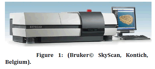
Figure 1. Figure 1: (Bruker© SkyScan, Kontich, Belgium).
Samples wrapped in aluminum foil inserted in the putty impression (Figure 2). After the putty impression sets, the aluminum foil peeled off. Its space in the block was replaced with silicone impression material (Genie-Sultan Healthcare) to simulate the PDL. The tooth returned into the block, and the excess impression material cut off using a surgical blade, and whole tooth placed in mounting model (Figure 3).
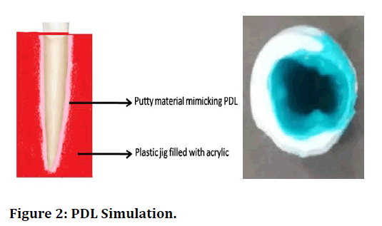
Figure 2. PDL Simulation.
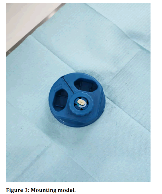
Figure 3. Mounting model.
The samples randomly divided into four groups (Figure 4).
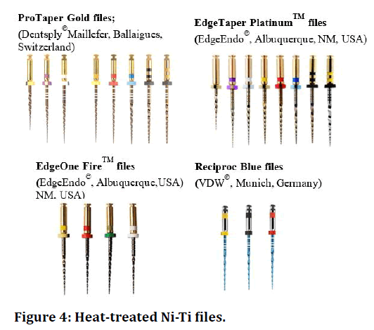
Figure 4. Heat-treated Ni-Ti files.
Group 1A(n=10) instrumented by ProTaper® GOLD files (SX, S1, S2, F1, F2, F3 & F4). (Dentsply© Maillefer, Ballaigues, Switzerland) (Continuous system).
Group 1B(n=10) instrumented by EdgeTaper PlatinumTM files (SX, S1, S2, F1, F2, F3 & F4). (EdgeEndo©, USA) (Continuoussystem).
Group 2A (n=10) instrumented by EdgeOne FireTM files (small, primary, medium, and large). (EdgeEndo©, USA) (Reciprocating system).
Group 2B (n=10) instrumented by Reciproc® Blue files (R25 & R40). (VDW©, Munich, Germany) (Reciprocating system).
A single operator with knowledge and experience in all systems performed all root canal instrumentation. According to manufacturers ' instructions, the rotary machine Dentsply© X-smart plus motor, force, torque set, and instrumentation performed in each group. Each file prepared three root canals maximum, and the instrument flutes cleaned before inserting the file into the next root canal. Canals irrigated with 2.25% sodium hypochlorite using a 30G needle inserted 1mm shorter than working length. Subsequently, teeth rinsed with 1 ml 17% EDTA accompanied by 1 ml 2.25 % sodium hypochlorite for 1 minute, respectively, between the use of each instrument or after three packing motions. Finally, the canals rinsed with 1 ml of normal saline and sterile paper point to dry the canals.
The scanning performed the same way as done preoperatively. After scanning, a reconstruction of the projected images conducted using the N-Recon® program version 1.6.9.4 (Bruker© Skyscan, Kontich, Belgium) to produce reconstructed cross-section images. Numerical parameters needed to obtain the best image results were checked and adjusted.
A ring artifact reduction of 5 for nonuniformity of the background image taken by the x-ray camera; 25% beam hardening compensation to prevent the specimen from appearing artificially more dense at or near its surface and less dense at its central parts; and a smoothing of 2 using Gaussian kernel was applied. A 16- bit TIF file format was the choice for saving the images because of the variety of densities comprising the specimen.
Reconstructed images were loaded to the Dataviewer® (Bruker© Skyscan, Kontich, Belgium) software to determine image quality, reorient, resize, and visually inspect defects and potential new cracks and after instrumentation.
Finally, 3D visualization and production of color-coded images of the samples obtained using CTVol© 2.3.2.0 (Bruker© Skyscan, Kontich, Belgium).
Definition of dentinal microcracks
Cracks identified as: (No defect) no cracks, (Type I) cracks on the inner canal wall not reaching the outer surface of the canal, (Type II) cracks from the outer surface of the root but not reaching the inner canal wall, and (Type III) cracks extending from the inner canal wall and reaching the outer surface, and types recorded in an alphabetic code as shown in Figure 5. (Pictures from micro-CT).
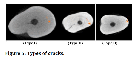
Figure 5. Types of cracks.
Dentinal defects evaluation
Reconstructed images transferred to the software DataViewer (version 1.5.6.2, Bruker© micro-CT). Threedimensional re-imaging was performed on each sample to obtain coronal, sagittal, and trans-axial axis images. The cross-sectional images of the samples before and after preparation were analyzed.
Distribution of microcracks
Using software DataViewer (version 1.5.6.2, Bruker© micro-CT), samples are marked horizontally from the apex(5.6mm) till coronal, and the number of microcrack in each group analysed (Figure 6).
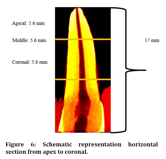
Figure 6. Schematic representation horizontal section from apex to coronal.
Cutting efficiency evaluation
The recorded images were processed using the software DataViewer (version 1.5.6.2, Bruker© micro-CT) to calculate quantitative parameters and construct visual 3D models. The volume of interest for each specimen was noted by integrating regions of interest in all crosssections. The grayscale range was required to recognize the dentin before and after instrumentation was determined by using a density histogram with the global threshold method. Accuracy of the segmentation ensured using a comparison between original segmented scans. Results expressed as the change in volumetric analysis.
Percentage of untreated wall
The recorded images were processed using the software DataViewer (version 1.5.6.2, Bruker© micro-CT) to calculate the volume of untreated walls. Results expressed in terms of the percentage of untreated walls per sample.
Statistical analysis
Statistical Packages for Software Sciences (SPSS) (version 21 Armonk, New York, IBM Corporation) perform all statistical analyses for this project. Data are shown as mean and standard deviation and as numbers and percentages. Between comparison, Fischer Exact tests applied, P-value <0.05 accepted as the significant level for all statistical tests.
More removal of tooth structure was in Reciproc Blue files (12.1 ± 2.98). However, no statistically significant differences were seen between the E.T.P, PTG, Reciproc®Blue, and EdgeOneFireTM with pre-shaping (p=0.427), after shaping (p=0.865), and the untreated wall (Table 1).
| Variables | EdgeTaper Platinum Mean ± SD | ProTaper Mean ± SD | Reciproc blue Mean ± SD | Eugene fire Mean ± SD | P-value § |
|---|---|---|---|---|---|
| Before shaping ability | 8.63 ± 3.29 | 7.99 ± 2.11 | 9.86 ± 2.89 | 8.39 ± 1.95 | 0.427 |
| After shaping ability | 11.6 ± 3.16 | 11.7 ± 2.52 | 12.1 ± 2.98 | 11.0 ± 2.62 | 0.865 |
| Untreated wall | 0.55 ± 0.38 | 0.44 ± 0.55 | 0.49 ± 0.54 | 0.70 ± 0.98 | 0.828 |
Table 1: Inter-comparison between edgetaper platinumtm, protaper® gold, reciproc®blue and edgeone firetm of shaping ability using new heat-treated ni-tisystems.
In comparison between continuous and reciprocating motion files, slightly more removal of teeth structure was seen in reciprocating motion files but was not statistically significant difference before in shaping (p=0.331) after shaping (p=0.899) and untreated wall (p=0.614) (Table 2 and Figure 7).
| Variables | Continuous Mean ± SD | Reciprocal Mean ± SD | P-value § |
|---|---|---|---|
| Before shaping ability | 8.31 ± 2.72 | 9.13 ± 2.51 | 0.331 |
| After shaping ability | 11.7 ± 2.78 | 11.6 ± 2.79 | 0.899 |
| Untreated wall | 0.49 ± 0.47 | 0.60 ± 0.78 | 0.614 |
Table 2: Comparison between reciprocal motion and continuous motion of shaping ability using new heattreated ni-tisystems.
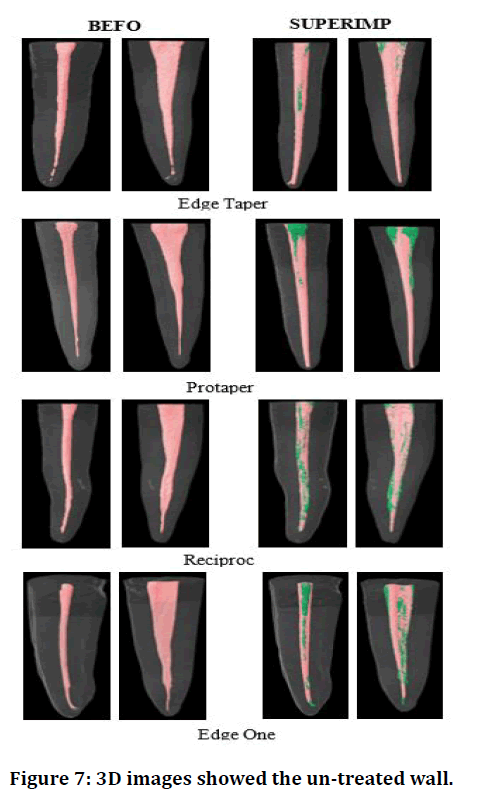
Figure 7. 3D images showed the un-treated wall.
No defect seen in Reciproc Blue and Edge One Fire files, and only 10% of Type I defects observed in Edge Taper Platinum and Protaper Gold files.
In the middle third, Reciproc Blue files showed 90% No defect, followed by Edge one Fire files (60%), Protaper gold (50%), Edge taper platinum (30%). 50% of Type I defects were observed in Edge Taper Platinum files, followed by Protaper Gold (40%), Reciproc Blue (10%). Type I and II defects were found together in 20% of Edge Taper Platinum files.
In the apical third, 70% of Type I defects in Reciproc Blue and Protaper Gold files, followed by 60% and 50% ofType I defect seen in Edge Taper Platinum and Edge one fire files.
However, there were no statistically significant differences between E.T.P. PTG, Reciproc®Blue, and Edge OneFireTM to coronal 3rd, middle third, and apical third (all p>0.05) (Table 3).
| Variables | Edge taper platinum N (%) | Protaper gold N (%) | Reciproc blue N (%) | Eugene fire N (%) | P-value § |
|---|---|---|---|---|---|
| Coronal 3rd | |||||
| No defect | 09 (90.0%) | 09 (90.0%) | 10 (100%) | 10 (100%) | 1 |
| Type I | 01 (10.0%) | 01 (10.0%) | 0 | 0 | |
| Middle 3rd | |||||
| No defect | 03 (30.0%) | 05 (50.0%) | 09 (90.0%) | 06 (60.0%) | 0.084 |
| Type I | 05 (50.0%) | 04 (40.0%) | 01 (10.0%) | 04 (40.0%) | |
| Type III | 0 | 01 (10.0%) | 0 | 0 | |
| Type I and II | 02 (20.0%) | 0 | 0 | 0 | |
| Apical 3rd | |||||
| No defect | 03 (30.0%) | 03 (30.0%)0 | 03 (30.0%) | 04 (40.0%) | 0.949 |
| Type I | 06 (60.0%) | 07 (70.0%) | 07 (70.0%) | 05 (50.0%) | |
| Type II | 0 | 0 | 0 | 01 (10.0%) | |
| Type I and II | 01 (10.0%) | 0 | 0 | 0 | |
Table 3: Comparison between edgetaper platinum, protaper gold, reciproc blue, and edgeonefire after using new heat-treated Ni-Ti systems.
In comparison between Continuous and reciprocations motion files, reciprocating files produced No defect, and 10% of files showed Type I defect.
In the middle third, 75% of No defect was seen in Reciprocating files, while 45% of Type I defect seen in continuous files. Type I defect was seen in the Apical third, using both continuous (65%) and reciprocation (60%) motion files. However, statistically, the defects are seen between continuous and reciprocal motions among coronal 3rd, middle third, and apical third were not positively significant (p>0.05)(Table 4 and Figure 8).
| Variables | Continuous N (%) | Reciprocal N (%) | P-value § |
|---|---|---|---|
| Coronal 3rd | |||
| No defect | 18 (90.0%) | 20 (100%) | 0.487 |
| Type I | 02 (10.0%) | 0 | |
| Middle 3rd | |||
| No defect | 08 (40.0%) | 15 (75.0%) | 0.07 |
| Type I | 09 (45.0%) | 05 (25.0%) | |
| Type III | 01 (05.0%) | 0 | |
| Type I and II | 02 (10.0%) | 0 | |
| Apical 3rd | |||
| No defect | 06 (30.0%) | 07 (35.0%) | 1 |
| Type I | 13 (65.0%) | 12 (60.0%) | |
| Type II | 0 | 01 (05.0%) | |
| Type I and II | 01 (05.0%) | 0 | |
§ P-value calculated using Fischer Exact test.
Table 4: Comparison between Reciprocal motion and continuous motion after using new heattreated Ni-Ti systems.
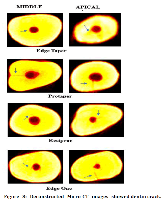
Figure 8. Reconstructed Micro-CT images showed dentin crack.
Discussion and Conclusion
One of the most important objectives during root canal instrumentation is the complete elimination of debris and microorganisms from the root canal system [1]. The present study evaluated the shaping ability, incidence, and Type of micro-cracks formed using four newly advanced endodontic heat-treated NiTi (superelastic) files with two different motions on extracted human lower premolar teeth, using Micro-CT.
Study methodologies by Hin, et al. Milani, et al. and Arias, et al. [8-10] indicates that the lack of periodontal ligament and bone around the teeth could change the clinical aura and the internal forces applied on root structure during shaping procedures [11]. Hence, in this present study, putty was used to simulate periodontal ligament and mimic the clinical environment. The comparison between two motions and intercomparison between files showed no statistically significant differences before and after instrumentation, like the studies done by Stringheta et al. Serefoglu et al. and Hwang et al. [12-14]. However, Reciproc® Blue showed more removal of dentin volume, corresponding to the study done by Busquim et al. [15], where their observations showed the same as the present study. In untreated walls, anatomical challenges like the isthmus, recesses, and flattened root canals, bacterial retention can serve as a source of persistent infection [3]. Thus it required suboptimal mechanical action, such as enhancing the efficiency of the irrigating solution, the physical action of ultrasonic activation, and intracanal dressing for complete debridement and disinfection of the canal.
Certain studies showed that dentinal defects could be affected by numerous factors like the physical properties of teeth [16], instrumentation with rotary and reciprocating Ni-Ti instruments causing dentinal cracks [17], and vertical root fractures [18,19].
High stress concentration in the walls of the root canal system caused by NiTi rotary instruments during root canal preparation increased the risk of dentinal damage [20], which explains the increased formation of micro-cracks apical third followed by the middle third, then in the coronal third.
According to Yoldas et al.[6], the formation of dentinal microcracks could be due to the tip of rotary instruments, the cross-section geometry, the taper type (constant or gradual), constant or variable step, and finally, the form of the cannelure.
However, in the present study, E.T.P and PTG (triangular cross-sections [21], Reciproc® Blue (Sshaped cross-sections [22], and EdgeOne FireTM (parallelogram with two cutting edges [23] showed no statistically significant differences in the formation of dentinal cracks despite their different cross-sectional designs or the sequence of rotary and reciprocating instrument files used.
The taper of the files could be a contributing factor in dentinal crack formation [24], and a greater taper means reduced dentinal thickness. Wilcox [18] mentioned that the likelihood of root fracture increased with increased removal of tooth structure. Root canal instrumentation with larger instruments is associated with crack development and propagation of existing cracks [25], which explains the increased dentinal cracks in the apical third due to its less dentinal thickness compared to the middle and coronal third. Burklein et al. [26] used Reciproc® files up to size 40/0.06, whereas Liu et al. [17] used up to 25/ 0.08, which must have contributed to their conflicting results. In our study, the files chosen had similar taper, and the canals instrumented up to size 40, 0.06; using E.T.P and EdgeOne FireTM (taper 0.05), and PTG and Reciproc® Blue files (0.06 taper).
According to Kansal et al. [27], a reciprocating motion produced fewer dentinal cracks than the conventional continuous rotation, which resulted in a high level of stress concentrations on root canal walls [26] than the reciprocating motion, which targeted towards the center of the canal, facilitating equal stress distribution by Franco et al. and Yared et al. [28,29].
In this study, continuous rotation showed more crack formation with Type II defect at the apical third but not positively significant, as observed in most samples (65% continuous and 60% reciprocating). In Accordance with Wilcox classification, the present study showed that the E.T.P. and PTG had the same Type II defect in the apical third (in 6 and 7 samples respectively) and comparing between Reciproc® Blue and EdgeOneFireTM, Type II defect observed at the apical third (7 and 5 samples, respectively) as in the previous studies done [30,31]; and no significant differences observed between rotary and reciprocating systems [27,32,33].
In another study by Liu et al. [17], Reciproc® files working in reciprocating movement caused cracks in only 5% of teeth, whereas OneShapeTM and ProTaper® files working in continuous rotation caused cracks in 35% and 50% of teeth, respectively, which in accordance to our study revealed that continuous motion showed more dentin micro-crack at the apical third. Thus, all the newer NiTi rotary instrumentation systems studied resulted in dentine damage and cracks in varying degrees due to the preparation procedures.
Increased dentin cracks were observed in the apical third (Type II), irrespective of the file system, with PTG and Edge Taper showing the highest crack formation and least by EdgeOneFireTM followed by Reciproc® Blue file system.
Our findings showed no significant difference between rotary and reciprocating Ni-Ti file systems, and the null hypothesis proved.
Future research comparing these systems in severely curved canals and incidence of microcracks after other endodontic procedures such as filling, retreatment, and post preparations reviewed.
References
- González-Rodrı́guez MP, Ferrer-Luque CM. A comparison of profile, hero 642, and k3 instrumentation systems in teeth using digital imaging analysis. Oral Surg Oral Med Oral Pathol Oral Radiol Endodont 2004; 97:112-115.
- Shen Y, Coil JM, Zhou H, et al. H y F lex nickel–titanium rotary instruments after clinical use: metallurgical properties. Int Endod J 2013; 46:720-9.
- Ricucci D, Siqueira JF. Apical actinomycosis as a continuum of intraradicular and extraradicular infection: case report and critical review on its involvement with treatment failure. J Endod 2008; 34:1124-9.
- De Melo Ribeiro MV, Silva-Sousa YT, Versiani MA et al. Comparison of the cleaning efficacy of self-adjusting file and rotary systems in the apical third of oval-shaped canals. J Endod 2013; 39:398-401.
- Peters OA, Laib A, Göhring TN .Changes in root canal geometry after preparation assessed by high-resolution computed tomography. J Endodont 2001; 27:1-6.
- Yoldas O, Yilmaz S, Atakan G .Dentinal microcrack formation during root canal preparations by different NiTi rotary instruments and the self-adjusting file. J Endodont 2012; 38:232-5.
- Marciano MA, Duarte MA, Ordinola-Zapata R. Applications of micro-computed tomography in endodontic research. Current microscopy contributions to advances in science and technology. Badajoz, Spain. Formatex Research Center 2012; 782-788.
- Hin ES, Wu MK, Wesselink PR, et al. Effects of self-adjusting file, mtwo, and protaper on the root canal wall. J Endodont 2013; 39:262-4.
- Milani AS, Froughreyhani M, Rahimi S, et al. The effect of root canal preparation on the development of dentin cracks. Iranian Endodont J 2012; 7:177.
- Arias A, Lee YH, Peters CI, et al. Comparison of 2 canal preparation techniques in the induction of microcracks: A pilot study with cadaver mandibles. J Endodont 2014; 40:982-985.
- Shemesh H, Bier CA, Wu MK, et al. The effects of canal preparation and filling on the incidence of dentinal defects. Int Endodont J 2009; 42:208-13.
- Stringheta CP, Bueno CE, Kato AS. Micro‐computed tomographic evaluation of the shaping ability of four instrumentation systems in curved root canals. Int Endodont J 2019; 52:908-16.
- Serefoglu B, Piskin B. Micro-computed tomography evaluation of the self-adjusting file and protaper universal system on curved mandibular molars. Dent Mater J 2017; 28:2016-55.
- Hwang YH, Bae KS, Baek SH. Shaping ability of the conventional nickel-titanium and reciprocating nickel-titanium file systems: A comparative study using micro-computed tomography. J Endodont 2014; 40:1186-9.
- Busquim S, Cunha RS, Freire L, et al. A micro‐computed tomography evaluation of long‐oval canal preparation using reciprocating or rotary systems. Int Endodont J 2015; 48:1001-6.
- Ertas H, Sagsen B, Arslan H. Effects of physical and morphological properties of roots on fracture resistance. Eur J Dent 2014; 8:261.
- Liu R, Hou BX, Wesselink PR. The incidence of root microcracks caused by three different single-file systems versus the ProTaper system. J Endodont 2013; 39:1054-6.
- Wilcox LR, Roskelley C, Sutton T. The relationship of root canal enlargement to finger-spreader induced vertical root fracture. J Endodont 1997; 23:533-4.
- Tsesis I, Rosen E, Tamse A. Diagnosis of vertical root fractures in endodontically treated teeth based on clinical and radiographic indices: a systematic review. J Endodont 2010; 36:1455-8
- Kim HC, Lee MH, Yum J .Potential relationship between the design of nickel-titanium rotary instruments and vertical root fracture. J Endodont 2010; 36:1195-9.
- Braga LC, Silva AC, Buono VT . Impact of heat treatments on the fatigue resistance of different rotary nickel-titanium instruments. J Endodont 2014; 40:1494-7.
- Burklein S, Benten S, Schafer E. Quantitative evaluation of apically extruded debris with different single-file systems: Reciproc, F360 and OneShape versus Mtwo. Int Endodont J 2014; 47:405–9.
- Gambarini G, Galli M, Di Nardo D . Differences in cyclic fatigue lifespan between two different heat-treated NiTi endodontic rotary instruments: Waveone gold vs. edgeone fire. J Clin Exp Dent 2019; 11:e609.
- Bier CA, Shemesh H, Tanomaru-Filho M. The ability of different nickel-titanium rotary instruments to induce dentinal damage during canal preparation. J Endodont 2009; 35:236-8.
- Çapar İD, Uysal B, Ok E. Effect of the size of the apical enlargement with rotary instruments, single-cone filling, post space preparation with drills, fiber post removal, and root canal filling removal on apical crack initiation and propagation. J Endodont 2015; 41:253-6.
- Bürklein S, Hinschitza K, Dammaschke T, et al. Shaping ability and cleaning effectiveness of two single‐file systems in severely curved root canals of extracted teeth: Reciproc and waveone versus mtwo and protaper. Int Endodont J 2012; 45:449-61.
- Kansal R, Rajput A, Talwar S. Assessment of dentinal damage during canal preparation using reciprocating and rotary files. J Endodont 2014; 40:1443-6.
- Franco V, Fabiani C, Taschieri S. Investigation on the shaping ability of nickel-titanium files when used with a reciprocating motion. J Endodont 2011; 37:1398-401
- Yared G. Canal preparation using only one Ni‐Ti rotary instrument: Preliminary observations. Int Endodont J 2008; 41:339-44.
- Karataş E, Gunduz HA, Kırıcı DÖ, et al. Dentinal crack formation during root canal preparations by the twisted file adaptive, protaper next, protaper universal, and waveone instruments. J Endodont 2015; 41:261-264.
- Pop I, Manoharan A, Zanini F, et al. Synchrotron light-based μCT to analyse the presence of dentinal microcracks post-rotary and reciprocating NiTi instrumentation. Clin Oral Investigation 2015; 19:11-6.
- Ashwinkumar V, Krithikadatta J, Surendran S, et al. Effect of reciprocating file motion on microcrack formation in root canals: An SEM study. Int Endodont J 2014; 47:622-7.
- Çiçek E, Koçak MM, Sağlam BC, et al. Evaluation of microcrack formation in root canals after instrumentation with different NiTi rotary file systems: A scanning electron microscopy study. Scanning 2015; 37:49-53.
Author Info
Abdulhakim Al Gamdi and Shibu Thomas Mathew*
Department of Endodontics, Riyadh Elm University, Riyadh, Kingdom of Saudi ArabiaCitation: Abdulhakim Al Gamdi, Shibu Thomas Mathew,Micro-Computed Tomographic Evaluation of Shaping Ability and Dentinal Microcracks Formation After Using New Heat-Treated Nickel-Titanium Systems , J Res Med Dent Sci, 2021, 9(6): 346-354
Received: 31-May-2021 Accepted: 21-Jun-2021
