Case Report - (2022) Volume 10, Issue 5
Morphological Variation of Formation of Median and Musculocutaneous Nerve with Relation to the Axillary and Brachial Artery
Jayakumari S*, rishnaveni Sharth and Asimitha Ramu
*Correspondence: Jayakumari S, Department of Anatomy, Sree Balaji Medical College and Hospital, Bharath Institute of Higher Education and Research, Chromepet, Chennai, Tamil nadu, India, Email:
Abstract
The knowledge of variant formation of brachial plexus and its branches are important for the surgeon and orthopedician pertaining to trauma and surgical procedures of the upper extremity. Current study was aimed to report variations in the formation of the median nerve with relation to the brachial artery. And also, the variation in the branching pattern of Musculo Cutaneous Nerve (MCN). Careful observation was made to note the formation and branching pattern of Median (MN) and Musculo Cutaneous Nerve (MCN) with the relation of axillary and brachial artery of formalin embalmed cadaver. In the present study, we noted the MN thus formed by 2 roots; lying behind the brachial artery. The coracobrachialis muscle was innervated by the direct branch from the lateral cord of brachial plexus. Unusual variant in the formation of median nerve in relation to the brachial artery may be important diagnoses to neurologist and surgeons.Keywords
Median nerve, Musculocutaneous nerve, Axillary artery, Brachial artery, Profunda brachial arteryIntroduction
Brachial plexus is a group of nerves which innervates the muscles of the back and the upper limb. The ventral rami of the lower four cervical (C5,C6,C7,C8) and first Thoracic (T1) spinal nerves unite together and form the plexus [1]. The lateral cord is formed by the anterior divisions of the upper and middle trunks and the anterior division of the inferior trunk continues as a medial cord. From the Lateral Cord (LC) there are three branches arises they are, Lateral Pectoral Nerve (LPN), Musculocutaneous Nerve (MCN) and lateral root of median nerve. The LPN supplies the pectoral muscles. The MCN pierces the coracobrachialis muscle and also innervate the three muscles of the arm. The lateral root of median nerve (roots C5, C6 and C7) and medial root of median nerve (roots C8 T1) join to form the median nerve. These two roots are joins anterior to the axillary artery. Then it’s decent down word lateral to the axillary artery. In the middle of the arm, it runs medial to the brachial artery [2].
But in the present study the MCN is not piercing the coracobrachialis muscle; neither the lateral and medial roots are not encircling the axillary artery. The MN formed by encircling profunda brachial artery lying posterior to the brachial artery.
Purpose of this study to highlight the variant formation of MCN and MN its relation with brachial artery. These variations are more vulnerable during the surgical producers of axilla and upper arm. Complex nature of the brachial plexus and its relation with brachial artery has been interesting area for the anatomists as well as for clinicians. So, this observation will be use full for orthopaedic and vascular surgeon.
Case Presentation
During routine dissection, an abnormal formation of MN and different branching pattern of MN was observed incidentally in left upper limb of a 55 years old male cadaver assigned to the anatomy department of the Sree Balaji Medical College and hospital, Chennai.
The course and branching pattern of the MN and MCN in the arm and fore arm were carefully dissected.
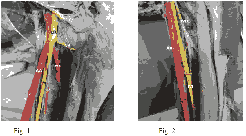
Variation in formation of MN and MCN were documented and appropriate photographs were taken (Figure 1).
Figure 1: lateral root and medial root surrounds the profunda Brachial Artery. LR-Lateral Root, Axillary Artery, CB-branch to Coraco Brachialis muscle, PBA-Profunda Brachial Artery, M-Median nerve,MCN-Musculo Cutaneous Nerve
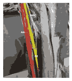
In our case on the left side of the arm, we observed medial root of median cord and lateral root of lateral cord encircling the profunda brachial artery to form the MN. It is formed approximately 2.8 cm distal to the pectorals minor muscle and lying lateral to the anterior circumflex humeral artery. The profunda brachii artery arises 4.2 cm distal to the lower border of pectoralis minor muscle. Then the MN was the brachial artery till cubital fossa. It receives an accessary branch from the MCN before reaching the cubital fossa. The communicating branch was 1.5 cm long and had an oblique course. The brachial artery runs to the median nerve. Generally musculocutaneous nerve enters arm by piercing into the coracobrachialis muscle. In this case the MCN instead of piercing the coracobrachialis muscle, it adhered to the MN for certain distance the arm. The nerve is lying medial to the coracobrachialis muscle. However, the motor branch to the coracobrachialis muscle arose from the lateral cord (Figure 2).
Figure 2: Median nerve receives a branch from musculocutaneous nerve, MCN-Musculo Cutaneous nerve, AA-Axillary artery, M Median nerve.
The MCN divided into three branches innervating the brachialis and biceps brachii muscle of the arm. It also communicates with the median nerve by an oblique branch. In the forearm and hand the course and branching pattern of the two nerves was normal. Thebranching pattern and course of the median and musculocutaneous nerve were normal in right side in every aspect
Results and Discussion
An interesting finding which was observed in the present case was relationship between MN with the brachial artery. Normally the medial root of median nerve and lateral root of median nerve unite to the axillary artery to form the MN Experts [3]. Observed the medial cord lying behind the axillary artery. It gives ulnar nerve and medial root of the median nerve which emerged on the medial side of the artery.
In our study the medial cord is normal in position. Ulnar nerve is medial side of the axillary artery and brachial artery. But the medial root of median nerve and the lateral root of MN join together behind the brachial artery to form the MN. These two roots are encircling the profunda brachial artery. The MN was lying posterior to the brachial artery throughout the course in the arm.
A number of variations in the course and distribution of the MCN nerve have been reported. Usually, the MCN arises from the lateral cord of brachial plexus, pierces the coracobrachialis muscles and then pass down ward between the biceps and brachialis muscles. Near to the cubital fossa, MCN comes to the surface laterally to the biceps brachii muscle and anterior to the brachialis muscle, known as the lateral cutaneous nerve of forearm. Then it descends along the lateral margin of the forearm and gives cutaneous branches to the lateral surface of the forearm [1].
Experts found an unusual variation in MCN nerves in 6.25% cases and its absence has been reported with prevalence ranging from 1.7% to 15% [4]. Absences of the MCN have been described in many reports and experts reported these variations in 65% of the population [5]. But in our case the coracobrachialis muscle was supplied by the direct branch from the lateral cord. Brachialis and the biceps brachii muscle were supplied by the MCN.
According to Grey H, the MCN sends an independent branch to the coracobrachialis muscle instead of perforating [6].
The remaining branches of the nerve runs parallel to the MN by a variable distance until it pass under the biceps brachii. In our study instead of piercing the coracobrachialis muscle, the nerve adheres to the median nerve for some distance down in the arm and has several branches, passes between the biceps and brachialis muscle to supply muscles.
A small twig arises from the trunk supply the coracobrachialis muscle. Number of literatures are available regarding the variations of brachial plexuses; there have been attempts to classify such variations. In this case, our variation like Type III, in which neither the nerve nor the communicant branch perforate the coracobrachialis muscle [5].
Conclusion
The present variation should be considered when the MN lying behind the brachial artery. It is encircling the profunda brachial artery. Moreover, knowledge of these variations is important in performing anaesthetic block to brachial plexus or in surgical procedures in the neck and upper arm region.
References
- Standring S, Ellis H, Healy JC, et al. Gray’s Anatomy In: Pectoral Girdle, Shoulder Region and Axilla, 41th Ed, Elsevier Churchill Livingstone. Phi. 2005; 846-848
- David J, Harold E. Gray’s Anatomy. In: Pectoral Girdle, Shoulder Region and Axilla, 40th ed, New York. Churchill Livingstone Elsevier. 2008;791-822
- Pandey SK, Shukla VK. Anatomical Variations of the Cords of Brachial Plexus and the Median Nerve. Clin Anat 2007; 20:150-156.
- Bhattarai C, Poudel PP. Unusual variation in musculocutaneous nerves in Nepalese. Kathmandu Univ Med J 2009; 7:408-410. [Crossref]
- Hansen K.B. Uber Varietaten Des Nervus Medianus Und Des Nervus Musculocutaneus Und Deren Beziehungen. Anat Anz 1955; 102:187-203.
- Gray H, Goss CM. Gray anatomia, 29a ed, Guanabara Koogan; Rio de Janeiro. 1977.
Author Info
Jayakumari S*, rishnaveni Sharth and Asimitha Ramu
Department of Anatomy, Sree Balaji Medical College and Hospital, Bharath Institute of Higher Education and Research, Chromepet, Chennai, Tamil nadu, IndiaCitation: Jayakumari S, Krishnaveni Sharth, Asimitha Ramu. Morphological Variation of Formation of Median and Musculocutaneous Nerve with Relation to the Axillary and Brachial Artery , J Res Med Dent Sci, 2022, 10 (5): 32-34.
Received: 23-Feb-2022, Manuscript No. 41495; , Pre QC No. 41495; Editor assigned: 25-Feb-2022, Pre QC No. 41495; Reviewed: 11-Mar-2022, QC No. 41495; Revised: 25-Apr-2022, Manuscript No. 41495; Published: 03-May-2022


