Research - (2020) Advances in Dental Surgery
Morphometric Analysis of Intracranial Clivus in South Indian Skulls
Thiru Kumaran and Yuvaraj Babu K*
*Correspondence: Yuvaraj Babu K, Department of Anatomy, Saveetha Dental College and Hospital, Saveetha Institute of Medical and Technical Sciences, Saveetha University, Chennai, India, Email:
Abstract
The clivus is a bony part of the cranium at the skull base, a shallow depression behind the dorsum sellae that slopes obliquely backward and downwards to the foramen magnum. It forms a gradual sloping process at the anterior most portion of the basilar occipital bone at its junction with the sphenoid bone. On axial planes, it sits just posterior to the sphenoid sinuses. The pons and medulla lie on the clivus. The aim of this study was to morphometrically analyse the significance of the inner surface of the clivus in the South Indian skulls, The study was done in 30 unsexed South Indian dry cranial cavities from the Department of Anatomy, Saveetha Dental College and Hospitals. The length and breadth of the internal surface of the clivus was measured using a vernier caliper and the angle was measured with the help of a goniometer. From our study the average breadth of the clivus was 24.05 ± 1.23 mm, average length was found to be 35.30 ± 3.24 mm and the average angle made by the clivus to the base of the skull was 38.15°± 2.54°. Our morphometric analysis of length, breadth and the angle formed may provide useful anatomical, anthropometrical, and surgical data of clivus for the surgeons in planning surgeries on the skull base as many important neurovascular structures are present in this region.
Keywords
Clivus, Intracranial clivus length, Intracranialclivus breadth, Basiocciput, Basisphenoid
Introduction
The clivus is a bony component present in the cranium of the skull base, a shallow depression behind the dorsum sellae which slopes obliquely towards backward and downwards to foramen magnum [1]. It forms a gradual sloping process at the anterior most portion of the membrane bone at its junction with the sphenoid [2]. On axial planes, it sits just posterior to the sphenoid air sinuses, the pons and medulla sit on the clivus along with the basilar artery and basilar plexus of vein [3]. The clivus may be an important landmark for checking for anatomical atlantooccipital alignment; the clivus, when viewed on a lateral Cervical spine X-ray, forms a line which, if extended, is believed to be Wackenheim'sclivus line. Just lateral to the clivus bilaterally is the foramen lacerum, the clivus forms the central skull base. It is formed by the synostosis of the basisphenoid and basioccipital [4].
During early development, the axial sclerotomes of the first somites are integrated into the skull base to create the basioccipital part of the clivus [4]. Neurenteric cysts of the clivus are uncommon developmental lesions that occur as a result of notochordaldysgenesis during embryonic development, they typically occur within the posterior fossa, occurring typically midline anterior to the brainstem or within the cerebellopontine angle [5,6]. Clival tumors are rare tumors that arise within the clivus, several bones at the underside of the skull between the occipital and sphenoid bones [7]. This area is surrounded by essential structures and nerves of the brainstem and important arteries, just like the internal carotid arteries and basilar arteries [7,8]. For several decades neurosurgeons are challenged by lesions of skull base especially involving the clivus as various neurovascular structures are related to this complex anatomical region It is important to know the precise morphometry of the skull base to facilitate surgical approaches and neurosurgical operations in this area.
With a rich case bank established over 3 decades we have been able to publish extensively in our domain [9-19]. Based on this inspiration we aim to morphometricallyanalyse the significance of the inner surface of the clivus in the South Indian skulls, we measured length and breadth in mm and also measured the angle formed by the clivus to the skull base.
Materials and Methods
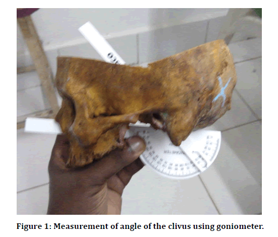
The study was done in 30 unsexed South Indian dry cranial cavities from the Department of Anatomy, Saveetha Dental College and Hospitals. The length and breath of the internal surface of the clivus was measured using a vernier caliper and the angle of the clivus was measured with the help of a goniometer (Figure 1). Length was measured from the top of the Clivus till the anterior margin of foramen magnum and breadth was measured from one side to another side at the top of the clivus. All measurements were tabulated and statistically analysed.

Figure 1: Measurement of angle of the clivus using goniometer.
Results and Discussion
Range and average measurements of length, breadth, and angle of clivus are mentioned in Table 1.
| Range | Average | |
|---|---|---|
| Breadth in mm | 22-31 | 24.05 ± 1.23 |
| Length in mm | 29-45 | 35.30 ± 3.24 |
| Angle in Degree | 31°-47° | 38.15° ± 2.54° |
Table 1: Range and average measurements of length, breadth, and angle of clivus.
From our study the average breadth of clivus was 24.05 ± 1.23 mm, average length was found to be 35.30 ± 3.24 mm and the average angle made by clivus to the base of skull was 38.15° ± 2.54°. In the study done by Ji et al. the average clival length, the widest and narrowest breadth of the clivus were 26, 33 and 19 mm, respectively [20]. The clivus lies at an angle of 45° from the vertical which is higher than the average angle 38.15° from our study [21]. It is clear from the study that Clivus in most of the skulls are approximately of the same range,the parameters obtained in the present study will be helpful for anyone contemplating the use of clival screws. Lot of especially important neurovascular structures and cranial nerves are related to the inner aspect of the clivus; hence surgeons need to be overly cautious not to damage any of these structures during any surgical procedures in this area.
Conclusion
Our morphometric analysis of length, breadth and the angle formed may provide useful anatomical, anthropometrical, and surgical data of clivus for the surgeons in planning surgeries on the skull base as many important neurovascular structures are present in this region.
Acknowledgement
We acknowledge the department of anatomy for allowing us to use skulls from their collection for our study.
Conflict of Interest
The author declares that there is no conflict of interest in the present study.
References
- Cannizzaro D, Tropeano MP, Milani D, et al. Microsurgical versus endoscopic trans-sphenoidal approaches for clivus chordoma: a pooled and meta-analysis. Neurosurg Rev 2020; 43:3.
- Labidi M, Watanabe K, Bouazza S, et al. Clivus chordomas: A systematic review and meta-analysis of contemporary surgical management. J Neurosurg Sci 2016; 60:476–484.
- Samii M, Knosp E. Introduction: The clivus. Approaches to the clivus. Springer 1992; 1–6.
- Samii M, Knosp E. Dorsolateral approach to the clivus and foramen magnum. Approaches to the clivus. 1992; 117–128.
- Khan N, Zumstein B. Transverse clivus fracture: Case presentation and significance of clinico-anatomic correlations. Surg Neurol 2000; 54:171–177.
- Burkart CM, Theodosopoulos PV, Keller JT, et al. Endoscopic transnasal approach to the clivus: A radiographic anatomical study. Laryngoscope 2009; 119:1672–1678.
- https://radiologyassistant.nl/
- Chan JYW, Wong STS, Wei WI. Surgical salvage of recurrent T3 nasopharyngeal carcinoma: Prognostic significance of clivus, maxillary, temporal and sphenoid bone invasion. Oral Oncol 2019; 91: 85–91.
- Abdul Wahab PU, Nathan PS, Madhulaxmi M, et al. Risk factors for post-operative infection following single piece osteotomy. J Maxillofacial Oral Surg 2017; 16:328–332.
- Eapen BV, Baig MF, Avinash S. An assessment of the incidence of prolonged postoperative bleeding after dental extraction among patients on uninterrupted low dose aspirin therapy and to evaluate the need to stop such medication prior to dental extractions. J Maxillofacial Oral Surg 2017; 16:48–52.
- Patil SB, Durairaj D, Kumar GS, et al. Comparison of extended nasolabial flap versus buccal fat pad graft in the surgical management of oral submucous fibrosis: A prospective pilot study. J Maxillofacial Oral Surg 2017; 16:312–321.
- Jain M, Nazar N. Comparative evaluation of the efficacy of intraligamentary and supraperiosteal injections in the extraction of maxillary teeth: A randomized controlled clinical trial. J Contemporary Dent Prac 2018; 19:1117–1121.
- Marimuthu T, Devadoss P, Kumar SM. (2018) Prevalence and measurement of anterior loop of the mandibular canal using CBCT: A cross sectional study. Clin Implant Dent Related Res 2018; 20:531–534.
- Marimuthu M, Andiappan M, Wahab A, et al. Canonical wnt pathway gene expression and their clinical correlation in oral squamous cell carcinoma. Indian J Dent Res 2018; 29:291–297.
- Wahab PA, Madhulaxmi M, Senthilnathan P, et al. Scalpel versus diathermy in wound healing after mucosal incisions: A split-mouth study. J Oral Maxillofacial Surg 2018; 76:1160–1164.
- Abhinav RP, Selvarasu K, Maheswari GU, et al. The patterns and etiology of maxillofacial trauma in South India. Annals Maxillofacial Surg 2019; 9:114–117.
- Ramadorai A, Ravi P, Narayanan V. Rhinocerebral mucormycosis: A prospective analysis of an effective treatment protocol. Annals Maxillofacial Surg 2019; 9:192–196.
- Senthil Kumar MS, Ramani P, Rajendran V, et al. Inflammatory pseudotumour of the maxillary sinus: Clinicopathological report. Oral Surg 2019; 12:255–259.
- Sweta VR, Abhinav RP, Ramesh A. Role of virtual reality in pain perception of patients following the administration of local anesthesia. Annals Maxillofacial Surg 2019; 9:110–113.
- Ji W, Tong J, Huang Z, et al. A clivus plate fixation for reconstruction of ventral defect of the craniovertebral junction: a novel fixation device for craniovertebral instability. Eur Spine J 2015; 24:1658–1665.
- Erdem H, Kizilkanat ED, Boyan N, et al. Anatomy and clinical importance of the extracranial clivus and surrounding structures. Int J Morphol 2018; 557–562.
Author Info
Thiru Kumaran and Yuvaraj Babu K*
Department of Anatomy, Saveetha Dental College and Hospital, Saveetha Institute of Medical and Technical Sciences, Saveetha University, Chennai, IndiaCitation: Thiru Kumaran, Yuvaraj Babu K, Morphometric Analysis of Intracranial Clivus in South Indian Skulls, J Res Med Dent Sci, 2020, 8 (7): 147-149.
Received: 15-Sep-2020 Accepted: 16-Oct-2020 Published: 23-Oct-2020
